http://journals.tubitak.gov.tr/medical/ © TÜBİTAK
doi:10.3906/sag-1507-193
Effects of different drug treatments on the proliferation of human ovarian carcinoma cell
line MDAH-2774
Şule AYLA1,2,*, Ayhan BİLİR3, Şenol ERTÜRKOĞLU4, Gamze TANRIVERDİ4, B. Cem SONER5, Kenan SOFUOĞLU6, Laura GHISOLFI7, Gülperi ÖKTEM8
1Department of Histology and Embryology, School of Medicine, İstanbul Medipol University, İstanbul, Turkey 2Regenerative and Restorative Medical Research Center (REMER), İstanbul Medipol University, İstanbul, Turkey
3Department of Histology and Embryology, School of Medicine, İstanbul Aydın University, İstanbul, Turkey 4Department of Histology and Embryology, Cerrahpaşa Medical Faculty, İstanbul University, İstanbul, Turkey
5Department of Medical Pharmacology, Faculty of Medicine, Necmettin Erbakan University, Konya, Turkey 6Medistate Hospital, İstanbul, Turkey
7Beth Israel Deaconess Medical Center, Harvard Medical School, Boston, MA, USA 8Department of Histology and Embryology, School of Medicine, Ege University, İzmir, Turkey
1. Introduction
Ovarian cancer is called ‘the silent killer’ because it does not have any obvious symptoms such as fatigue, weight change, abdominal distention, and pain. The lack of efficient and early detection is the reason for its high mortality rate (1). Ovarian cancer is the fourth leading cause of cancer death in women and has the highest mortality rate among all gynecological cancers. The lifetime risk of ovarian cancer is one in 60 women in industrial countries, but it is less common among Asian and African women. Similar to other solid tumors, the growth and metastasis of ovarian cancer requires a chain of events. These events include deterioration of the intercellular structure, changes in cellular adhesion, cell migration, invasion, multiplication, and new blood vessel formation (2).
Because of vague symptoms and inadequate screening methods in the early stages of ovarian cancer, more than 60% of patients are diagnosed at an advanced stage (3). Surgical and cytotoxic treatments often show limited benefits. Thus, agents that sensitize ovarian cancer cells to cytotoxic treatments have been investigated. For example, gemcitabine is a pyrimidine analog of deoxycytidine. Its anticancer effect results from preventing DNA synthesis through inhibition of polymerase and ribonucleotide reductase enzymes (4). Gemcitabine is one of the most commonly used chemotherapeutic agents in epithelial ovarian cancer (5). Gemcitabine increases the sensitivity of tumor cells to chemotherapy and prolongs the duration of inhibition of DNA synthesis (4). In non-small-cell lung cancer, gemcitabine and the combination of etoposide and
Background/aim: In this study, the effects of resveratrol as a natural polyphenol compound, gemcitabine as an antimetabolite that
has nucleoside structure analogous to deoxycytidine, and para-aminophenol-derived paracetamol were investigated with single and combined applications in monolayers of the MDAH-2774 human ovarian cancer cell line.
Materials and methods: Drugs were evaluated in cell culture with respect to cell proliferation, cell cytotoxicity (trypan blue dye
exclusion test), synthesis phase of cell cycle, and cell structure in 24, 48, 72, and 96 h.
Result: Resveratrol and gemcitabine diminished both cell proliferation and cell cycle synthesis phase indication in monolayer cell
cultures (P < 0.05). All combination groups showed similar effects that were mainly more effective in respect to single usage of resveratrol and gemcitabine in monolayer cell cultures.
Conclusion: The effects of gemcitabine, resveratrol, and paracetamol were investigated in monolayers of the MDAH-2774 human
ovarian cancer cell line and a decrease in cell number in cell cycle synthesis phase, prevention of cell proliferation, and destruction of cell structure were observed.
Key words: Ovarian cancer, gemcitabine, paracetamol, resveratrol
Received: 31.07.2015 Accepted/Published Online: 11.01.2018 Final Version: 30.04.2018
cisplatin show similar tumor inhibition efficacy. Twenty percent of ovarian cancer patients with acquired resistance to cisplatin respond to treatment with gemcitabine.
Paracetamol/acetaminophen (N-acetyl-para-aminophenol) is used classically to relieve pain, fever, and malaise (6,7). This drug has also been found to have some beneficial effects against cancer. High-dose paracetamol can enhance chemotherapy activity against different cancer cells in vitro and in vivo (8–10).
Various natural and synthetic materials are used in cancer therapy to inhibit cell proliferation and trigger apoptosis. Resveratrol (trans-3,4’-trihydroxystillbene), a naturally occurring polyphenolic antioxidant found in grapes and red wine, elicits diverse biochemical responses and shows antiaging, antiinflammatory, and antiproliferative effects in several cell types (11,12). The molecular mechanisms of resveratrol’s inhibitory effect on cellular signaling pathways have been described previously (13).
The aim of the current study is to evaluate the in vitro effects of gemcitabine, paracetamol, and resveratrol alone and in combination on the time- and dose-dependent proliferation and survival of human ovarian carcinoma cell line MDAH-2774.
2. Materials and methods
2.1. Cell culture conditions and reagents
Human MDAH-2774 ovarian carcinoma cells (CRL No: 10303; American Type Culture Collection, Manassas, VA, USA) were cultured in RPMI-1640 medium (Biological Industries, Beit Haemek, Israel) supplemented with 10% heat-inactivated fetal calf serum (GIBCO, Invitrogen Co., Paisley, UK), 100 units/mL penicillin (Sigma Chemical Co., St Louis, MO, USA), and 100 µg/mL streptomycin (Sigma Chemical Co.). Semiconfluent cells were harvested from flasks using 0.05% trypsin (Sigma Chemical Co.), following the addition of RPMI-1640 for trypsin inactivation, and resuspended in culture medium.
2.2. IC50 and cytotoxicity
MDAH-2774 cells were seeded (5 × 10⁵ cells/well) in
six-well plates and cultured for 24, 48, 72, or 96 h in the presence of gemcitabine at 0.1, 1, 10, and 100 µg/mL and resveratrol at 0.1, 1, 10, and 100 µM in 5 mL of media. Paracetamol was added at 40 µg/mL. At the end of each culturing period, cells were trypsinized (0.05%) and centrifuged at 1500 rpm. Cell viability was determined by the trypan blue exclusion assay. Cells (5 × 10⁵ cells/well
with 100% vitality) were then plated in six-well culture plates containing 5 mL of RPMI-1640 medium. The concentrations of each drug required for 50% inhibition (IC50) at 96 h were 10 µg/mL for gemcitabine, 10 µM for resveratrol, and 40 µg/ml for paracetamol.
MDAH-2774 cells were seeded at 5 × 10⁵ cells/well in
six-well plates and treated with gemcitabine (10 µg/mL), resveratrol (10 µM), paracetamol (40 µg/mL), gemcitabine + resveratrol (10 µg/mL + 10 µM), or gemcitabine + paracetamol (10 µg/mL + 40 µg/mL) for 24, 48, 72, and 96 h. A buffer-treated control group was also included. At each time point, cells were harvested and counted. The number of dead cells was assessed using the trypan blue exclusion assay.
2.3. Immunocytochemistry and cell proliferation
MDAH-2774 cells (1 × 10⁵ cells/well) were seeded on
cover slips in 24-well plates. After 2 h, cells were treated with 10 µM resveratrol, 10 µg/mL gemcitabine, 40 µg/mL paracetamol, gemcitabine + resveratrol (10 µg/mL +10 µM), or gemcitabine + paracetamol (10 µg/mL + 40 µg/ ml). After 24, 48, 72, and 96 h, the cells were incubated with BrdU at 37 °C for 1 h and then in PBS for 15 min. Cells were fixed in 70% ethanol at –20 °C for 30 min and then prepared for analysis.
Cells were rehydrated for 10 min in PBS and then incubated in 0.5% H2O2 in methanol for 20 min in the dark. A 4 N HCl solution was then applied to the samples for 30 min and the cells were rinsed with distilled water, followed by three washes with PBS. After blocking 15 min in Ultra-V-Block (Thermo Fisher Scientific, Marietta, OH, USA), coverslips were incubated with a primary antibody solution (NCL-BrdU mouse monoclonal 1:100, Santa Cruz Biotechnology, Santa Cruz, CA, USA) for 1 h in a humidified environment at room temperature. After rinsing with PBS, a secondary antibody solution (Santa Cruz Biotechnology) was applied for 30 min. The samples were rinsed with PBS and incubated with streptavidin peroxidase for 30 min in a humidified environment. Coverslips were then rinsed with PBS, incubated in substrate-chromogen (AEC) for 20 min, and washed with distilled water. Cells were stained with Mayer’s hematoxylin (Sigma Chemical Co.) and the coverslips were mounted (Ultramount, Dako, Carpinteria, CA, USA) on slides.
2.4. Electron microscopy
MDAH-2774 cells (1 × 10⁵ cells/well) were seeded on
coverslips in 24-well plates. After 2 h, cells were treated with 10 µM resveratrol, 10 µg/mL gemcitabine, 40 µg/ mL paracetamol, gemcitabine + resveratrol (10 µg/mL + 10 µM), or gemcitabine + paracetamol (10 µg/mL + 40 µg/mL) in 1 mL. The drugs were removed after 24 or 96 h, and a 2.5% glutaraldehyde solution was added. After incubation for 30 min at 4 °C, samples were washed twice with phosphate solution and incubated in 0.5 mL of osmium tetroxide for 60 min. This was followed by another wash with phosphate solution. Cells were then successively incubated in 30%, 50%, 70%, and 90% ethanol for 10 min each. This was followed by two incubations of 10 min each in 100% ethanol. Cells were next incubated
successively for 10 min in a solution of 1:3 amyl acetate/ alcohol, 1:1 amyl acetate/alcohol, 3:1 amyl acetate/alcohol, and 100% amyl acetate for 1 h. Coverslips were air-dried at room temperature and cells were visualized using a JMS 5200 scanning electron microscope (JEOL, Peabody, MA, USA).
2.5. Statistical analysis
SPSS 10.0 was used for all analyses. Because the data did not show a homogeneous distribution, the nonparametric Kruskal–Wallis test was used to calculate significance between more than two independent groups. Further analysis of the significant groups was performed using the Mann–Whitney U test. P < 0.05 was considered significant.
3. Results
3.1. Cell proliferation
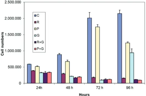
To study the effect of different types of drug treatments on the number of viable ovarian cancer cells, MDAH-2774 cells were treated with resveratrol, paracetamol, gemcitabine, gemcitabine + paracetamol, or gemcitabine + resveratrol for 24, 48, 72, or 96 h and then stained with trypan blue. As expected, the total number of MDAH-2774 cells increased with time. As shown in Figure 1, treatment with resveratrol or paracetamol alone had no effect on cell proliferation within the first 24 h (P > 0.05), but it decreased cell proliferation at the later time points when compared to the control group (P < 0.05). Treatment with gemcitabine alone significantly inhibited cell proliferation at each time point when compared to the control group (P < 0.05). Treatment with gemcitabine + paracetamol, or gemcitabine + resveratrol also significantly inhibited cell
proliferation at each time point when compared to the control group (P < 0.05) (Figure 1).
3.2. Cytotoxicity data
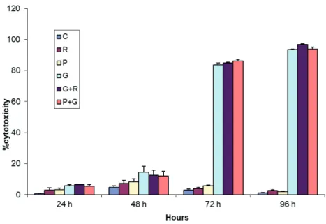
MDAH-2774 cells treated with resveratrol, paracetamol, gemcitabine, gemcitabine + paracetamol, or gemcitabine + resveratrol for 24, 48, 72, or 96 h were stained with trypan blue to evaluate the effects of the drug treatments on viability. In the first 24 h of treatment, none of the drugs applied alone or in combination induced significant cell death (P > 0.05) (Figure 2). However, significantly fewer viable cells (unstained cells) were detected after 48, 72, and 96 h in all treatment groups compared to the control. Gemcitabine, applied alone or in combination with resveratrol or paracetamol, caused the greatest cytotoxicity (P < 0.05) (Figure 2).
3.3. BrdU labeling index
To understand more precisely the effect of the different drug treatments, proliferating MDAH-2774 cells on coverslips were treated with resveratrol, paracetamol, gemcitabine, gemcitabine + paracetamol, or gemcitabine + resveratrol for 24, 48, 72, or 96 h and stained with BrdU (Figures 3a–3l). As expected, many cells in the control group were in the active DNA synthesis phase (BrdU-positive cells) (Figures 3a and 3g). Resveratrol treatment had a small effect (P > 0.05) on proliferation in the first 24 h followed by a significant decrease of the BrdU labeling index (BrdU-LI) at 48, 72, and 96 h (P < 0.05) compared to the control group (Figures 3b, 3h, and 4). Interestingly, in samples treated with paracetamol, the BrdU-LI percentage was significantly lower than in the control group (P < 0.05) only at 24 h, while it was higher at the remaining time points (Figures 3c, 3i, and 4).
Figure 1: Cell proliferation (C: control, R: resveratrol, P: paracetamol, G: gemcitabine, R
+ G: resveratrol + gemcitabine, P + G: paracetamol + gemcitabine). Data are expressed as mean ± SD.
Incubation of MDAH-2774 cells with gemcitabine (Figures 3d and 3j), alone or in combination with resveratrol (Figures 3e and 3k) or paracetamol (Figures 3f and 3l), strongly inhibited cell proliferation at all time points when compared to the control group (P < 0.05) (Figure 4).
3.4. Effect of different drug treatments on cell morphology
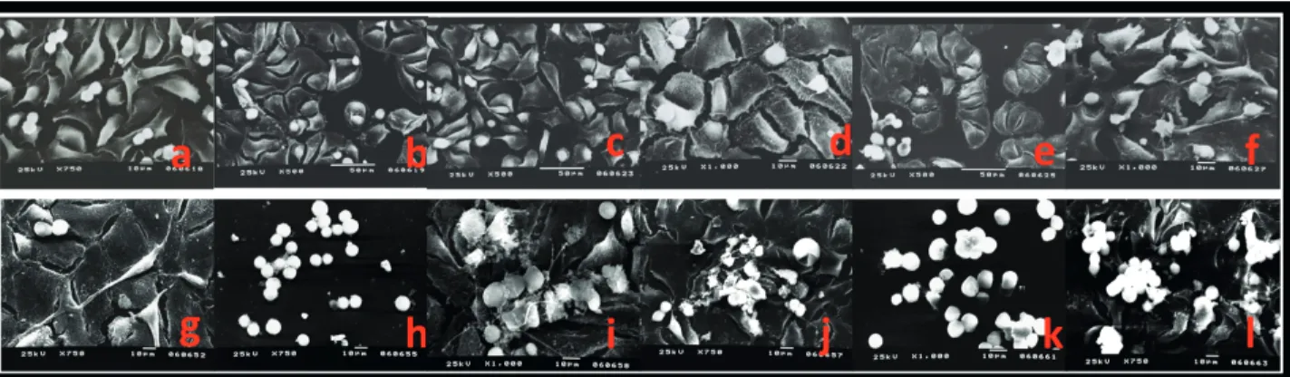
MDAH-2774 cells treated with resveratrol, paracetamol, gemcitabine, gemcitabine + paracetamol, or gemcitabine + resveratrol were collected after 24 or 96 h and processed for scanning electron microscopy (Figures 5a–5l). At 96
h, the cells in the control group appeared healthy, many of them in mitosis, with a flat shape and exhibiting normal interactions with neighboring cells. Intact membranes with long thin cytoplasmic extensions and microvilli were observed (Figures 5a and 5g). Cells treated with resveratrol for 24 h were morphologically similar to those in the control group but showed a decreased number of cytoplasmic extensions. Additionally, many cells were blocked in mitosis. After 96 h, cells treated with resveratrol were loosely attached with a preapoptotic appearance (Figures 5b and 5h). In the paracetamol treatment group,
Figure 2: Cytotoxicity (viability) (C: control, R: resveratrol, P: paracetamol, G:
gemcitabine, G + R: resveratrol + gemcitabine, P + G: paracetamol + gemcitabine). Data are expressed as mean ± SD.
Figure 3. Immunocytochemistry (BrDU). All at magnification of 40×. a–f: 24 h; g–l: 96 h; a: 24 h control group, where a large number of
MDAH-2774 ovary tumor cells marked with BrDU (red color) were observed (unmarked cells are blue); b: resveratrol; c: paracetamol; d: gemcitabine, with fewer marked cells than the control; e: resveratrol + gemcitabine; f: paracetamol + gemcitabine; g: 96 h control group, where results are similar to those of the 24 h control group; h: resveratrol, with fewer marked cells than at 24 h; i: paracetamol; j: gemcitabine, with fewer marked cells than the control group; k: resveratrol + gemcitabine, with fewer marked cells than the control group; l: paracetamol + gemcitabine.
cells showed an apoptotic phenotype that was especially evident at 96 h (Figures 5c and 5j). Treatment with gemcitabine alone disrupted the cell morphology with a loss of cytoplasmic extensions and dull villi. This phenotype was particularly evident at 96 h. Cells also presented membrane blebbing, a sign of apoptosis (Figures 5d and 5j). Combination treatment of resveratrol and gemcitabine caused the loss of cellular cytoplasmic extensions and a transition to a prespheroid shape at 24 h. At 96 h, more round and aggregated cells, loosely attached to the surface and without cytoplasmic extensions, were observed. In addition, compared to the control group, many fewer mitotic cells were recorded (Figures 5e and 5k). In the cells treated with paracetamol and gemcitabine, major changes were observed in the cell morphology. The vast majority of
the cells had lost their microvilli and the connections with other cells were decreased. The number of apoptotic cells was higher than in the control group (Figures 5f and 5l).
In summary, all treatments caused a loss of cytoplasmic extensions, a decrease in mitotic cells, and an increase in apoptotic cells.
4. Discussion
The use of natural products including medicinal plants has become more and more important in primary health care especially in developing countries. Many pharmacognostical and pharmacological investigations are carried out to identify new drugs or to find new lead structures to develop novel therapeutic agents for the treatment of human diseases such as cancer (14).
Figure 4: BrDU labeling index (C: control, R: resveratrol, P: paracetamol, G: gemcitabine, G + R:
resveratrol + gemcitabine, P + G: paracetamol + gemcitabine). Data are expressed as mean ± SD.
Figure 5. Scanning electron microscopy. All magnification except that of k is 750×. a–f: 24 h; g–l: 96 h; a: 24 h control group; b: resveratrol;
c: paracetamol; d: gemcitabine; e: resveratrol + gemcitabine; f: paracetamol + gemcitabine; g: 96 h control group; h: resveratrol, where degenerated tumor cells with a round appearance are evident; i: paracetamol tumor cells appear more degenerated compared to 24 h; j: gemcitabine, where tumor cells are more damaged compared to 24 h; k: resveratrol + gemcitabine, where damaged and rounded cells are evident, 1000×; l: paracetamol + gemcitabine, where damaged, rounded, and apoptotic cells are evident.
Our results show that treatment of MDAH-2774 cells with resveratrol, paracetamol, and gemcitabine, separately or in combination, significantly inhibited cell proliferation at all the considered time points, although to a lesser extent in the first 24 h. In addition, all treatments, applied alone or in combination, induced significant cell death at 48, 72, and 96 h, with gemcitabine showing the strongest effect either alone or combined with resveratrol or paracetamol.
Recent studies have shown that agents like resveratrol, found in high concentrations in grape seeds and skin and well known for its antioxidant properties, may contribute to cancer prevention (15). It was shown that grape seed extract and resveratrol at concentrations of 1 to 100 µM have varying degrees of cytotoxic and proapoptotic activity in several cancer cell lines (15). Other researchers showed that resveratrol may decrease the proliferation of tumor cells by inducing apoptosis (16–18).
In the current study, we showed that 10 µM resveratrol significantly inhibited the proliferation of MDAH-2774 cells and induced death at all time points tested. Inside a cell, resveratrol prevents the activation of cyclooxygenase-1, cyclooxygenase-2, and NF-κB while activating p53, bax, and caspase (13). NF-κB, a transcription factor involved in the regulation of responses to different stimuli, also regulates cell proliferation and survival (19,20), linking it to cancer development. The inhibition of NF-κB by resveratrol has been described in many cancer cell lineages such as epithelial (HeLa), lymphoid (Jurkat), and myeloid (U937) (12,21,22). In this study, we showed that resveratrol also inhibited the proliferation of MDAH-2774 ovarian cancer cells.
In a randomized study comparing the effects of 5-fluorouracil and gemcitabine treatments on the 5-year survival of pancreatic cancer patients, the latter drug, which prevents DNA replication and repair, increased the rate of survival with the induction of fewer side effects. Furthermore, gemcitabine increased the sensitivity of tumor cells to radiotherapy and, when used in combination with radiotherapy, prolonged the inhibition of DNA synthesis. Therefore, it has been suggested that gemcitabine should be used as a first-line treatment of pancreatic cancer (23). In our study, gemcitabine was a highly effective drug against MDAH-2774 ovarian tumor cells, as shown by inducing significantly lower proliferation, BrdU labeling, and viability. This is consistent with previous reports in other tumor models (24). Gemcitabine treatment significantly reduced cell proliferation and decreased the BrdU-LI at 24 and 48 h. At later time points (72 and 96 h), while significantly inhibiting cell proliferation, gemcitabine increased the number of cells in the S-phase (BrdU-positive). It has been observed in breast cancer that a heterogeneous population of tumor cells is formed due to rapid cell cycling and cell variability (10). Some of these cells are sensitive to gemcitabine and will die. Others
are resistant and, with time, begin to cycle, divide, and proliferate.
Several molecular mechanisms have been suggested to explain why paracetamol has inhibitory effects on the growth of ovarian cancer. In cells, paracetamol is converted to N-acetyl-p-benzoquinone (NAPQ1) by P450, which is then detoxified by conjugation with glutathione (GSH). This eliminates a thiol redox control mechanism essential for the survival of ovarian tumor cells. Once GSH is depleted, NAPQ1 can damage essential cellular proteins including Ca+2-ATPase on the nuclear membrane. This is followed by
accumulation of Ca+2 in the nuclei, mitochondrial oxidative
stress, caspase-3-independent DNA fragmentation, and cell death (25,26). In addition, the paracetamol-glutathione conjugate inhibits the activity of glutathione S-transferase, an enzyme that participates in the mechanism of resistance to some anticancer drugs such as cisplatin and carboplatin. This effect is consistent with the results of a study by Bilir et al., who reported that paracetamol increased the sensitivity of MDAH-2774 human ovarian cancer cells to carboplatin (9) .
In the current study, a very strong effect was recorded in the combination treatment group for resveratrol + gemcitabine or paracetamol + gemcitabine, starting after 72 h. Treatment of MDAH-2774 cells with these combinations caused a significant decrease in both cell proliferation and the BrdU-LI, as well as a strong increase in cytotoxicity. This was a result of paracetamol sensitizing cells to gemcitabine treatment. Paracetamol alone promoted the proliferation of MDAH-2774 cells, as evidenced by a higher number of cells in the S-phase and a higher BrdU-LI. This suggests that the increased sensitivity of these cells to gemcitabine, which acts by binding to newly synthetized DNA, could be promoted by paracetamol-induced cell proliferation.
As an alternative mechanism, the combination of gemcitabine and paracetamol could act by increasing the number of apoptotic cells and accelerating the apoptosis rate. Our data suggest that the high cytotoxicity in the resveratrol + gemcitabine and paracetamol + gemcitabine groups is most likely mediated by this mechanism. In both combination treatment groups, there was only a slight increase in cytotoxicity at 96 h compared to the previous time point. The effect of the combination of paracetamol and gemcitabine on inhibiting DNA synthesis, particularly at 72 and 96 h, was stronger than the effect of gemcitabine alone.
Despite the efficacy of standard chemotherapy drugs, tumors often relapse and show resistance to new therapeutic approaches. This multiple drug resistance effect may be due to a decrease in intracellular drug accumulation (due to lower penetration of drugs into the cells or higher extrusion of drugs from the cells), a lower ability of the drug to reach its target, an increase in the cells detoxification ability, or a change in drug distribution (27).
Many mechanisms responsible for drug resistance in tumors have been identified. In 1976, P-glycoprotein (P-gp), a 170-kDa cell membrane protein, was identified as being responsible for a decrease in the intracellular accumulation of many drugs (27). A study by Draper et al. showed that the development of resistance through P-gp activity could be inhibited by NSAIDs such as indomethacin (27). In our study, we showed that paracetamol + gemcitabine and resveratrol + gemcitabine significantly decreased both cell proliferation and the BrdU-LI, as well as increased cell cytotoxicity.
Despite increasing knowledge of the molecular mechanisms driving tumor development, cancer remains a deadly disease. For this reason, continued investigations into the processes regulating cancer formation and progression are of great importance. A deeper understanding of tumor biology is the basis for developing more advanced diagnostic approaches and effective treatments.
Acknowledgment
The authors would like to thank Prof Dr Melek Öztürk for the critical review.
References
1. Marczak A, Denel M. Trabectedin as a single agent and in combination with pegylated liposomal doxorubicin – activity against ovarian cancer cells. Contemp Oncol (Pozn) 2014; 18: 149-152.
2. Liotta LA, Steeg PS, Stetler-Stevenson WG. Cancer metastasis and angiogenesis: an imbalance of positive and negative regulation. Cell 1991; 64: 327-336.
3. Di J, Duiveman-de Boer T, Figdor CG, Torensma R. Aiming to immune elimination of ovarian cancer stem cells. World J Stem Cells 2013; 5: 149-162.
4. Kaufmann M, von Minckwitz G. Gemcitabine in ovarian cancer: an overview of safety and efficacy. Eur J Cancer 1997; 33: 31-33.
5. Chanpanitkitchot S, Tangjitgamol S, Khunnarong J, Thavaramara T, Pataradool K, Srijaipracharoen S. Treatment outcomes of gemcitabine in refractory or recurrent epithelial ovarian cancer patients. Asian Pac J Cancer Prev 2014; 15: 5215-5221.
6. Kristensen DM, Mazaud-Guittot S, Gaudriault P, Lesne L, Serrano T, Main KM, Jegou B. Analgesic use – prevalence, biomonitoring and endocrine and reproductive effects. Nat Rev Endocrinol 2016; 12: 381-393.
7. Jégou B. Paracetamol-induced endocrine disruption in human fetal testes. Nat Rev Endocrinol 2015; 11: 453-454.
8. Ozdemirler G, Aykac G, Uysal M, Oz H. Liver lipid peroxidation and glutathione-related defence enzyme systems in mice treated with paracetamol. J Appl Toxicol 1994; 14: 297-299.
9. Bilir A, Altinoz MA, Attar E, Erkan M, Aydiner A. Acetaminophen modulations of chemotherapy efficacy in MDAH 2774 human endometrioid ovarian cancer cells in vitro. Neoplasma 2002; 49: 38-42.
10. Smith HS, Wolman SR, Hackett AJ. The biology of breast cancer at the cellular level. Biochim Biophys Acta 1984; 738: 103-123.
11. Eo SH, Cho H, Kim SJ. Resveratrol inhibits nitric oxide-induced apoptosis via the NF-kappa B pathway in rabbit articular chondrocytes. Biomol Ther (Seoul) 2013; 21: 364-370.
12. Wenzel E, Somoza V. Metabolism and bioavailability of trans-resveratrol. Mol Nutr Food Res 2005; 49: 472-481.
13. Aggarwal BB, Shishodia S. Molecular targets of dietary agents for prevention and therapy of cancer. Biochem Pharmacol 2006; 71: 1397-1421.
14. Engel N, Falodun A, Kühn J, Kragl U, Langer P, Nebe B. Pro-apoptotic and anti-adhesive effects of four African plant extracts on the breast cancer cell line MCF-7. BMC Complement Altern Med 2014 ; 14: 334.
15. Cakir Z, Saydam G, Sahin F, Baran Y. The roles of bioactive sphingolipids in resveratrol-induced apoptosis in HL60: acute myeloid leukemia cells. J Cancer Res Clin Oncol 2011; 137: 279-286.
16. Aluyen JK, Ton QN, Tran T, Yang AE, Gottlieb HB, Bellanger RA. Resveratrol: potential as anticancer agent. J Diet Suppl 2012; 9: 45-56.
17. Benitez DA, Pozo-Guisado E, Alvarez-Barrientos A, Fernandez-Salguero PM, Castellon EA. Mechanisms involved in resveratrol-induced apoptosis and cell cycle arrest in prostate cancer-derived cell lines. J Androl 2007; 28: 282-293. 18. Riles WL, Erickson J, Nayyar S, Atten MJ, Attar BM, Holian
O. Resveratrol engages selective apoptotic signals in gastric adenocarcinoma cells. World J Gastroenterol 2006; 12: 5628-5634.
19. Luque I, Gelinas C. Rel/NF-kappa B and I kappa B factors in oncogenesis. Semin Cancer Biol 1997; 8: 103-111.
20. Sharma HW, Narayanan R. The NF-kappaB transcription factor in oncogenesis. Anticancer Res 1996; 16: 589-596. 21. Pervaiz S. Chemotherapeutic potential of the chemopreventive
phytoalexin resveratrol. Drug Resist Updat 2004; 7: 333-344. 22. Manna SK, Mukhopadhyay A, Aggarwal BB. Resveratrol
suppresses TNF-induced activation of nuclear transcription factors NF-kappa B, activator protein-1, and apoptosis: potential role of reactive oxygen intermediates and lipid peroxidation. J Immunol 2000; 164: 6509-6519.
23. Yip-Schneider MT, Sweeney CJ, Jung SH, Crowell PL, Marshall MS. Cell cycle effects of nonsteroidal anti-inflammatory drugs and enhanced growth inhibition in combination with gemcitabine in pancreatic carcinoma cells. J Pharmacol Exp Ther 2001; 298: 976-985.
24. Grunewald R, Kantarjian H, Du M, Faucher K, Tarassoff P, Plunkett W. Gemcitabine in leukemia: a phase I clinical, plasma, and cellular pharmacology study. J Clin Oncol 1992; 10: 406-413.
25. Meric A, Bilir A. Inflammation, Nonsteroid and steroid anti-inflammatory agents and ovarian cancer. In: Bardos AP, editor. Treatment of Ovarian Cancer. New York, NY, USA: Nova Biomedical Books; 2004. pp. 101-137.
26. Bilir A, Guneri AD, Altinoz MA. Acetaminophen and DMSO modulate growth and gemcitabine cytotoxicity in FM3A breast cancer cells in vitro. Neoplasma 2004; 51: 460-464.
27. Draper MP, Martell RL, Levy SB. Indomethacin-mediated reversal of multidrug resistance and drug efflux in human and murine cell lines overexpressing MRP, but not P-glycoprotein. Br J Cancer 1997; 75: 810-815.