by Oldenbourg Wissenschaftsverlag, München
Characterization of Sr
2+uptake on natural minerals of kaolinite
and magnesite using XRPD, SEM
/EDS, XPS, and DRIFT
By T. Shahwan1,∗and H. N. Erten2 1
Department of Chemistry, Izmir Institute of Technology, 35430 Urla, Izmir, Turkey
2 Department of Chemistry, Bilkent University, 06800 Bilkent, Ankara, Turkey
(Received August 26, 2004; accepted in revised form October 26, 2004)
Sr2+/ Kaolinite / Magnesite
Summary. The sorption behavior of Sr2+ ions on natural
minerals rich in kaolinite and magnesite was studied using SEM/EDS, XPS, XRPD, AAS/AES and DRIFT techniques. Quantitative analysis of the XPS data shows that magnesite is more effective in Sr2+ uptake than kaolinite. DRIFT spectra
and XRPD patterns indicate that the structures of both min-erals were not affected upon Sr2+ sorption. Intercalation of
DMSO in kaolinite lamellae aiming at increasing the inter-layer space did not significantly enhance the sorption capacity of the clay towards Sr2+ probably due to the lack of a
nega-tive charge on the accessible sites. EDS mapping indicated that while the sorbed Sr is equally distributed on surface of natural kaolinite, it was associated – to a larger extent – with the regions richer in Mg in the case of natural magnesite. Comparing the uptake mechanisms of natural magnesite with that of pure MgCO3, it was seen that while natural magnesite
sorbed Sr2+mainly through an ion exchange type mechanism,
the formation of SrCO3 coprecipitate was detected on the
surface of the MgCO3 at higher loadings.
Introduction
Disposal of radionuclides that are potentially harmful to the biological environment into underground repositories re-quires a thorough assessment of radiological hazards that might arise from the long term storage of the radioactive waste in geological formations. Soil forms a natural bar-rier against the migration of radionuclides into the bio-environment and therefore studies regarding interactions be-tween various radionuclides and soil fractions are essential in establishing an understanding of the extent of retardation of radionuclides migration.
From radioactive waste management view point, 90Sr
(t1/2= 29.1 y) possesses a particular importance due to its
long half life and high yield (5.77%) during fission [1]. Various studies have been carried out to examine different aspects of the sorption behavior of Sr on a variety of solid materials [2–13]. Whereas the sorption behavior of Sr2+
on kaolinite was investigated in a limited number of stud-ies [14–17], our literature survey did not yield information *Author for correspondence (E-mail: [email protected]).
regarding the uptake of Sr2+by magnesite, although it is one
of the most important carbonates on Earth’s crust.
In general, the sorption reactions on kaolinite are ex-pected to occur rapidly with ion exchange reported to be the retention mechanism taking place mostly at the surface and edge positions of the clay. This stems from the structural properties of kaolinite which possesses a relatively tight in-terlayer structure held by H-bonds. This makes sorption by interlayer sites improbable and as a result limits the sorption capacity of kaolinite compared with the sorption capacity of other expandable clays like montmorillonite. It is reported that the cation exchange capacity of kaolinite is about one-tenth of that of montmorillonite [18]. In a detailed study using SEM/EDS, XANES and EXAFS techniques, it was observed that the dominant exchange sites in kaolinite have an octahedral geometry and that Sr2+binds to the kaolinite
surface by exchanging into the relatively abundant octahe-dral sites found near the surface and – less commonly – at the edges of the clay [15].
In this study, the sorption of Sr2+ on two natural soil
fractions originating from Turkey was investigated. The soil fractions used in this work were obtained from natu-ral deposits and were rich in kaolinite and magnesite. The surface sensitive technique of X-ray photoelectron spec-troscopy (XPS) was used to reveal the amount of Sr that was sorbed by each soil fraction and identify the quantities of various ions – originally present in the soil structure – prior to and following Sr2+sorption. The amount of Sr2+
re-maining in solution at the end of sorption was also measured using atomic absorption spectroscopy (AAS). Diffuse re-flectance infrared spectroscopy (DRIFT) and powder X-ray diffraction (XRPD) were used to characterize the natural minerals and elucidate any structural change occurring in the solid matrices as a result of sorption. Back scattered electron images were recorded and elemental contents of the min-erals were analyzed using scanning electron microscope-energy dispersive X-ray spectroscopy (SEM/EDS).
Experimental
Sorption experiments
The natural soil fractions were obtained from the Turkish General Directorate of Mineral Research and Exploration
(MTA). The samples were dry sieved and the particle size of the samples used in the experiments were< 38 µm. Batch method was used throughout the study. The sorption ex-periments were carried out by shaking 100.0 mL aliquots of 1.0 × 10⁻², 1.0 × 10⁻⁴, and 1.0 × 10⁻⁶M of SrCl2
solu-tions with 1.0 g of each of the mineral fractions for 48 hours using a magnetic stirrer. The samples were then filtrated and dried overnight at 90◦C. Throughout this work, no at-tempt was made to adjust the pH or fix it to a certain value, as we were attempting to mimic the situation in the natu-ral systems which allows for pH fluctuations in the course of interaction between aqueous solutions of metals and the soil fractions. During the sorption experiments of Sr2+ on
kaolinite, the pH range was 5.7–7.3. In the case of sorption on natural magnesite and pure MgCO3, the pH ranges were
8.2–7.6 and 10.5–8.3, respectively. Within the pH of our ex-periments, the chemical form of Sr in aqueous solution is expected to be Sr2+. This element belongs to group II which
contains elements of low charge densities, in particular those of higher atomic weight like Sr. At neutral pH values or even in moderately alkaline media, alkaline earth elements show little tendency to hydrolyze, the thing that makes hydroxyl, SrOH+, or hydroxide, Sr(OH)2, specie soluble in aqueous
media [19].
The intercalation of dimethylsulfoxide (DMSO) within the kaolinite lamellae was achieved by stirring a suspen-sion composed of 20 g of the clay in 100 ml DMSO for 24 hours at 65◦C. The redundant DMSO was then removed by sedimentation repeated washes with methanol and decanting over a period of 5 days [20].
Flame–atomic absorption/emission spectroscopy (AAS/ AES) were used for bulk analysis of the filtrates by using a Thermo Elemental SOLAAR M6 Series atomic absorp-tion spectrometer with air-acetylene flame (or nitrous oxide-acetylene, as in the case of Si analysis) being used as oxidant-fuel.
Diffuse-reflectance (DRIFT) technique was used to record the spectra of the minerals in the middle IR region using a Nicole Magna 550 type instrument. The samples were introduced as powders diluted with KBr and the spectra were recorded in the range 400–4000 cm−1. The resolution was 4 cm−1 and a total of 32 scans were recorded for each spectrum. The software used to process the results was Omnic 1.3.
The solid samples were sprinkled onto adhesive car-bon taps supported by circular metallic disks. The sam-ples were then analyzed using a Philips XL-30S FEG type SEM/EDS instrument. Images of the sample surfaces were recorded at different magnifications with the highest being
×80 000. EDS elemental analysis was performed at five
dif-ferent points of the surface in order to minimize any possible anomalies arising from the heterogeneous nature of the ana-lyzed surface. EDS mapping was conducted at magnification of×500 and a voltage of 18 kV under vacuum conditions of 3.5 × 10⁻⁵mbar.
The XPS spectra of the mineral samples prior to and following sorption of Sr2+ were recorded using a VG
Sci-entific Escascope instrument – located at the interface an-alysis centre at University of Bristol – with Mg Kα X-rays
(hν = 1253.6 eV) as a source. Wide and regional spectra were recorded with step scans of 40 eV, 30 eV and step sizes
of 1.0 and 0.1 eV, respectively. Samples were mounted as freshly ground powders pressed onto adhesive copper tape. Pressure was kept below 1× 10⁻⁸mbar during analysis. C 1s line (B.E = 284.8 eV) originating from the adventitious hy-drocarbons at the surface of the samples was used as the reference line. Sensitivity corrections were done using Wag-ner sensitivity factors and quantification was performed via a VG Scientific VGS5250 software.
Samples of natural-, and Sr-sorbed mineral fractions were first ground, mounted on rectangular glass holders, then introduced for X-ray diffraction analysis. A Rigaku miniflex X-ray diffractometer was used. The source con-sisted of unfiltered Cu Kαradiation, generated in a tube
op-erating at 30 kV and 15 mA. Spectra were recorded with 2 theta values ranging from 2 to 60 degrees – depending on the type of the analyzed sample – in steps of 0.02 degree and dwell times of 10 s per step. Analysis was performed at ambient temperature.
Results and discussion
Characterization of the natural minerals
The XRPD analysis showed that the natural kaolinite sam-ples were composed of kaolinite and quartz as an impu-rity (Fig. 1a). In the figure, kaolinite is characterized by the marked 001 and 002 features and the presence of quartz in the sample is evident from the well known 101 and 100 peaks. The XPRD analysis of the natural magnesite samples indicated the presence of some quartz, calcite, and kaoli-nite in addition to the major MgCO3 component (Fig. 2a).
Magnesite is characterized mainly by the 104 reflection in addition to other less intense features as shown in the corres-ponding figure.
The DRIFT spectra of natural kaolinite and natural mag-nesite are given in Figs. 3 and 4, respectively. Kaolins can be readily distinguished from other clays by differences in position and relative intensities of their OH stretching bands. The OH stretchings occurring around the 3700–3620 cm−1 doublet are characteristic for kaolin clays. The feature near 3700 cm−1lies well separated from those of most other
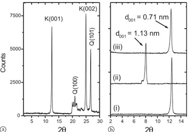
min-Fig. 1. XRPD diagrams of: (a) natural kaolinite, (b) the d001reflection
in (i) natural kaolinite, (ii) DMSO-intercalated kaolinite, (iii) Sr sorbed (DMSO-intercalated) kaolinite. K: kaolinite, Q: quartz.
Fig. 2. XRPD diagrams of: (a) natural magnesite, (b) Sr-sorbed
magne-site (1.0 × 10⁻²M). K: kaolinite, Q: quartz, M: magnesite.
Fig. 3. DRIFT spectrum of: kaolinite.
Fig. 4. DRIFT spectrum of: (a) natural magnesite, (b) pure MgCO3.
eral bands. The OH deformation bands near 938–916 cm−1 are also typical for the kaolin group minerals and arise from vibrations of the inner and inner surface OH groups within the clay matrix [21]. Other features appearing in the spec-trum belong to a variety of Al−O and Si−O stretching and bending vibrations [22]. A detailed assignment of all the bands arising in the kaolinite spectrum is given in Table 1.
The DRIFT spectra of natural magnesite and pure MgCO3are shown in Fig. 4a and b, respectively. The FTIR
Table 1. Assignments of the bands appearing in the DRIFT spectrum
of natural kaolinite.
Band Position (cm−1) Vibration Assignment
3700 Inner surface−OH stretching vibration 3620 Inner−OH stretching vibration 1112, 1038, 1010 Si−O bending vibration 938, 918 Al−OH bending vibration 792, 754 Si−O−Al compound vibrations
692 Si−O stretching vibration
540 Si−O−Al compound vibrations
471, 433 Si−O vibrations
spectra of carbonate minerals contains modes arising from out-of-plane bending (ν2), the asymmetric stretching (ν3),
the in-plane-bending (ν4), and the combination modes (ν1+ ν3) and (ν1+ ν4). The symmetric stretching mode (ν1) is
usually very weak and hardly detected [23] and its pres-ence is referred to a disorder in the symmetry of carbonate group. Complete assignment of the bands is given in Table 2. The bands arising from the vibrational modes of carbonate in natural magnesite coincide with the bands belonging to pure MgCO3as shown in the Fig. 2. On the other hand, the
bands appearing near 3700 and 3620 cm−1 in the spectrum of natural magnesite belong to the kaolinite impurity in the magnesite mineral.
Natural kaolinite and magnesite samples were also ana-lyzed using SEM/EDS technique. Typical SEM microim-ages of powder samples of the two minerals are shown in Fig. 5a and b, respectively. The characteristic hexagonal plates and the layered nature of kaolinite structure are ev-ident in Fig. 5a. The heterogeneous nature of magnesite surface is seen from Fig. 5b, which also shows the mag-nesite crystals to possess a rod-like morphology. The EDS analysis (reported as mole percentages) revealed that natu-ral kaolinite was composed of 64.2% SiO2, 33.5% Al2O3,
and minor quantities of K2O and CaO, probably
originat-ing from small amounts of associated non-kaolinitic mineral impurities. Natural magnesite samples were composed of 29.3% CO2, 22.6% MgO, 27.9% SiO2, 17.8% Al2O3, and
2.4% CaO. The values represent an average of five meas-urements that were taken at different points of the mineral surface. The maximum deviation of the major components from the average value in the measurements performed on natural kaolinite was less than 10%, whereas the same de-viation in the case of magnesite amounted in some cases to
Table 2. Assignments of the bands appearing in the DRIFT spectrum
of natural magnesite.
Band Position (cm−1) Vibration Assignment
3700a Inner surface−OH stretching vibration
3620a Inner−OH stretching vibration
1461 Asymmetric stretching
884 Out-of-plane bending
748 In-plane-bending
2539 Combination modes (ν1+ ν3)
1824 Combination modes (ν1+ ν4)
a: Bands arising from the presence of some kaolinite as an impurity in the natural magnesite sample.
Fig. 5. SEM microimages of the surfaces of: (a) natural kaolinite,
(b) natural magnesite.
about 50% verifying the heterogeneous nature of the magne-site surface.
Characterization of the Sr2+-sorbed minerals
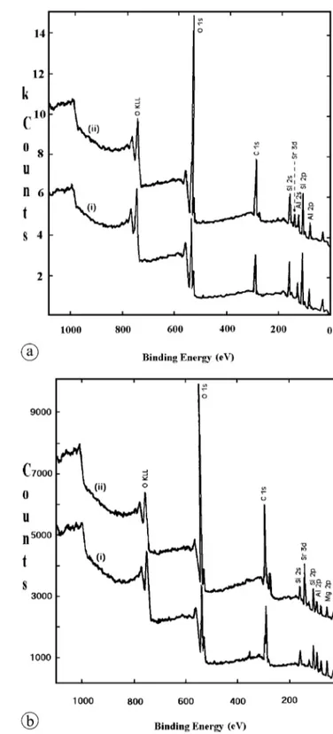
XPS is an inherent surface sensitive technique that has been applied in various studies for qualitative and quantitative characterization of sorption [24–30]. In this work, the XPS measurements were used to elucidate the elemental contents of the natural and Sr-sorbed mineral samples. Typical spec-tra for kaolinite and magnesite minerals before and after Sr2+ sorption are given in Fig. 6a and b, respectively. The
XPS data were corrected using the corresponding sensitiv-ity factors of the elements, and then the resulting amounts were normalized to the sum of Al and Si amounts. Here it was assumed that the quantities of Al and Si did not change in the course of the sorption process, as it was unlikely for such skeletal elements to take part in any exchange process. In order to verify this, blank experiments were performed in which kaolinite was mixed with de-ionized water for the same period applied in the sorption experiments. The results of the AAS measurements indicated that the concentration of both of Al and Si in solution are below the detection limit of the instrument (order of magnitude of ppb). The same was
Fig. 6. Typical XPS spectra of: (a) natural kaolinite (i), and Sr-sorbed
kaolinite (ii), (b) natural magnesite (i), and Sr-sorbed magnesite (ii).
also verified in solutions of Sr2+obtained following the
sorp-tion experiments.
In the case of sorption on kaolinite, the pH was in the approximate range 5.7–7.3 depending on the initial concen-tration of Sr2+ solutions. This value is above the zero point
of charge of kaolinite (∼ 3.5), the thing that ensures that the kaolinite surface is negatively charged. In general, the pH increased as the initial concentration of Sr2+in solution
decreased. The ratios of all the detected elements in the nat-ural samples of kaolinite and magnesite (excluding C and O) prior to and following Sr2+ sorption are given in Tables 3
Table 3. The percentage elemental composition of natural kaolinite
be-fore and after Sr2+sorption and the element/(Al+Si) ratios calculated
using the XPS data.
% Composition Element/(Al+Si) Element Kaolinite Sr-Kaolinite Kaolinite Sr-Kaolinite
Al+Si 97.63 95.41 1.000 1.000
Ca 1.49 N.D. 0.015 0.000
K 0.88 N.D. 0.009 0.000
Sr 0.00 4.59 0.000 0.048
N.D.: not detected.
Table 4. The percentage elemental composition of natural magnesite
before and after Sr2+ sorption and the element/(Al+Si) ratios
calcu-lated using the XPS data.
% Composition Element/(Al+Si) Element Magnesite Sr-Magnesite Magnesite Sr-Magnesite
Al+Si 69.59 71.11 1.000 1.000
Mg 27.32 18.12 0.393 0.255
Ca 3.09 N.D. 0.044 0.000
Sr 0.00 10.77 0.000 0.151
N.D.: not detected.
and 4, respectively. It is evident from the tables that the en-richment of Sr2+ on natural kaolinite is accompanied with
depletion in the amounts of K+and Ca2+initially present in
the clay structure.
Similarly, the uptake of Sr2+ by natural magnesite leads
to a decrease in the amounts of Mg2+ and Ca2+. The
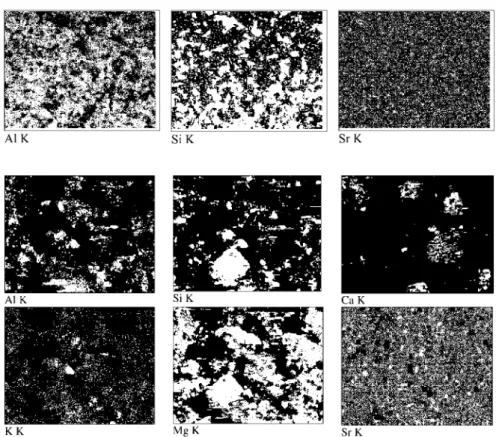
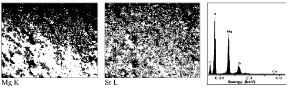
vari-Fig. 7. EDS mapping of Al, Si, and Sr on the
surface of natural kaolinite.
Fig. 8. EDS mapping of Al, Si, Ca, K, Mg,
and Sr on the surface of natural magnesite. ation in bulk concentration of K, Ca, and Mg was verified using AES measurements. The concentration of these elem-ents was measured in Sr2+-solutions prior to and following
the sorption experiments. At the start of the experiments, the concentration of these elements was much below 1 ppm, resulting from the fact that de-ionized water was used in preparing the solutions of Sr2+. The AES measurements of
these elements after sorption showed an increase in their concentration, indicating that an ion-exchange took place between them and the Sr2+fixed by the clay.
Comparing the amounts of Sr2+ scavenged by natural
kaolinite and natural magnesite it is clear that the latter is more effective in Sr2+ fixation. EDS mapping results of
the Sr-sorbed kaolinite and magnesite samples are given in Figs. 7 and 8, respectively. The maps were recorded at a magnification of 500×, corresponding to an approximate area of 200× 200 µm2
. Comparing the image of Sr to those of Al and Si (Fig. 7) it can be seen that Sr seems to be “ho-mogeneously” distributed on kaolinite surface. Here it must be noted that as far as EDS analysis of Sr on aluminosil-icates is concerned, that analysis must be based on the K line of Sr as the L line seemed to overlap with the K line of Si which possesses a close energy. EDS mapping results of Sr-sorbed natural magnesite surface (Fig. 8) revealed the heterogeneous nature of the mineral. According to images, Sr seems to be more associated with regions richer in Mg, in-dicating that the magnesite regions of the natural mineral are more effective in Sr fixation than the kaolinite constituents within the same mineral.
DRIFT spectra of the Sr-sorbed kaolinite revealed that Sr sorption did not cause any change in the various stretching and bending vibrations of the active modes of the natural sample given in Fig. 3. The XRPD studies on natural and Sr-sorbed kaolinite showed also that the structure of the clay
was retained upon sorption. The fact that the XRPD peak intensities, peak shapes, and peak positions – which reflect the extent of crystallinity of the clay – along the primary 001 and 002 planes in the clay matrix were almost unaf-fected, is indicative that sorption has taken place on the surface and edge positions of kaolinite. This suggests that the interlayer positions of the clay were not involved in Sr2+
fixation due to the relatively stronger binding forces among the kaolinite sheets compared to other clays like montmo-rillonite, where interlayer sites are much more effective in sorption than the outer sites. In order to examine the effect of expanding the interlayer space of kaolinite on the amount of adsorbed Sr2+, the clay was intercalated with
dimethyl-sulfoxide, DMSO ((CH3)2SO). Intercalation of DMSO in
kaolinite provides a method for the incorporation of alkali and alkaline metal salts into the kaolinite structure by re-placement of DMSO [31]. DMSO molecules are bound into the kaolinite structure by hydrogen bonding of the S=O to the gibbsite-like hydroxyls and by a coordination of the sul-fur to the oxygens of the siloxane surface [31] . Intercalation of DMSO within the kaolinite lamellae caused a change in position of the primary 001 and 002 reflections to higher
dhkl values. Fig. 1b shows the increase of d001from 0.72 nm
to 1.13 nm The figure shows also that DMSO molecules were readily removed from kaolinite upon the exposure of the clay to Sr2+solutions as indicated by the re-appearance
of the XRPD reflections at their original spacing. The bulk analysis of the Sr2+ solutions using AES at the end of
the mixing period indicated that the difference in the Sr2+
concentration between the solutions that were exposed to natural kaolinite and those exposed to DMSO-intercalated kaolinite was small. In both cases, the percentage sorption was calculated to be around 39, 56, and 62% for solutions with initial Sr2+concentration of 1.0 × 10⁻², 1.0 × 10⁻⁴, and
1.0 × 10⁻⁶M, respectively. This can be probably explained by assuming that Sr2+which displaced DMSO in the
inter-layer region of kaolinite have experienced a fast sorption step, which lead to displacing DMSO molecules, followed by a desorption step during the later stages of mixing, lead-ing to the removal of most of Sr2+ ions from the interlayer
region. The forces of interaction during the sorption step could be weak van der Waals type forces, the thing that might have facilitated desorption under the effect of mix-ing. The interlayer region of kaolinite is known to be tightly packed, and unless there is an isomorphous substitution in this region, no permanent negative charge will develop on the internal surfaces like it is the case in expandable clays such as montmorillonite. The presence of a negative charge on accessible sites is essential for a stable binding of positive cations like Sr2+.
The XPRD characterization of magnesite before and after Sr2+ sorption has also shown that the matrix of the
min-eral did not undergo structural changes at different load-ings. Fig. 2b shows the XRPD diagram of Sr-loaded nat-ural magnesite prepared at an initial Sr2+ concentration of
1.0 × 10⁻²M. The same observation is evident also from the DRIFT characterization, which showed that the vibrational bands shown in Fig. 4 were all unaffected. As previously mentioned, no pH adjustment was made and the pH ranged from 8.2–7.6 during the mixing process of Sr2+ ions with
natural magnesite.
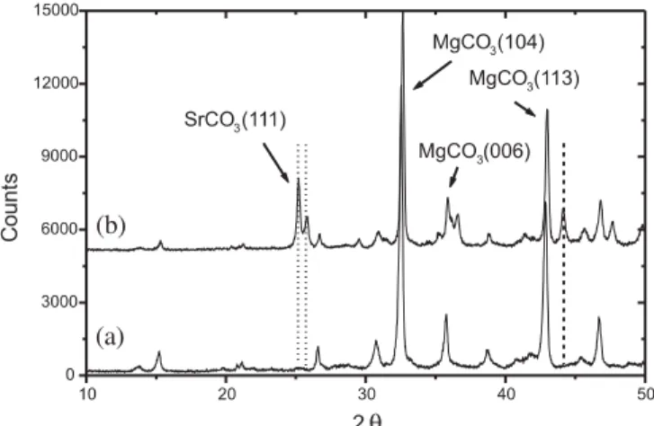
Fig. 9. XRPD diagram of: (a) pure MgCO3, (b) Sr-sorbed MgCO3.
The sorption of Sr2+ ions was also studied on pure
MgCO3samples. Unlike the case in natural magnesite, the
XRPD analysis of the Sr-sorbed samples revealed the for-mation of strontianite (SrCO3) as a coprecipitate on the
MgCO3 samples treated with an initial Sr2+ concentration
of 1.0 × 10⁻²M (Fig. 9). Here also there was no pH con-trol but the pH in these experiments ranged from 10.5–8.3, higher than the range reported above for natural magnesite, thus indicating that the medium was richer in CO3
2− ions.
In both cases, the decrease in the pH at the end of mixing could plausibly be caused by the removal of carbonate ions from the medium either upon formation of SrCO3or the
re-precipitation of CaCO3. It is not clear what is the exact
rea-son that makes natural magnesite less soluble than MgCO3,
but it might be considered that the presence of impurities within magnesite, in particular near the surface, could have kinetically inhibited its solubility. In a study on calcite [32], it was reported that some metal ions encountered within the surface of this mineral at even small concentration could lead to inhibiting the solubility of the mineral. According to the same study, in solid–liquid interaction, barriers against mixing might develop, leading to kinetic stabilization of the reaction far from thermodynamic stabilization.
The formation of strontianite was characterized by an enhanced ability of MgCO3towards the uptake of Sr2+
com-pared to a much lower ability at the lower Sr2+
concentra-tions of 1.0 × 10⁻⁴ and 1.0 × 10⁻⁶M, for which no stron-tianite formation was detected. The EDS results for the pure MgCO3 samples prior to and following Sr2+ sorption are
given in Table 5. The data were collected from 5 different
Table 5. The chemical analysis (% Mol) of pure MgCO3and Sr-sorbed
MgCO3obtained from the EDS analysis at different initial
concentra-tion of Sr2+.
Constituent MgCO3 Sr-sorbed MgCO3
1.0 × 10⁻⁶M 1.0 × 10⁻⁴M 1.0 × 10⁻²M CO2 57.8 58.7 58.4 59.6 MgO 40.3 39.6 39.2 35.3 CaO 0.8 0.7 0.7 0.6 Others 1.1 0.9 0.6 0.1 SrO − N.D. 1.1 4.4 N.D.: not detected.
Fig. 10. EDS mapping of Mg and Sr on the
surface of Sr-sorbed MgCO3. A typical EDS
spectrum is also shown. points and the values given in the table represent the average
with maximum deviations from the average values of 2.9%, 4.2%, and 8.7% for CO2, MgO and SrO, respectively. The
EDS mapping of Mg and Sr on MgCO3is given in Fig. 10.
The figure shows Sr to be spread all over the surface but at higher concentrations in certain regions than the others. Spot analysis of the brighter regions on Sr map verified that the larger concentration of the ion was associated with the presence of strontianite.
Sorption mechanisms of Sr2+ions
Sorption on natural kaolinite
According to the XPS findings, K+ and Ca2+, initially
present in natural kaolinite, were depleted upon sorption of Sr2+ ions. Such ions are normally not encountered in the
structure of pure kaolinite, and it seems that they were as-sociated with an impurity that exists in minor quantity in the natural clay, or were initially attached to vacant sites on the clay surface. The removal of these elements upon fixation of Sr2+is indicative of the contribution of ion exchange
mech-anism to the sorption process. Furthermore, the fact that the total quantities of both of K+ and Ca2+ were only one half
the quantity of sorbed Sr2+ (see Table 3), points to the fact
that ion exchange is not the sole mechanism of uptake. As outlined above, the XRPD characterization of natural and Sr-loaded kaolinite showed no change in the basic re-flections of the clay upon sorption. Moreover, expanding the interlayer space of kaolinite by intercalation of DMSO, did not lead to a significant enhancement of Sr2+ uptake by the
clay. This can be an indication that sorption of Sr2+on
kaoli-nite occurs mainly on the outer surface and edges of kaolin-ite. During the sorption experiments on this clay, pH was in the approximate range 5.7–7.3 depending on the initial con-centration of Sr2+solutions. This pH range is above the zero
point of charge of kaolinite (∼ 3.5), the thing that ensures that the kaolinite surface is negatively charged. Hence, in addition to ion-exchange discussed above, another possible mechanism of Sr2+sorption might be operating through
in-teractions with the AlO− and SiO− sites created as a result of the deprotonation of the acidic hydroxyl groups. The ex-tent of such protonation is pH-dependent and is referred to as ‘hydrolytic adsorption’ that can be described by the follow-ing Eq. (1):
surface−OH + Sr2+→ surface−O−Sr++ H+.
It is reported that the external charge on kaolinite surface can be attributed to silanol groups as well as amphoteric alumi-nol groups at the crystal edges which deprotonate yielding SiO−and AlO−surface complexes at moderate and high pH
values [33]. Hence, in the absence of impurities that might give rise to ion-exchange in natural kaolinite, hydrolytic sorption will be the primary mechanism that will determine the cation exchange capacity of pure kaolinite.
Sorption on natural magnesite and pure MgCO3
The surface of carbonate minerals possesses a dynamic na-ture and, as a result, the fixation of ‘foreign’ ions can take place via a variety of mechanisms. These mechanisms in-cludes ion exchange, burial of the ions within the lattice of carbonate upon recrystallization, binding of the ions to co-ordinative unsaturated carbonate groups (or specie) at the surface of the mineral, precipitation in a distinct phase as metal carbonate, metal hydroxide, or both, and solid state diffusion (negligible for low residence times of less than 100 years [34]). The nature of solution medium (concentra-tion, pH, and temperature) and the type of surface speciation taking place at the solid–liquid interface will determine the type of fixation. According to our results, in the case of natural magnesite, in the absence of any SrCO3 formation
(as revealed by XRPD findings) and based on the compar-ison of the quantity of fixed Sr2+ to that of depleted Mg2+
(as obtained from XPS data; Table 4), ion exchange seems to be the primary process of sorption. Similar observations were reported for Sr2+ sorption on calcite [15, 35, 36]. The
fact that the Sr2+ neighboring structural environment in
Sr-sorbed calcite was similar to calcite rather than to stron-tianite was used as an evidence that the uptake of Sr2+ ions
occurred via an ion exchange mechanism [36].
On the other hand, in the case of MgCO3, fixation seems
to be dominated by SrCO3 formation, the thing possibly
stemming from the higher concentration of carbonate in the reaction medium as discussed previously. Other sorp-tion mechanisms could have done some contribusorp-tion to the amount of scavenged Sr2+ions, but their role seems to be
in-significant. It is reported that precipitation mechanism leads to enhancement of removal of ions from solution and greatly immobilize these ions by ‘freezing’ them in the host min-eral structure [37], the thing inline with our results which revealed a higher ability of MgCO3to fix Sr2+compared to
natural magnesite.
Conclusion
Natural kaolinite and magnesite used in this study were of heterogeneous nature. Based on XPS findings, natural mag-nesite showed higher sorption capacity compared to natural kaolinite. The fact that the structure of kaolinite was re-tained upon Sr2+uptake indicates that surface and edge sites
are more effective in sorption. The EDS mapping of kaoli-nite surface indicated that Sr2+ was equally distributed on
that surface. The absence of any precipitate formation in the case of Sr2+ fixation by natural magnesite, and based
on XPS quantification analysis, the uptake mechanism on this mineral seems to occur primarily via ion exchange. The high depletion of Mg2+ upon Sr2+ sorption supported
this observation, the thing also clear from the EDS maps which showed that Sr was localized in a larger extents at regions richer in Mg. Unlike the case of natural magne-site, the sorption mechanism of Sr2+ by pure MgCO
3
in-volved a SrCO3precipitate formation, the thing referred to
the higher pH of the aqueous MgCO3medium. Precipitate
formation lead to an enhanced removal of Sr2+ compared
to the situations where Sr2+is removed by an ion-exchange
type mechanism.
Acknowledgment. The authors would like to thank Dr. K. Hallam at the
Interface Analysis Centre/University of Bristol for his help in the XPS measurements. Ms. Duygu Oguz and Mr. Gokhan Erdogan at the Cen-ter of MaCen-terial Research/Izmir Institute of Technology for their help in the SEM/EDS measurements.
References
1. Lieser, K. H.: Radionuclides in the geosphere: sources, mobil-ity, reactions in natural waters and interactions with solids. Ra-diochim. Acta 70/71, 355 (1995).
2. Hsu, C., Liu, D., Chuang, C.: Equilibrium and kinetic sorption be-haviors of cesium and strontium in soils. Appl. Radiat. Isotope 45, 981 (1994).
3. Wang; X., Chen; Y., Wu, Y.: Sorption and desorption of ra-diostrontium on powdered bentonite: Effect of pH and fulvic acid. J. Radioanal. Nucl. Chem. 261, 497 (2004).
4. Khan, S. A.: Sorption of the long-lived radionuclides cesium-134, strontium-85 and cobalt-60 on bentonite, J. Radioanal Nucl. Chem. 258, 3 (2003).
5. Iwaida, T., Nagasaki, S., Tanaka, S.: Sorption study of stron-tium onto hydrated cement phases using a sequential desorption method. Radiochim. Acta 88, 483 (2000).
6. Khan, S. A., Rehman, R., Khan, M. A.: Sorption of strontium on Bentonite. Waste Management 15, 641 (1995).
7. Liang, T.: The influence of cation concentration on the sorption of strontium on mordenite. Appl. Radiat. Isotopes 51, 527 (1999). 8. Karasyova, O. N., Ivanova, L. I., Lakshtanov, L. Z., Lövgren, L.:
Strontium sorption on hematite at elevated temperatures. J. Col-loid Interf. Sci. 220, 419 (1999).
9. Bors, J., Dultz, St., Riebe, B.: Retention of radionuclides by organophilic bentonite. Eng. Geol. 54, 195 (1999).
10. Eriksen, T. E., Jansson, M., Molera, M.: Sorption effects on cation diffusion in compacted bentonite. Eng. Geol. 54, 231 (1999). 11. Zhu, C.: Estimation of surface precipitation constants for sorption
of divalent metals onto hydrous ferric oxide and calcite. Chem. Geol. 188, 23 (2002).
12. Nakashima, Y.: Diffusivity measurement of heavy ions in Wyoming montmorillonite gels by X-ray computed tomography. J. Contam. Hydrol. 61, 147 (2003).
13. Juang, R., Lin, S., Wang, T.: Removal of metal ions from the com-plexed solutions in fixed bed using a strong-acid ion exchange resin. Chemosphere 53, 1221 (2003).
14. Sahai, N., Carroll, S. A., Roberts, S., O’Day, P. A.: X-Ray absorp-tion spectroscopy of strontium(II) coordinaabsorp-tion: II. sorpabsorp-tion and precipitation at kaolinite, amorphous silica, and goethite surfaces. J. Colloid Interf. Sci. 222, 198 (2000).
15. Parkman, R. H., Charnock, J. M., Livens, F. R., Vaughan, D. J.: A study of the interaction of strontium ions in aqueous solution
with the surfaces of calcite and kaolinite. Geochim. Cosmochim. Acta 62, 1481 (1998).
16. Nordstrom, D. K., McNutt, R. H., Puigdomènech, I., Smellie, J. A. T., Wolf, M.: Ground water chemistry and geochemical mod-eling of water–rock interactions at the Osamu Utsumi mine and the Morro do Ferro analogue study sites, Poços de Caldas, Minas Gerais, Brazil. J. Geochem. Explor. 45, 249 (1992).
17. Cole, T., Bidoglio, G., Soupioni, M., O’Gorman, M., Gibson, N.: Diffusion mechanisms of multiple strontium species in clay. Geochim. Cosmochim. Acta 64, 385 (2000).
18. Bergaya, F., Vayer, M.: CEC of clays: Measurement by adsorption of a copper ethylenediamine complex. Appl. Clay Sci. 12, 275 (1997).
19. Baes Jr., C. F., Mesmer, R. E.: The Hydrolysis of Cations. John Wiley & Sons Inc. (1976).
20. Patakfalvi, R., Oszko, A., Dekany I.: Synthesis and characteri-zation of silver nanoparticle/kaolinite composites. Colloid Sur-face A 220, 45 (2003).
21. Wilson, M. J.: Clay Mineralogy: Spectroscopic and Chemical De-termination Methods. Chapman & Hall, London (1994). 22. Suraj, G., Iyer, C. S. P., Rugmini, S., Lalithambika, M.: The effect
of microionization on kaolinites and their sorption behavior. Appl. Clay Sci. 12, 111 (1997).
23. Böttcher, M. E., Gehlken, P., Steele, D. F.: Characterization of in-organic and biogenic magnesian calcites by Fourier Transform infrared spectroscopy. Solid State Ionics 101–103, 1379 (1997). 24. Shahwan, T., Suzer, S., Erten, H. N.: Sorption studies of Cs+
and Ba2+ cations on magnesite. Appl. Radiat. Isotopes 49, 915
(1998).
25. Ning, X., Hochella Jr., M. F., Brown Jr., G. E., Parks, G. A.: Co(II) sorption at the calcite–water interface: I. X-ray photoelectron spectroscopic study. Geochim. Cosmochim. Acta 60, 2801 (1996) 26. Drot, R., Simoni, E., Alnot, M., Ehrhardt, J. J.: Structural envi-ronment of uranium(VI) and europium(III) species sorbed onto phosphate surfaces: XPS and optical spectroscopy studies. J. Col-loid Interf. Sci. 205, 410 (1998).
27. Dutta, N. C., Iwasaki, T., Ebina, T., Hayashi, H.: A combined X-ray photoelectron and auger electron spectroscopic study of ce-sium in variable-charge montmorillonites. J. Colloid Interf. Sci. 216, 161 (1999)
28. Merdy, P., Guillon, E., Aplincourt, M., Dumonceau, J., Vezin, H.: Copper sorption on a straw lignin: experiments and EPR charac-terization. J. Colloid Interf. Sci. 245, 24 (2002).
29. Garc´ıa-S´anchez, A., ´Alvarez-Ayuso, E.: Sorption of Zn, Cd and Cr on calcite. Application to purification of industrial wastewaters. Miner. Eng. 15, 539 (2002).
30. Lomenech, C., Simoni, E., Drot, R., Ehrhardt, J. J., Mielczarski, J.: Sorption of uranium(VI) species on zircon: structural investiga-tion of the solid/solution interface. J. Colloid Interf. Sci. 261, 221 (2003).
31. Frost, R. L., Kristof, J., Paroz, G. N., Kloprogge, J. T.: Molecular structure of dimethyl sulfoxide intercalated kaolinite. J. Phys. Chem B 102, 8519 (1998).
32. Hay, M. B., Workman, R. K. Manne, S.: Mechanisms of metal ion sorption on calcite: composition mapping by lateral force mi-croscopy. Langmuir 19, 3727 (2003).
33. Coppin, F., Berger, G., Bauer, A., Castet, S., Loubet, M.: Sorption of lanthanides on smectite and kaolinite. Chem. Geol. 182, 57 (2002).
34. Hoffman, U., Stipp, S. L. S.: The behavior of Ni2+on calcite
sur-face. Geochim. Cosmochim. Acta 65, 4131 (2001).
35. Zachara, J. M., Cowan, C. E., Resch, C. T.: Sorption of divalent metals on calcite. Geochim. Cosmochim. Acta 55, 1549 (1991). 36. Pingitore Jr., N. E., Lytle, F. W., Davies, B. M., Eastman, M. P.,
Eller, P. G., Larson, E. M.: Mode of incorporation of Sr2+in
cal-cite: Determination by X-ray absorption spectroscopy. Geochim. Cosmochim. Acta 56, 1531 (1992).
37. Wang, Y., Xu, H.: Prediction of trace metal partitioning between minerals and aqueous solutions: a linear free energy correlation approach. Geochim. Cosmochim. Acta 65, 1529 (2001).