original article
C
entral venous cannulation is a necessary invasive procedure for fluid management, hemodynam-ic monitoring and vasoactive drug therapy. The preferred site for cannulation is the right internal jugular vein (RIJV). The reasons for this preference include the RIJV being the shortest route to the superior vena cavaComparison of the Trendelenburg position
versus upper-limb tourniquet on internal
jugular vein diameter
Pelin Karaaslan,
aBanu Vural Gokay,
bMuhammet Ahmet Karakaya,
aKamil Darcin,
cAfak Durur
Karakaya,
dTugrul Ormeci,
dEmine Arzu Kose
aFrom the aDepartment of Anesthesiology and Reanimation, Istanbul Medipol University, Istanbul, Turkey, bDepartment of Anesthesiology and Reanimation, Acibadem Universitesi, Istanbul, Turkey; cDepartment of Anesthesiology and Reanimation, Koc University, Istanbul, Turkey, dDepartment of Radiology, Istanbul Medipol University, Istanbul, Turkey
Correspondence: Dr. Pelin Karaaslan · Department of Anesthesiology and Reanimation, Istanbul Medipol University, TEM Avrupa Otoyolu Goztepe, Cikisi No: 1, Bagcilar, Istanbul 34078, Turkey · T: +902127607831 · [email protected] · http://orcid.org/0000-0002-5273-1871
Ann Saudi Med 2017; 37(4): 308-312 DOI: 10.5144/0256-4947.2017.308
BACKGROUND: Central venous cannulation is a necessary invasive procedure for fluid management, haemodynamic monitoring and vasoactive drug therapy. The right internal jugular vein (RIJV) is the pre-ferred site. Enlargement of the jugular vein area facilitates catheterization and reduces complication rates. Common methods to enlarge the RIJV cross-sectional area are the Trendelenburg position and the Valsalva maneuver.
OBJECTIVE: Compare the Trendelenburg position with upper-extremity venous return blockage using the tourniquet technique.
DESIGN: Prospective clinical study.
SETTING: University hospital.
SUBJECTS AND METHODS: Healthy adult volunteers (American Society of Anesthesiologists class I) aged 18–45 years were included in the study. The first measurement was made when the volunteers were in the supine position. The RIJV diameter and cross-sectional area were measured from the apex of the triangle formed by the clavicle and the two ends of the sternocleidomastoid muscle, which is used for the conven-tional approach. The second measurement was performed in a 20° Trendelenburg position. After the drain-age of the veins using an Esbach banddrain-age both arms were cuffed. The third measurement was made when tourniquets were inflated.
MAIN OUTCOME MEASURE(S): Hemodynamic measurements and RIJV dimensions.
RESULTS: In 65 volunteers the diameter and cross-sectional area of the RIJV were significantly widened in both Trendelenburg and tourniquet measurements compared with the supine position (P<.001 for both measures). Measurements using the upper extremity tourniquet were significantly larger than Trendelenburg measurements (P=.002 and <.001 for cross-sectional area and diameter, respectively).
CONCLUSION: Channelling of the upper-extremity venous return to the jugular vein was significantly supe-rior when compared with the Trendelenburg position and the supine position.
LIMITATIONS: No catheterization and study limited to healthy volunteers.
(SVC), not being on the side of the thoracic duct, and being far from the pleura, thus leading to lower com-plication rates. Several studies on adults have shown that the use of ultrasonography (USG) both improves the success rate while shortening the cannulation time and reduces the complication rate. Complications of
original article
TRENDELENBURG POSITION
IJV cannulation include carotid puncture, nerve dam-age, hydrothorax, haemothorax, pseudoaneurysm and arteriovenous fistula.1,2 Internal jugular vein (IJV)
can-nulation is routinely performed with USG in our clinic. Gordon and colleagues demonstrated that enlarge-ment of the jugular vein cross-sectional area facili-tates catheterization and reduces complication rates.3
Methods for expanding the cross-sectional area are the Trendelenburg position and the Valsalva manoeu-vre. The Valsalva manoeuvre increases intrathoracic pressure and thus increases RIJV diameter and area. 4-7 However, the maneuver is not perfect —it can lead
to haemodynamic instability, arterial rupture and local haematoma. Hypotension, bradycardia and other com-plications can be observed at pressures exceeding 20 cm H2O. Positive intrathoracic pressure and positive
end-expiratory pressure (PEEP) have also been shown to be effective.4,8-10
In cannulation, the Trendelenburg position is recom-mended for increasing the vein diameter, which increas-es the succincreas-ess rate, and reducing the risk of air embo-lism. Ely et al demonstrated in their survey that 91% of clinicians routinely prefer an upside-down position in their central venous cannulation practice.11 Similarly, the
Trendelenburg position is used in all CVP applications in our clinic, as long as there is no contraindication. The Trendelenburg position increases cerebral, arterial, ve-nous, and intraocular pressures.12,13 In addition, there
may be a decrease in the capacity of the respiratory system due to the movement of the diaphragm towards the thorax.13 It cannot be applied in cases where these
effects are not wanted in the patient or when the bed cannot be positioned. This position may make catheter-ization difficult and may increase the rate of complica-tions. The aim of this study was to develop an alterna-tive to the Trendelenburg position. The Trendelenburg
position was compared with upper-extremity venous return blockage by using a tourniquet technique.
SUBJECTS AND METHODS
After informed consent from volunteers and approval from the University’s Ethics Committee were obtained, healthy (American Society of Anesthesiologists class I, a measure of physical status) adult volunteers aged 18–45 years were enrolled prospectively. The power analysis for the study was conducted with G Power 3.1.9.2, and the total sample size for 95% actual power was determined on 52 volunteers, based on previous field measurements. The 52 volunteers lay on a posi-tionable bed. Initial systolic and diastolic blood pres-sure values, heart rates, and saturations were recorded as basal values. Subjects whose pressure values were within the normal range according to the American Heart Association were included in the study; subjects with hypertension were excluded.
For the present study, the first measurement was made with the volunteer lying in the supine position. The RIJV diameter and cross-sectional area were mea-sured from the apex of the triangle formed by the clavicle and the two ends of the sternocleidomastoid muscle, which is used for the conventional approach. In all measurements, the same roll was placed under the shoulder of the patients to achieve a slight head extension and the head position was held 30° to the left using a protractor in all measurements. For the sec-ond measurement, the bed was then placed in a 20° Trendelenburg position, and after waiting for 3 minutes, the head was turned 30° in the opposite direction. After the visualization of the RIJV by ultrasonography (USG), the diameter and area were recorded (Figure 1A). Except for the Trendelenburg position, no maneuvers were made that could have had an effect on the IJV area
Figure 1. (A) RIJV measurement in the Trendelenburg position; (B) RIJV measurement with upper-limb tourniquet inflated.
original article
TRENDELENBURG POSITION measurement, such as Valsalva or liver compression.After the measurements in the Trendelenburg position were completed, the bed position was restored, and after waiting for 10 minutes, both arms were lifted from the heart level and left in this position for 3 minutes. After the drainage of the veins using an Esbach ban-dage both arms were cuffed. Tourniquets were then in-flated bilaterally so that the cuff pressure was 50 mm Hg above the systolic pressure and the third measurement of the RIJV diameter and area was performed (Figure 1B). Blood pressure, oxygen saturation and pulse rate were recorded at each different measurement point. A 12-MHz linear probe (LOCIQ 700; GE, Milwaukee, WI, USA) USG device was used for the measurements. The RIJV area measurements were performed on the frozen images of the area. Cross-sectional area measurements were conducted automatically with the program stored in the software of the USG device. The measurements were taken as follows:
1) The RIJV diameter and area measurement in the supine position at 0°;
2) The RIJV diameter and area measurement in 20° Trendelenburg position;
3) The RIJV diameter and area measurement after achieving a cuff pressure 50 mm Hg above the basal systolic pressure value after upper-extremity venous drainage
Table 1. Demographic data for the 65 healthy volunteers.
Age 26.0 (5.21) [18-45]
Weight 65.75 (9.16) [48-85]
Height 167.32 (8.97) [148-187]
Data are mean (standard deviation), [range].
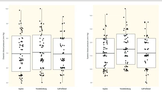
Figure 2. Mean (SD, range) diastolic (left) and systolic (right) blood pressure in the three positions (statistically significant differences between supine, inflated cuff and Trendelenburg positions by one-way ANOVA for mean diastolic (type III sum of squares=9.1x10⁵, F=2449.2, P<.001) and systolic blood pressures (type III sum of squares=2.4x10⁶, F=7150.8,
P<.001). No post-hoc tests.
The statistical analysis was carried out the software program SPSS 16.0. The repeated-measures ANOVA test was used for the statistical evaluation of the mea-surements and P<.05 was considered statistically sig-nificant.
RESULTS
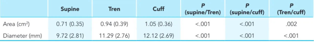
Sixty-five healthy volunteers were enrolled (Table 1). The systolic and diastolic pressures differed significant-ly among the three positions (Figure 2). However, the heart rate and oxygen saturation values were similar at all measurement times. In the supine position, the RIJV mean (standard deviation) anterior-posterior (AP) diam-eter was 9.7 (2.81) mm, the mean (SD) cross-sectional area was 0.71 (0.35) cm2. The mean AP diameter and
cross-sectional area of the volunteers placed in the Trendelenburg position were 11.29 (2.76) mm and 0.94
original article
TRENDELENBURG POSITION
(0.39) cm2, respectively. Compared with the supine
po-sition, the diameter and area of RIJV were significantly widened (P<.001). When the measurements were per-formed in the supine position using a tourniquet in the upper extremity, these results were 12.12 (2.69) mm and 1.05 (0.36) cm2, respectively. These
measure-ments were significantly higher than measuremeasure-ments performed in the supine position and Trendelenburg position (Table 2).
DISCUSSION
Internal jugular vein catheterization is easy and com-plications are decreased when cross-sectional area is increased.14-16 Various methods have been
attempt-ed to achieve cross-sectional area expansion. The Trendelenburg position is the most preferred because it increases venous return and IJV diameter.17 It also
re-duces the risk of air embolism.1,5,8 Apart from this, the
Valsalva manoeuvre and hepatic compression are used to increase the venous return and IJV diameters.5,9,14 We
found no studies showing the effect of taking advan-tage of upper-extremity venous drainage on IJV diam-eter and cross-sectional area. In this study, according to these measurements, the channelling of the upper-extremity venous return to the jugular vein was signifi-cantly superior when compared with the Trendelenburg position and the supine position.
During the Trendelenburg position, the abdominal organs push the diaphragm and the vital capacity de-creases. This can trigger hypoxia and hypercarbia. In the elderly and infirm additional complications may ensue even when the Trendelenburg position is held for a short time.3,18 The upside-down position
increas-es cardiac load, blood princreas-essure and venous princreas-essure.3
Malignant cardiac arrhythmias and mitral valve insuffi-ciencies can be seen.10,19 Even a 20° upside-down
posi-tion lasting 3 minutes will cause right ventricular stress.20
Marcus et al showed that Trendelenburg degrees over 20° could be both harmful to the patient and make the approach difficult.10 The Trendelenburg position is not
recommended especially in cases of respiratory dis-tress, unstable haemodynamics and obesity. Another
Table 2. Cross-sectional area and diameter measurements with the Trendelenburg position and after upper-extremity venous drainage in 65 healthy adults.
Supine Tren Cuff (supine/Tren)P (supine/cuff)P (Tren/cuff)P
Area (cm2) 0.71 (0.35) 0.94 (0.39) 1.05 (0.36) <.001 <.001 .002
Diameter (mm) 9.72 (2.81) 11.29 (2.76) 12.12 (2.69) <.001 <.001 <.001
Data are mean (standard deviation). Tren: Trendelenburg position; cuff= cuff-inflated (after upper-extremity venous drainage)
problem that may arise is increased intraocular and in-tracranial pressure.3,8,21 In some patients (i.e., decreased
cranial compliance, narrow-angle glaucoma), both in-creased pressures can lead to unwanted conditions. In addition, secretions can accumulate in the nasopharynx and leakage of secretions can trigger aspiration pneu-monia in this upside-down position. A more than 25ľ tilt cannot be tolerated by an awake patient so in the pres-ent study we preferred a 20ľ Trendelenburg.
Additionally, the Trendelenburg position cannot be used when the operation tables are not positionable, especially when anesthesia is performed outside of the operating room Based on the results of this study, it can be recommended that the upper-extremity ve-nous drainage method can be used in this situation. Gok et al studied the effect of leg-raising on the IJV size and found effects similar on vessel size simi-lar to the Trendelenburg position. Both leg raise and Trendelenburg were superior to the supine position, but there were no significant differences between the two. In our study, upper-extremity venous drainage re-sulted in significantly larger vein diameters when com-pared to the supine and Trendelenburg positions.22
The area width of the IJV depends on compliance and transmural pressure (the venous pressure and the surrounding tissue pressure). Intravascular pres-sure is the major component of the transmural value. Therefore, increasing intravascular fullness expands the area. We think that channelling upper-extremity ve-nous return to the jugular vein may have been effective through this pathway in the expansion of the RIJV cross-sectional area. In light of findings by Gwak et al, where carotid and RIJV overlap, the probability is increased when turning the head to the left, we have taken, as a basis, a 30 degree contralateral head rotation, as sug-gested in the given study.23 Forty degree head rotation
was not exceeded, as it has been indicated that pro-gressive head rotation increases overlap risk. This de-creases the probability of arterial puncture.
There are a few limitations in this study. Firstly, this study was limited to healthy volunteers. Therefore, he-modynamic changes were not reflected and
general-original article
TRENDELENBURG POSITION1. Dincyurek GN, Mogol EB, Turker G,
Yavas-caoglu B, Gurbet A, Kaya FN, et al. The ef-fects of the Trendelenburg position and the Valsalva manoeuvre on internal jugular vein diameter and placement in children. Singa-pore Med J. 2015;56(8):468-471.
2. Hilty WM, Hudson PA, Levitt MA, Hall JB.
Real-time ultrasound-guided femoral vein catheterization during cardiopulmonary re-suscitation. Ann Emerg Med. 1997;29:331-336.
3. Gordon AC, Saliken JC, Johns D, Owen
R, Gray RR. US-guided puncture of the in-ternal jugular vein: complications and ana-tomic considerations. J Vasc Interv Radiol. 1998;9:333-338.
4. Beddy P, Geoghegan T, Ramesh N,
Buck-ley O, O’Brien J, Colville J, et al. Valsalva and gravitational variability of the internal jugular vein and common femoral vein: ultrasound assessment. Eur J Radiol. 2006;58:307-309.
5. Lobato EB, Florete OG Jr, Paige GB,
Mo-rey TE. Cross-sectional area and intravascu-lar pressure of the right internal juguintravascu-lar vein during anesthesia: effects of Trendelenburg position, positive intrathoracic pressure, and hepatic compression. J Clin Anesth. 1998;10:1-5.
6. Armstrong PJ, Sutherland R, Scott DH. The
effect of position and different manoeuvres on internal jugular vein diameter size. Acta Anaesthesiol Scand. 1994;38:229-231.
7. Yildirim I, Yüksel M, Okur N, Okur E, Kilic
MA. The sizes of internal jugular veins in Turk-ish children aged between 7 and 12 years. Int J Pediatr Otorhinolaryngol. 2004;68:1059-1062.
8. Botero M, White SE, Younginer JG, Lobato
EB. Effects of Trendelenburg position and positive intrathoracic pressure on internal jugular vein cross-sectional area in
anesthe-tized children. J Clin Anesth. 2001;13:90-93.
9. Verghese ST, Nath A, Zenger D, Patel RI,
Kaplan RF, Patel KM. The effects of the simu-lated Valsalva maneuver, liver compression, and/or Trendelenburg position on the cross-sectional area of the internal jugular vein in infants and young children. Anesth Analg. 2002;94:250-254.
10. Marcus HE, Bonkat E, Dagtekin O, Schier
R, Petzke F, Wippermann J, et al. The im-pact of Trendelenburg position and posi-tive end-expiratory pressure on the internal jugular cross-sectional area. Anesth Analg. 2010;111:432-436.
11. Ely EW, Hite RD, Baker AM, Johnson MM,
Bowton DL, Haponik EF. Venous air embo-lism from central venous catheterization: a need for increased physician awareness. Crit Care Med. 1999;27(10):2113-2117.
12. Grebenik CR, Boyce A, Sinclair ME, Evans
RD, Mason DG, Martin B. NICE guidelines for central venous catheterization in children. Is the evidence base sufficient? Br J Anaesth. 2004;92(6):827-830.
13. Hewer CL. The physiology and
complica-tions of the Trendelenburg position. Canad M.A.J. 1956;74: 285-288.
14. Lee JG, Park HB, Shin HY, Kim JD, Yu SB,
Kim DS, et al. Effect of Trendelenburg po-sition on right and left internal jugular vein cross-sectional area. Korean J Anesthesiol. 2014;67(5):305-309.
15. Armstrong PJ, Cullen M, Scott DH. The
‘SiteRite’ ultrasound machine-an aid to in-ternal jugular vein cannulation. Anaesthesia. 1993;48:319-323.
16. Tryba M, Kleine P, Zenz M.
Sonograph-ic studies for optimizing the cannulation of the internal jugular vein. Anaesthesist. 1982;31:626-629.
17. Schettini ST, Martins de Oliveira LFY,
Henao HR, Lederman HM. Ultrasound evalu-ation of techniques for internal jugular vein puncture in children. Acta Cirurgica Brasilei-ra. 2008;23(5):469-472.
18. Sulek CA, Gravenstein N, Blackshear
RH, Weiis L. Head rotation during inter-nal jugular vein cannulation and the risk of carotid artery puncture. Anesth Analg. 1996;82:125-128.
19. Fahy BG, Barnas GM, Nagla SE, Flowers
JL, Njoku MJ, Agarwal M. Effects of Tren-delenburg and reverse TrenTren-delenburg pos-tures on lung and chest wall mechanics. J Clin Anesth. 1996;8(3):236-244.
20. Reich DL, Konstadt SN, Raissi S,
Hub-bard M, Thys DM. Trendelenburg position and passive leg raising do not significantly improve cardiopulmonary performance in the anesthetized patient with coronary artery disease. Crit Care Med. 1989;17(4):313-317.
21. Mavrocordatos P, Bissonnette B,
Ra-vussin P. Effects of neck position and head elevation on intracranial pressure in an-aesthetized neurosurgical patients: pre-liminary results. J Neurosurg Anesthesiol. 2000;12(1):10-14.
22. Gok F, SarkilarG, Kilicaslan A, Yosunkaya
A, Uzun ST. Comparison of the effect of the Trendelenburg and passive leg rais-ing positions on internal jugular vein size in critically ill patients. Int J Clin Exp Med. 2015;8(10):19037-43. eCollection 2015.
23. Gwak MJ, Park JY, Suk EH, Kim DH.
Ef-fects of head rotation on the right internal jugular vein in infants and young children. Anaesthesia. 2010;65:272-276.
24. Bellazzini MA, Rankin PM, Gangnon
RE, Bjoernsen LP. Ultrasound validation of maneuvers to increase internal jugular vein cross-sectional area and decrease com-pressibility. AJEM. 2009 May;27(4):454-459.
REFERENCES
ized to patients who have comorbidities such as dia-betes mellitus and cardiovascular diseases. It is obvi-ous that the position and blockage of upper-extremity venous return may lead to different responses in he-modynamics in patients with comorbid diseases and in different age groups. The second limitation was that we only performed measurements without catheterization. Therefore, we cannot provide data on the success rate and complications. In a future study, we will perform area measurements in patients to be catheterised and compare success and complication rates after catheter-ization. We also did not compare maneuvers, such as Valsalva and hepatic compression, where the effects on area measurement have been proven. Bellazzini et
al found the Valsalva maneuver to be superior to the Trendelenburg position.24 In a future study, the effect
of the Valsalva maneuver can be compared with the model of channelling upper-extremity venous return to the IJV.
In conclusion, the results of this study indicated that both the Trendelenburg position and the upper extremity venous return model were able to increase the RIJV diameter and cross-sectional area. Upper-extremity venous return was significantly superior to the Trendelenburg position.
Conflict of interest