ARAÞTIRMALAR (Research Reports)
Özet
Feokromositoma adrenal medullanýn kromaffin hücrelerinden köken alan ve katekolamin salgýlayan bir tümördür. Sessiz feokromositomalar anestezi sýrasýnda ortaya çýkan hipertansif ataklarla kendilerini belli edebilirler. Bu olguda plazma ve idrar katekolamin seviyeleri normal olan, hipertansiyon hikayesi olmayan ve cerrahi sýrasýnda taný konan bir feokromositma vakasý sunulmaktadýr. Tüm yeni ilaç ve tekniklere raðmen taný konmamýþ feokromositmanýn anestetik yönetimi anestezist için beklenmedik sorunlar oluþmaktadýr. Bu vakada hipertansif krize raðmen herhangi bir komplikasyonla karþýlaþýlmamýþ olmasý büyük bir þanstýr.
Anahtar Sözcükler: Adrenal Bez Tümörleri; Katekolaminler; Feokromositom. Abstract
Pheochromocytoma is a catecholamine secreting tumor which originates in the chromaffin cells of adrenal medulla. Silent pheochromocytomas can become evident with hypertensive attack during anaesthesia. Here is presented a case of pheochromocytoma without high plasma or urinary levels of catecholamines, and without history of hypertension which was detected with hypertensive attack during surgical extirpation. With all the newest medications and techniques that have been developed the anesthetic management of undiagnosed pheochromocytomas are still sources of unexpected problems for the anesthetists. In this case it was a great chance that there were no complications despite the hypertensive crisis. Key words: Adrenal Gland Neoplasm; Catecholamines; Pheochromocytoma.
OLGU SUNUMU(Case Reports)
Submitted : May 04, 2009 Revised : October 16, 2009 Accepted : December 23, 2010
Taný Almamýþ Feokromositomalý Hastada Hipertansif Kriz
Hypertensive Crisis in a Patient With an
Undiagnosed Pheocromocytoma
Corresponding Author: Yard. Doç. Dr. Züleyha Kazak Ufuk Üniversitesi Týp Fakültesi, Anesteziyoloji ve Reanimasyon Anabilim Dalý, Ankara- Türkey.
Phone : +90 - 3122044098 e-mail : [email protected]
Züleyha Kazak
Asist. Professor of Anesthesiology and Reanimation Ufuk University
Kamil Darcin
M.D., Anesthesiology and Reanimation Ufuk University
Kansu Kazbek
M.D., Anesthesiology and Reanimation Ufuk University
Sumru Sekerci
Professor of Anesthesiology and Reanimation Ufuk University
Hikmet Süer
Professor of Anesthesiology and Reanimation Ufuk University
This study was presented at the XLIIth National Anesthesiology and Reanimation Congress, 29 October -02 December 2008, Antalya, Turkey.
Introduction
Adrenal incidentalomas are adrenal masses detected during imaging performed for other than adrenal pathologies reasons (1). The incidence of adrenal incidentalomas in autopsy series varies between 1 and 8.7% (2). Statistically, the 1% incidence for the age of 30 rises up to 7% for the age of 70 (3). Pheochromocytoma is a tumour that secretes catecholamine and originates in the chromaffin cells of adrenal medulla (4). The incidence of pheochromocytoma is about 1-4/1,000,000 per year (5). Silent pheochromocytomas may occur with a hypertensive attack during surgery or anaesthesia. Mortality in hypertensive attack due to pheochromocytomas can be as high as 80% (6). Here is a pheochromocytoma case detected with hypertensive attack during surgical extirpation which had normal plasma or urinary levels of catecholamines and without a history of hypertension.
Case Report
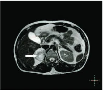
A 38 years old male patient, presenting with dyspeptic complaints, applied to our general surgery department. Oesophagogastroduodenoscopy showed normal findings. As the abdominal ultrasonography yielded a hyperechogenic solid mass of 50x59 mm at the right adrenal gland, magnetic resonance imaging done and a 60x55 mm size solid mass with regular borders and showing heterogeneous contrast uptake was detected (Figure 1). The patient who had no history of hypertension, palpitation, tremor or loss of weight complaints had a normal physical examination. There was nothing to indicate pheochromocytoma in the family history.
In the laboratory tests, serum biochemical examination, electrolyte levels and complete blood count were found within normal limits. The plasma levels of cortisol, aldosteron and ACTH, plasma renin activity; and amounts of VMA, metanephrine and 5-HIAA in 24 hour urinary output were all within normal range (cortisol: 6.80 µg/dl [5-25 µg/dl], Plasma renin activity: 1.90 ng/ml/h [0.98-4.18 ng/ml/h], ACTH: 27.5 pg/ml [9-52 pg/ml] aldosteron: 2.66 pg/ml [2-16 pg/dl], VMA in 24 hour urinary output:7.55 mg/day [1-10 mg/day], metanephrine: 0.78 mg/day [0-1 mg/day], 5-HIAA: 6.99 mg/day [2-10 mg/day]). ECG revealed normal sinus rhythm, 80 beats /min and no axis deviation, ST - T wave changes or premature ventriculer beats. Chest radiogram was normal. After endocrinologic and urologic consultations, the diagnosis of incidentaloma was made and surgical operation was planned by the urology department.
The patient was accepted to the operating room and monitorization of ECG, noninvasive blood pressure and pulse oximetry was started. Induction of anaesthesia was done with 6mg/kg thiopental sodium, 75 µg fentanyl and 1 mg/kg vecuronium. The vitality signs of the patient were observed normal during the induction and intubation. Sevoflurane 2% in 50% O2 and 50% N2O was given for
the maintenance of anaesthesia. Invasive blood pressure monitorization via radial artery was done all through the operation. On the position of the left lateral decubitus, upon opening the right retroperitoneal region, the blood pressure of the patient rose to 180/100 mmHg and the surgery was paused as a result. 100 mg propofol, 100 µg fentanyl, 2 mg vecuronium and a total of 30 mg glyceryl trinitrate (Nitro)® were given. Infusion of glyceryl trinitrate was started at a rate of 21µg/kg/min but blood pressure control couldnt be achieved, and then Sodium nitroprussid (Nipruss)® infusion was started at a rate of 1µg/kg/min. As the blood pressure could be lowered to 150/80 mmHg surgeons were allowed to resume the operation. To increase the depth of anaesthesia inhaled, sevoflurane concentration was increased. Despite the antihypertensives used, the blood pressure raised up to 330/120 mmHg when the tumor was touched by surgical team. As it was considered as a pheochromocytoma attack, the operation was not allowed to go on. Infusion of esmolol at a rate of 300µg/kg/min was started in addition to the prior antihypertensive treatments. The patient was extubated when the blood pressure was 140/80 mmHg and the heart rate was 86 beats/min. The patient was transferred to the intensive care unit (ICU) with antihypertensive infusions. During the postoperative period, the 24 hour urine measurements of VMA: 44 mg/day, metanephrine: 3.28 mg/day, 5-HIAA: 48.8 mg/day and HVA: 60 mg/day confirmed the diagnosis of pheochromocytoma. The patient was supervised in the ICU until his blood pressure was within normal limits for 20 hours and then he was discharged with the prospects of preparing for the surgery. But patient did not return to hospital for pheochromocytoma excision. His good health status was learned through phone consultation.
Hypertensive Crisis in a Patient With an Undiagnosis Pheocromocytoma
Effective premedication will reduce the risks of peroperative hypertension and tachycardia due to possible high levels of circulating catecholamines. While 50% of pheochromocytomas are diagnosed postmortem, some are diagnosed with high mortality hypertensive attacks. It should be kept in mind that the high plasma catecholamine levels may lead to vasoconstriction during operation and therefore patients can need volume replacement postoperatively.
The fact that the patients with adrenal masses are examined in detail with appropriate tests preoperatively and that they are prepared for the operation are crucial in terms of lowering the mortality and morbidity. In this patient, even though there was no evidence of pheochromocytoma in the preoperative tests, during the operation a life threatening hypertensive attack resistant to antihypertensive medications was encountered. Despite all the newest medications and techniques that have been developed, t h e a n e s t h e t i c m a n a g e m e n t o f u n d i a g n o s e d pheochromocytomas is still a source of unexpected problems for the anaesthetist. It was a great chance in this case that there were no complications in spite of the hypertensive crisis.
Figure 1. Abdominal MRI showes a solid mass of right
adrenal gland with regular borders and heterogeneous contrast uptake.
Discussion
In a study in which 2000 incidentalomas were examined, of these masses 82% were found out to be nonfunctional adenomas, 5.3% subclinical Cushing syndromes, 5.1% pheochromocytomas, 1% aldosteron secreting adenomas, 4.7% adrenocortical carcinomas and 2.5% metastatic diseases (7) The clinical suspicion for diagnosis of pheochromocytoma is of primary concern. For all adrenal masses, a detailed hormonal screening prior to surgery is n e c e s s a r y. E v e n t h o u g h t h e s y m p t o m s o f pheochromocytoma may vary, the triad of hypertension, headache and diaphoresis are present in 40-80% of the patients (8).
Pheochromocytoma can be diagnosed by documenting the increase of plasma and urinary catecholamines and their products. The presence of catecholamines and their metabolites in the plasma and urine is evidence for presence of pheochromocytoma with 95% certainty. The most sensitive test in diagnosis is the measurement of free metanephrine levels of plasma (9). As seen in Table I, metanephrine in urine is 95% diagnostic, but it was within normal limits in this patient. The aim in the preoperative preparation of pheochromocytoma is to bring the blood pressure, heart rate and intravascular volume to normal. Roizen criteria (10) can be used to evaluate the adequacy of the preoperative preparation.
Table I. Diagnostic test in pheochromocytoma (11)
Sensitivity Specifity Imaging criteria MRI 100% I-MIBG Scintigraphy 100% Computerised tomography 88% Ultrasonography 83% Biochemical criteria Plasma Free metanephrines 99% 89% Catecholamines 84% 81% Urine Fractioned metanephrines 97% 69% Catecholamines 86% 88% Total metanephrine 77% 93% VMA 64% 95%
Züleyha Kazak, Kamil Darcin, Kansu Kazbek, Sumru Sekerci, Hikmet Süer
References
1.Kloos RT, Gross MD, Francis IR, Korobkin M, Shapiro B. Incidentally discovered adrenal masses. Endocr Rev 1995; 16:460484.
2.Barzon L, Sonito N, Fallo F,ÊPalu G,ÊBoscaro M. Prevalence and natural history of adrenal incidentalomas. Eur J Endocrinol 2003; 149:273-285.
3.Ross NS, Aron DC. Hormonal evaluation of the patient with an incidentally discovered adrenal mass. N Engl J Med 1990; 323:1401-1405.
4.Pacak K, Eisenhofer G, Ahlman H, et al. Pheochromocytoma: recommendations for clinical practice from the First International Symposium. October 2005. Nat Clin Pract Endocrinol Metab 2007; 3:92102. 5.Ilias I, Pacak K. A clinical overview of pheochromocytomas/paragangliomas and carcinoid tumors. Nucl Med Biol. 2008; 35:27-34.
6.O'Riordan JA. Pheochromocytomas and anaesthesia. Int Anesthesiol Clin 1997; 35:99-127.
7.Young WF Jr. Management approaches to adrenal incidentalomas. Endocrinol Metab Clin North Am 2000, 29:159185.
8.Kaltsas GA, Papadogias D, Grossman AB. The clinical presentation (symptoms and signs) of sporadic and familial chromaffin cell tumours (phaeochromocytomas and paragangliomas). Front Horm Res 2004; 31:6175. 9.Lenders JW, Pacak K, Walther MM, et al. Biochemical diagnosis of pheochromocytoma: which test is best? JAMA 2002; 287:14271434.
10.Roizen MF, Horrigan RW, Koike M, et al. A prospective randomized trial of four anesthetic techniques for resection of pheochromocytoma. Anesthesiology 1982;57:43. 11. Myklejord DJ. Undiagnosed Pheochromocytoma:The Anesthesiologist Nightmare. Clin Med Res 2004; 1:59-62.
Hypertensive Crisis in a Patient With an Undiagnosis Pheocromocytoma