O
h
r
i
g
in
a
l
R
Turgay Aslan1, Özge Kurmuş1, Cemal Köseoğlu2, Ahmet Göktuğ Ertem2, Mehmet Erdoğan2, Tolga Han Efe2, Mehmet Bilge2 1Department of Cardiology, Ufuk University Faculty of Medicine, 2Department of Cardiology, Ankara Atatürk Training and Research Hospital, Ankara, Turkey
Aortic stiffness and hypertension
Aortic stiffness index and diurnal variability
(dipper/non-dipper) in hypertensive patients
DOI: 10.4328/JCAM.5547 Received: 30.11.2017 Accepted: 05.01.2018 Published Online: 07.01.2018 Printed: 01.03.2018 J Clin Anal Med 2018;9(2): 128-32 Corresponding Author: Turgay Aslan, Department of Cardiology, Ufuk University Faculty of Medicine, Ankara, Turkey.
GSM: +905064742819 E-Mail: [email protected]
Abstract
Aim: Aortic stiffness is an independent predictor of primary coronary events in hypertensive patients. In this study, we aimed to evaluate how aortic elasticity parameters [aortic distensibility (AD), aortic stiffness index (ASI), aortic strain (AS)] is affected by the diurnal rhythm of the blood pressure in hypertensive and normotensive individuals without a known cardiovascular disease. Material and Method: In this cross-sectional study, 58 hypertensive and 60 normotensive patients without known cardiovascular disease were enrolled. Ambulatory blood pressure monitoring was performed on hypertensive patients, and transtho-racic echocardiography was performed on all study participants. The AD, ASI, and AS were compared between controls, dippers, and non-dippers. Results: In our study, the highest “aortic stiffness index” value was detected in reverse dippers group followed by non-dippers group, dippers group, and control group with regard to age. Also, aortic strain and aortic distensibility values were highest in control group, respectively dippers group, non-dippers group and reverse dippers group. Discussion: We determine there is a relation with the diurnal rhythm of blood pressure and aortic stiffness parameters.
Keywords
Aortic Stiffness; Aortic Elasticity; Dipping Hypertension; Non-Dipping Hypertension; Nocturnal Blood Pressure
This study was previously presented as a poster at the
‘‘30th Turkish Cardiology Congress with International Participation’’
congress held on 23-26 October in Antalya.
| Journal of Clinical and Analytical Medicine
Aortic stiffness and hypertension
Introduction
Aortic stiffness is an independent predictor of all-cause and cardiovascular mortality. It occurs as a result of aging, smoking, hypercholesterolemia, diabetes mellitus and hypertension [1,2]. Increased aortic stiffness or decreased distensibility is indica-tive of the extensive involvement of atherosclerotic vascular system [3]. It has a decisive importance for vascular disorders such as renal disease, stroke, heart failure and myocardial in-farction [4,5].
Ambulatory blood pressure monitoring (ABPM) is a frequently used method to diagnose or follow up hypertension. It is consid-ered to be useful for patients with suspected white-coat hyper-tension, in elderly with resistant hypertension having symptoms related to hypotension due to antihypertensive drugs and in pa-tients who are believed to have important changes in nocturnal blood pressure.
Normal population studies revealed a nocturnal decrease in blood pressure (BP) in adults. If the average systolic BP (SBP) and diastolic BP (DBP) difference between waking and sleeping is 10–20%, then it is considered as dipping pattern; if the differ-ence is <%10 then it is considered as non-dipping pattern and also nocturnal blood pressure is high during the day it is con-sidered as reverse-dipping pattern. Patients having a difference >%20 were considered to have an extreme-dipping pattern.The underlying pathogenetic mechanisms of the non-dipping pat-tern are not fully understood. There are multiple possible un-derlying pathophysiological mechanisms in the impaired BP de-crease at night. Abnormal neurohormonal regulation, inde-creased dietary sodium intake, sedentary life and smoking of tobacco have been implicated in the blunted circadian rhythm of BP [6]. Certain diseases such as diabetes and chronic renal diseases also affect the circadian BP rhythm.
The aim of this study was to evaluate how aortic elasticity is affected by the diurnal rhythm of BP in hypertensive individuals without a known cardiovascular disease.
Material and Method
Study Design and Patient Population: In this study, 58 hyper-tensive patients who were previously diagnosed with essential hypertension and followed with medical therapy and 60 con-trols without hypertension were included. Patients with known or suspected coronary artery disease (CAD), reduced ejection fraction (EF) (<%60), more than mild valvular stenosis/regur-gitation, heart failure, cardiomyopathy, prosthetic heart valve, secondary hypertension, renal failure, history of cerebrovascu-lar accident, congenital or acquired aortic diseases, aortic an-eurysm, history of cardiovascular or aortic surgery, connective tissue disorders, low-quality echocardiographic and ultrasono-graphic images, conduction abnormalities, and atrial fibrillation on the electrocardiogram (ECG) were excluded. BP measure-ments were performed a week apart, at sitting position and after the rest for 5-10 minutes. Patients with a blood pressure of <140/90 were considered normotensive. ABPM was applied to the hypertensive group. Hypertensive patients were taking angiotensin-converting enzyme (ACE) inhibitors, angiotensin receptor blockers (ARB), thiazide diuretics group, and calcium channel blockers as a monotherapy or combination therapy.
Ambulatory Blood Pressure Recording: The recorder was pro-grammed to take BP measurements every 15 minutes during the daytime and every 30 minutes during the nighttime. The daytime period was defined as the interval between 6 AM and 12 PM and the nighttime period as between 12 PM and 6 AM. The patients were instructed to attend their usual day-to-day activities, but to keep still and relax their arms at the times of measurements. For the following conditions, the values were not taken into consideration: Systolic BP (SBP) >270 mmHg and <70 mmHg, for diastolic BP (DBP) >150 mmHg and <40 mmHg values; Equal measurements of SBP’s and DBP’s; >30 mmHg differences between the following 2 measurements of SBP’s or DBP’s. If the acceptable BP values were over 14 at daytime and over 7 at night, records were considered adequate.
Echocardiographic Examination: The echocardiographic exami-nations were obtained using “General Electric Vivid 7” with a multifrequency transducer (2.5–3.5 MHz) equipped with tissue Doppler imaging (TDI) technology (Vingmed Ultrasound, GE, Horten, Norway). All echocardiographic measurements were performed in five consecutive cycles, and their average was used for the calculations. Two-dimensional and standard M-Mode measurements were performed according to the Ameri-can Society of Echocardiography (ASE) recommendations [7]. The EF, end-diastolic volume, and end-systolic volume were analyzed by the Simpson method and Teichholz method. For the calculation of aortic stiffness, the diameter of the as-cending aorta was measured from the same view on the M-mode tracing at a level of 3 cm above the aortic valve. The systolic aortic diameter (AoS) was measured at the maximal anterior motion of the aorta, whereas the diastolic aortic di-ameter (AoD) was measured at the peak of the QRS complex on the simultaneously recorded ECG. Five consecutive measure-ments were performed, and their mean was calculated. At this time, brachial blood pressure was measured.
Assessment of Aortic Stiffness: Aortic elasticity was calculated according to the following three formulas:
‘’Aortic strain’’ (AS) = (AoS-AoD)/AoDx100,
“Aortic distensibility” (AD) = (2×Aortic strain/100)/(SBP-DBP) (10-³ cm2.dyn-¹)
“Aortic stiffness index” (ASI)= ln(SBP/DBP)/(AORTIC strain/100) AoD = aortic root diastolic diameter, AoS =aortic root end-systolic diameter
Statistical analysis were performed with SPSS 18.0 (PASW Sta-tistics for Windows, Version 18.0. Chicago: SPSS Inc.) software. Kolmogorov-Smirnov test was used to evaluate normal distri-bution. Continuous variables were expressed as mean ± SD or median; categorical variables were defined as numbers and percentages. Student’s t-test was used to compare continuous variables. Differences in the distribution of categorical vari-ables were assessed by chi-square analysis. One-way analysis of variance (ANOVA) was used to analyze the intragroup com-parisons. A p-value <0.05 was considered significant. Post-hoc multiple comparisons were made using Bonferroni correction.
Results
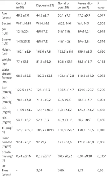
We enrolled 118 participants, and 23 (%39,65) were in the dip-pers group, 28 (%48,27) in the non-dipdip-pers group, 7 (%12,01) in the revers-dippers group, and 60 in the control group. Mean age of the hypertensive patients were 47,6 ±8,63 years and mean age of the control group were 48,5 ±7,0 years. Mean age was 44,5 ±9,7 years in dippers group, 50,1 ±7,7 in the non-dippers group, 47,5±5,7 in reverse dippers group. The majority of the partici-pants were women (%69,49). In the dippers group there were 14 women and 9 men, in the non-dippers group there were 22 women and 6 men, in reverse dippers group there were 4 wom-en and 3 mwom-en. The mean ambulatory systolic and diastolic blood pressure of hypertensive patients was 127,1±14,3/71,3±10,5 mmHg under drug treatment. It was 125,0±11,3/71,3±10,2 mmHg on dippers group, 126,3±14,7/69,5±9,5 mmHg on non dippers group, 134,0±20,0/78,5±13,7 on reverse dippers group. The mean clinical blood pressure of normotensive patients was 122,5±17,2/78,8±13,0 mmHg. Mean creatinine values of the control group were found to be lower than those of patient group although the patients who had impaired renal function tests were excluded, and there was no statistically significant difference between the groups in the subgroup analysis. The demographic and clinical characteristics of the patients are summarized in Table 1.
There was a significant difference between dippers group, non-dippers group, reverse group, and control group in terms of AS, AD, ASI. AS and AD was lowest in reverse dippers group, but ASI was highest in reverse dippers group (Table 2).
AS was significantly higher in control group compared to non-dippers, reverse dippers groups (p<0,01, p<0,01) respectively. There was no significant difference between the control and the dippers groups in terms of AS and ASI (p:0,87, p:0,23). AD was higher in control group than the dippers group, non dip-pers group, reverse dipdip-pers group (p<0,01, p<0,01, p<0,01) re-spectively. AS and AD were significantly higher in dippers group compared to non-dippers and reverse dippers group (p<0,01, p<0,01, p<0,01, p<0,01) respectively. ASI was significantly lower in dippers group compared to non-dippers and reverse group (p<0,01, p<0,01) respectively. There was no significant difference between non-dippers and reverse dippers groups in terms of AS and ASI (p=0,56, p=0,54). The AD was significantly higher in the non-dippers group than the reverse dippers group (p<0,01) (Table 3).
Discussion
In our study, the aortic elasticity parameters AS and AD were significantly lower, and ASI was higher in the non-dippers group than dippers group. Also, ASI was higher and AS, AD was lower in reverse dipper group than other 3 groups: normal subjects, the dippers, and the non-dippers group.
There is a clear, albeit complex, relationship between great ar-tery stiffness and atherosclerosis. Arterial stiffness and athero-sclerosis usually co-exist, and some studies have described a correlation between atherosclerotic burden and aortic stiffness. In a series of clinical studies, increased arterial stiffness was shown in patients with CAD compared to the ones without CAD [8-11].
Arterial stiffness is a predictor of future cardiovascular and coronary events [12]. Arterial stiffness was shown to increase with age in a group of patients without cardiovascular risk fac-tors [13]. After eliminating aging effects and other risk facfac-tors, increased arterial stiffness is an indicator of CAD, cerebrovas-cular and peripheral artery atherosclerosis [14,15]. Increasing arterial stiffness with advancing age, has been associated with a rise in SBP and pulse pressure (PP). An increase in SBP along with a decrease in DBP arising from the increase in PP—an in-dicator of the impaired elastic parameters of aorta—may lead to an elevation in left ventricle afterload and thereby impairing coronary perfusion [16,17]. Elastic parameters of aorta were shown to be a strong and independent risk factor for recurrent
Table 1. Clinical and demographic characteristics of patients Control
(n:60) Dippers(n:23) Non dip-pers(28) Revers dip-pers(n:7) P value Age (years) 48,5 ±7,0 44,5 ±9,7 50,1 ±7,7 47,5 ±5,7 0,077 Sex (n) W:41, M:19 W:14, M:9 W:22, M:6 W:4, M:3 0,505 DM (n,%) 12 (%20) 4(%17,3) 5(%17,8) 1(%14,2) 0,979 Smoke (n,%) 14(%23,3) 4(%17,3) 4(%14,2) 3(%42,8) 0,376 Height (cm) 162,1 ±8,9 163,6 ±7,8 162,5 ± 8,9 159,1 ±8,3 0,650 Weight (kg) 77 ±13,6 81,2 ±16,0 80,8 ±13,4 88,5 ±16,7 0,165 Waist circum-ference (cm) 98,2 ±12,3 102,3 ±13,8 102,1 ±12,8 110,5 ±14,0 0,073 SBP (mmHg) 122,5 ±17,2 125 ±11,3 126,3 ±14,7 134,0 ±20,7 0,290 DBP (mmHg) 78,8 ±13,0 71,3 ±10,2 69,5 ±9,5 78,5 ±13,7 0,001 LDL (mg/dl) 118,9 ±34,2 129,7 ±30,0 128 ±34,2 123,3 ±34,2 0,488 HDL (mg/dl) 54,7 ±16,7 52,3 ±9,3 49,9 ±11,6 50,7 ±8,9 0,480 TG (mg/ dl) 125,1 ±65,0 183,3 ±109,9 160,8 ±56,7 138,7 ±55,5 0,010 Blood Glucose (mg/dl) 92,4 ±26,7 92 ±9,7 121 ±67,6 121,0 ±40,0 0,006 Creati-nin (mg/ dl) 0,74 ±0,16 0,85 ±0,17 0,83 ±0,23 0,84 ±0,20 0,035* HT Time (years) - 3,04 3,86 2,71 0,63
* In subgroup analysis there was no significant difference between creatinine levels.
# DM: Diabetes mellitus, SBP: systolic blood pressure, DBP: diastolic blood pressure, LDL: low density lipoprotein, HDL: high density lipoprotein, TG: triglycerides, HT: hypertension
Table 2. Comparison of aortic elasticity between controls, dippers, non-dippers and reverse non-dippers
Control
(n:60) Dippers(n:23) Non dippers(n:28) Reverse dippers(n:7) P value AS 12,1 ±2,3 11,6 ±2,9 6,0 ±2,4 4,6 ±2,2 <0,001 ASI 3,6 ±1,0 5,2 ±1,8 11,7 ±5,9 13,5 ±5,7 <0,001 AD 6,0 ±1,9 4,3 ±1,1 2,1 ±0,9 1,7 ±0,9 <0,001 * AS: aortic strain, ASI: aortic stiffness index, AD: aortic distensibility
Aortic stiffness and hypertension
acute coronary events in patients with CAD [18]. Great artery stiffness was reported to be the major indicator of exercise-re-lated myocardial ischemia in patients with moderate CAD [19]. A nocturnal decrease in BP as a characteristic of diurnal BP al-teration was first put forward by Hill in 1898. Subsequently, less than 10% decrease in nocturnal BP was found to be related to left ventricle hypertrophy, cerebrovascular injury and cardiovas-cular events [20-22]. Kazuomi et al. detected a high occurrence of stroke in patients having reverse dipping and extreme dip-ping compared to the patients having dipdip-ping and non-dipdip-ping patterns. An extreme decrease in nocturnal SBP was proposed to cause silent cerebral infarcts through leading to cerebral hy-poxia in patients with “extreme dippers” pattern [23].
Physiological reduction in the night BP decreases with age. The possible reasons for this are suggested to be aging, decrease in great vessel distensibility values due to atherosclerosis, and dominance of sympathetic system-related vasoconstriction on parasympathetic system-related vasodilation as the result of the impairment in autonomic nervous system [24].
In accordance with our results, Bitigen et al. have also shown that there is a relation between diurnal BP pattern and aor-tic elasaor-tic properties. They reported that the highest ASI value was found in reverse dippers patients followed by non-dippers patients and the lowest in dippers patients. The ASI value of reverse dippers and non-dippers was higher than in the dippers group [25].
Our study has some limitations. One of the main limitations of this study is the small sample size. Further large-scale studies will be required to validate our results. Secondly, our study was a cross-sectional study so it couldn’t help to determine cause and effect. Another limitation is that the drug treatments used in the hypertension group are not homogeneous. It may have affected the diurnal rhythm of blood pressure. It may not be appropriate to establish a one-way causal link between two de-terminants; as nocturnal blood pressure decrease and arterial stiffness, because of they were influenced by many factors. In our study, the patients who had known cardiovascular diseases, which could influence the aortic elasticity properties, were ex-cluded.
Conclusion
We detected a relationship between two independent determi-nants of cardiovascular and cerebrovascular events develop-ment; “aortic stiffness parameters” and “inadequate nocturnal blood pressure decrease”. According to this relationship, aortic stiffness was observed highest in reverse dippers group fol-lowed by non-dippers, dippers and control groups. Aortic stiff-ness may be one of the early stage findings of atherosclerosis. Mortality and morbidity may be reduced through detection and proper treatment of increased aortic stiffness in the early pe-riod in hypertensive patients.
Scientific Responsibility Statement
The authors declare that they are responsible for the article’s scientific content including study design, data collection, analy-sis and interpretation, writing, some of the main line, or all of the preparation and scientific review of the contents and ap-proval of the final version of the article.
Animal and human rights statement
All procedures performed in this study were in accordance with the ethical standards of the institutional and/or national re-search committee and with the 1964 Helsinki declaration and its later amendments or comparable ethical standards. No ani-mal or human studies were carried out by the authors for this article.
Funding None
Conflict of interest
None of the authors received any type of financial support that could be considered potential conflict of interest regarding the manuscript or its submission.
References
1. Chae CU, Pfeffer MA, Glynn RJ, Mitchell GF, Taylor JO, Hennekens CH. Increased pulse pressure and risk of heart failure in the elderly. JAMA. 1999; 281(7): 634-9. 2. Franklin SS, Larson MG, Khan SA, Wong ND, Kannel WB, Levy D. Does the rela-tion of blood pressure to coronary heart disease risk change with aging? The Framingham Heart Study. Circulation. 2001; 103(9): 1245-9.
3. Blacher J, Guerin AP, Pannier B, Marchais SJ, Safar ME, London GM. Impact of aortic stiffness on survival in end-stage renal disease. Circulation. 1999; 99(18): 2434 -9.
4. Laurent S, Katsahian S, Fassot C, Tropeano Al, Gautier l, Laloux B, et al. Aortic stiffness is an independent predictor of fatal stroke in essential hypertension. Stroke. 2003; 34(5): 1203-6.
5. Mattace-Raso FU, van der Cammen TJ, Hofman A, van Popele NM, Bos ML, Schalekamp MA, et al. Arterial stiffness and risk of coronary heart disease and stroke: the Rotterdam Study. Circulation. 2006; 113(5): 657-63.
6. Kanbay M, Turgut F, Uyar ME, Akcay A, Covic A. Causes and mechanisms of non-dipping hypertension. Clin Exp Hypertens. 2008; 30(7): 585–97.
7. Sahn DJ, Maris A, Kisslo J, Weyman A. For the committee on M-mode standard-ization of the American Society of Echocardiography recommendation regard-ing quantitation in M-mode echocardiographic measurements. Circulation. 1978; 58(6): 1072-83.
8. Hirai T, Sasayama S, Kawasaki T, Yagi S. Stiffness of systemic arteries in pa-tients with myocardial infarction: a noninvasive method to predict severity of coronary atherosclerosis. Circulation. 1989; 80(1): 78–86.
9. Dart AM, Lacombe F, Yeoh JK, Cameron JD, Jennings GL, Laufer E, et al. Aortic distensibility in patients with isolated hypercholesterolemia, coronary artery dis-ease, or cardiac transplant. Lancet. 1991; 338(8762): 270-3.
10. Lehmann ED, Hopkins KD, Jones RL, Rudd AG, Gosling RG. Aortic distensibil-ity in patients with cerebrovascular disease. Clin Sci(Colch). 1995; 89(3): 247-53. 11. Gatzka CD, Cameron JD, Kingwell BA, Dart AM. Relation between coronary ar-tery disease, aortic stiffness, and left ventricular structure in a population sample. Hypertension. 1998; 32(3): 575-8.
12. Najjar SS, Scuteri A, Lakatta EG. Arterial aging: is it an immutable cardiovas-Table 3. Comparison of aortic elasticity parameters between subgroups
Co-Di Co-NonDi Co-RDi
Di-Non-Di Di-RDi Non-Di-RDi
AS Mean difffe-rence 0,46 6,09 7,44 5,62 6,97 1,35 p 0,87 <0,001 <0,001 <0.001 <0.001 0,56 AD Mean difffe-rence 1,61 3,81 4,27 2,19 2,65 0,45 p <0,001 <0,001 <0,001 <0,001 0,001 <0,001 ASI Mean difffe-rence -1,53 -8,05 -9,92 -6,52 -8,38 -1,86 p 0,23 <0,001 <0,001 <0,001 <0,001 0,54 *Co-Di: Control group and dippers group. Co-NonDi: Control group and non dippers group. Co-RDi: Control group and revers dippers group. Di-RDi: Dip-pers group and Revers dipDip-pers group. NonDi-RDi: Non dipDip-pers and revers dippers group
cular risk factor? Hypertension. 2005; 46(3): 454-62.
13. Gary FM, Helen P, Emelia JB, Martin GL, Michelle JK, Joseph AV, et al. Changes in arterial stiffness and wave reflection with advancing age in healthy men and women. Hypertension. 2004; 43(6): 1239-45
14. Galis ZS, Khatri JJ. Matrix metalloproteinases in vascular remodeling and ath-erogenesis: Circulation Research. 2002; 90(3): 251-62.
15. Benetos A, Safar M, Rudnichi A, Smulyan H, Richard JL, Ducimetieére P, et al. Pulse pressure: a predictor of long-term cardiovascular mortality in a French male population. Hypertension. 1997; 30(6): 1410 -5.
16. Franklin SS. Ageing and hypertension: the assessment of blood pressure indi-ces in predicting coronary heart disease. J Hypertens Suppl. 1999; 17(5): 29-36.
17. Safar ME. Pulse Pressure in essential Hypertension: clinical and therapeutical implications. J Hypertens. 1989; 7(10): 769-76.
18. Stefanadis C, Dernellis J, Tsiamis E, Stratos C, Diamantopoulos L, Michaelides A, et al. Aortic stiffness as a risk factor for recurrent acute coronary events in patients with ischemic heart disease. Eur Heart J. 2000; 21(5): 390-6.
19. Kingwell BA, Waddell TK, Medley TL, Cameron JD, Dart AM. Large artery stiff-ness predicts ischemic threshold in patients with coronary artery disease. J Am Coll Cardiol. 2002; 40(4): 773-9.
20. Palatini P, Penzo M, Racioppa A, Zugno E, Guzzardi G, Anaclerio M, et al. Clinical relevance of nighttime blood pressure and of daytime blood pressure variability. Arch Intern Med. 1992; 152(9): 1855-60.
21. Verdecchia P, Schillaci G, Gatteschi C, Zampi I, Battistelli M, Bartoccini C, et al. Blunted nocturnal fall in blood pressure in hypertensive women with future cardiovascular morbid events. Circulation. 1993; 88(3): 986-92.
22. Shimada K, Kawamoto A, Matsubayashi K, Nishinaga A, Kimura S, Ozawa T. Diurnal blood pressure variations and silent cerebrovascular damage in elderly patients with hypertension. J Hypertens. 1992; 10(8): 875-8.
23. Kazuomi K, Thomas GP, Takefumi M, Satoshi H, Joseph ES, Kazuyuki S. Stroke prognosis and abnormal nocturnal blood pressure falls in older hypertensives. Hy-pertension. 2001; 38(4): 852-7.
24. Kobrin I, Oigman W, Kumar A, Ventura HO, Messerli FH, Frohlich ED, et al. Diurnal variation of blood pressure in elderly patients with essential hypertension. J Am Geriatr Soc. 1984; 32(12): 896-9.
25. Bitigen A, Fotbolcu H, Güzet F, Karaahmet T, Başaran Y. Influence of diur-nal blood pressure rhythm on aortic elastic properties in hypertensive subjects. Acta Cardiol. 2006; 61(4): 417-20.
How to cite this article:
Aslan T, Kurmuş Ö, Köseoğlu C, Ertem AG, Erdoğan M, Efe TH, Bilge M. Aortic stiffness index and diurnal variability (dipper/non-dipper) in hypertensive patients. J Clin Anal Med 2018;9(2): 128-32.