See discussions, stats, and author profiles for this publication at: https://www.researchgate.net/publication/257751855
Platelet-Rich Plasma Injection Is More Effective than Hyaluronic Acid in the
Treatment of Knee Osteoarthritis.
Article in Acta chirurgiae orthopaedicae et traumatologiae Cechoslovaca · October 2013 Source: PubMed CITATIONS 60 READS 1,132 5 authors, including:
Some of the authors of this publication are also working on these related projects:
hybrid ceramics, universal adhesives, dental lasersView project Ferhat Say
Ondokuz Mayıs University, Samsun, Turkey
40PUBLICATIONS 198CITATIONS SEE PROFILE
Deniz Gürler
Samsun Training And Research Hospital
19PUBLICATIONS 117CITATIONS SEE PROFILE
Murat Bulbul
Istanbul Medipol University
135PUBLICATIONS 453CITATIONS SEE PROFILE
Melih Malkoc
46PUBLICATIONS 127CITATIONS SEE PROFILE
All content following this page was uploaded by Ferhat Say on 12 August 2014.
Platelet-Rich Plasma Injection Is More Effective
than Hyaluronic Acid in the Treatment of Knee
Osteoarthritis
Injekce plazmy obohacené destičkovým koncentrátem je efektivnější než kyselina
hyaluronová v léčbě gonartrózy
F. SAy1, D. GüRlER2, K. yEnER2, M. BülBül3, M. MAlKOç3
1 Department of Orthopaedics and Traumatology, Faculty of Medicine, Ondokuz Mayıs University, Samsun, Turkey
2 Department of Orthopaedics and Traumatology, Samsun Training and Research Hospital, Samsun, Turkey
3 Department of Orthopaedics and Traumatology, Faculty of Medicine, Medipol University, Istanbul, Turkey
ABSTRAcT
PURPOSE OF THE STUDy
There is increasing use of platelet-rich plasma (PRP) in orthopaedics as it is a simple, cheap and minimally invasive technique. This study aimed to compare the effects of the use of PRP and hyaluronic acid (HA) injections in the knee of patients diagnosed with and being followed-up for degenerative arthritis.
MATERIALS AnD METHODS
This prospective study included 90 patients with complaints of knee pain with findings of mild or moderate degenera-tive arthritis. In the PRP group (n = 45), one intra-articular injection was applied and in the HA group (n = 45), three doses of intra-articular injection were applied. Clinical evaluation was made by Knee Injury and Osteoarthritis Outcome Score (KOOS) and a visual pain scale.
RESULTS
no severe adverse events was observed. Statistically significant better results in the KOOS score and visual pain scale was determined in PRP group than HA group at 3 months and 6 months follow up. The cost of the application for the PRP group was lower than that of the HA group.
COnCLUSIOn
The results of this study have shown the application of single dose PRP to be a safe, effective and low-cost method for treating OA. However, further studies are required for a more clear result.
Key words: platelet-rich plasma, hyaluronic acid, knee osteoarthritis, growth factors.
279/
ACTA CHIRURGIAE ORTHOPAEDICAEET TRAUMATOLOGIAE ČECHOSL., 80, 2013
ORIGInAL PAPER
PůvODní PRáCE
InTRODucTIOn
Osteoarthritis (OA) is a progressive joint disease associated with cartilage degeneration which leads to loss of movement and thus impairs daily activities and quality of life. With increasing age, the frequency of OA increases (10). Although OA may affect all joints, the joints most often involved clinically are the knees (16).
Biomechanical and biochemical changes of OA in the joints results in failure of the repair process of torn cartilage. The first pathological changes which are seen are fibrillation and loss of joint cartilage together with thickening and reshaping of the subchondral bone. Fi-nally, the full layer betweeen the joint is lost (18). Start-ing from the weight-bearStart-ing area of the joint cartilage, subchondral bone, ligaments, synovium, joint capsule and and the muscles surrounding the joint are affected (14). OA results in clinical pain and restricted movement for the patients.
In current pharmacological treatment of OA, the medications used include analgesics, non-steroid anti-inflammatory drugs, corticosteroids, viscosupplemen-tation, glucosamine and chondroitin sulfate. These medications provide a symptomatic effect on OA and treatment choices are being researched which will modify cartilage degeneration and the disease. Growth factors, matrix-metalloproteinase inhibitors, cytokines, nitric oxide and gene therapy are the treatment choices being researched in this area (9).
Platelet Rich Plasma (PRP) is a treatment form stimu-lating natural healing steps through growth factors con-tained in the platelets. PRP applied to the wound area accelerates the physiological healing process, provides support for the connection of cells, reduces pain and has an anti-inflammatory and anti-bacterial effect (29). Ob-taining PRP growth factor is a simple, cheap and easy way (1). As it is autogenous in origin, easy to prepare and has an excellent reliability profile, it has opened the door to new treatment (23).
Studies in literature have reported the use of PRP in degenerative joint pathologies (8, 12, 13, 15, 21, 22, 26, 30, 31). There are 16 concentration systems which can be used to obtain PRP. Leukocytes and growth factor contained in PRP are obtained in different amounts from these systems (7). Apart from the concentration systems, PRP can be obtained manually from peripheral blood (4, 5). Unanswered questions remain related to the applica-tion of PRP, such as the ideal volume, apllicaapplica-tion fre-quency, application period and platelet activation (24).
The aim of this study was to determine the effects on pain and function of PRP obtained manually as a cheap and easy method in the treatment of knee osteoarthri-tis and to compare these data with that of viscosupple-mentation which is often used in clinical practice. The hypothesis was that a single dose of manually-prepared PRP would reduce pain associated with OA and increase function and that this effect would be superior to the frequently-used HA injection.
MATERIAl AnD METHODS
This study was conducted as a prospective, com-parative, clinical study. Informed consent was obtained from all patients and the study was approved by the Local Ethics Committee. Patients with a diagnosis of OA who had been followed-up and had not seen any benefit from analgesic and anti-inflammatory treatment over a period of at least three months were included in the study. The application was made to symptomatic knees in patients determined with bilateral gonarthro-sis. Patients were not included if they had any system-ic disease, active tumour or haematologsystem-ically malign disease, infection, a history of anticoagulant use, Hb
value < 11g/dl, thrombocyte count <150,000/mm3 or
radiologically gonarthrosis at Kellgren-Lawrence (20) Stage 4.
A total of 90 patients who conformed with the de-fined criteria were included in the study. All the pa-tients were evaluated with standing anterior posterior and lateral knee radiographs. Staging was calculated according to the Kellgren-Lawrence classification. The patients were separated into two groups of 45 as PRP group and hyaluronic acid (HA) group. A single dose of PRP was administered to the PRP group. Low molecu-lar weighted HA (730 with 900 kDa) at 25 mg/2.5 ml dosage was injected to the HA group once a week for a total of three doses.
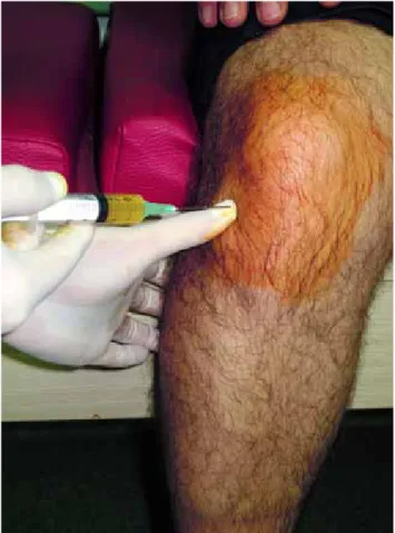
Fig. 1. PRP injection to the knee from the anterolateral portal.
The preparation and application of PRP was made by the same researcher (F.S.) to all the patients in that group under the same conditions. The method described by Anitua (4, 5) was used. A total of 30 cc peripheral blood was taken from antecubital re-gion of the patients into tubes containing 3.2% so-dium citrate. The tubes were centrifuges at 1800 rpm for eight minutes at room temperature. From the 3.5 ml PRP which was obtained, 1 ml was sent to the laboratory for bacteriological test and platelet count. After activation, the 2.5 ml PRP containing 5.5%
calcium chloride (CaCl2) (50 μl CaCl2 in 1 ml PRP)
was administered to the knee from the anterolateral portal under sterile conditions (Fig. 1). The patient remained in a supine position for 20 minutes follow-ing the administration. The result of the laboratory evaluation of the obtained PRP determined that the platelet count per mililitre increased by 400% com-pared to the thrombocyte count.
A standard rehabilitation programme was not applied to either group during follow-up. For the PRP group pa-tients, rest and not standing was recommended for the first day after the injection. Ice and paracetamol were recommended for the pain and swelling. Permission was given for the use of anti-inflammatory medication for seven days after PRP administration.
The patients were evaluated clinically pre-treatment and at the third and sixth month of follow-up. In the clinical evaluation, the Knee Injury and Osteoarthritis Outcome Score (28) (KOOS) and the Visual Analogue Pain Scale (VAS) were used. The patients were
ques-tioned with regard to side effects and subjective satis-faction.
Statistical evaluation
Results were stated as mean ± standard deviation (SD). Data were evaluated using SPSS software program (Windows Version 16.0). In the statistical evaluation of mean values beween groups, Student’s t-test, Chi-square test and MannWhitney U-test were used. The changes over time of the mean clinical scores of the groups were evaluated using Student’s t-test and Mann Whitney U-test. A value of p < 0.05 was accepted as statistically significant.
RESulTS
No complications were seen in any patient during the application or follow-up. In the PRP group, pain and mild swelling was determined in eight patients after the injection. All complaints were resolved with the applica-tion of ice and paracetamol.
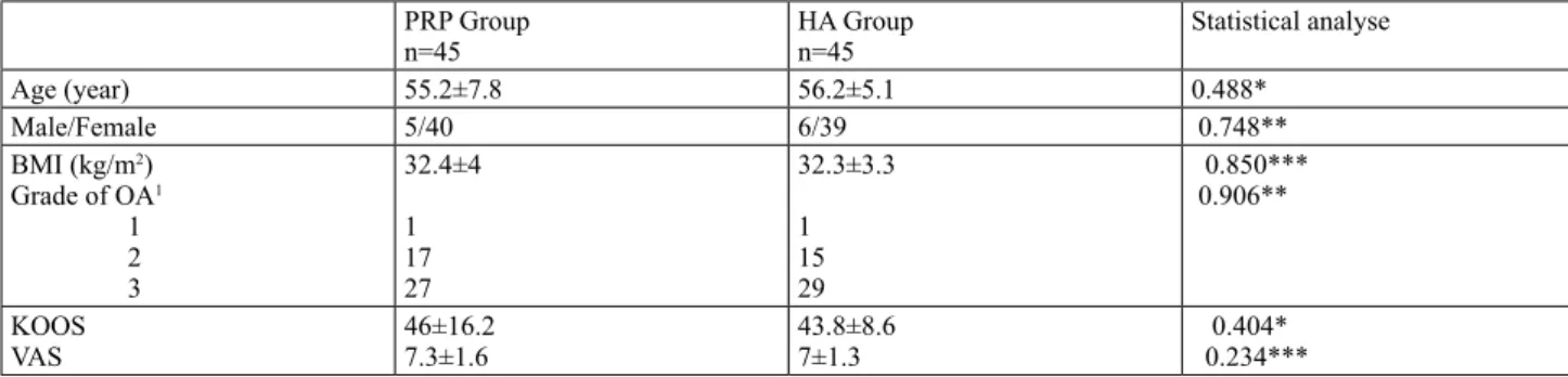
The patients of both groups were similar in terms of age, gender, body mass index, radiological osteoarthri-tis index and initial knee scores (Table 1). No staosteoarthri-tisti- statisti-cally significant difference was determined between the groups in terms of these factors.
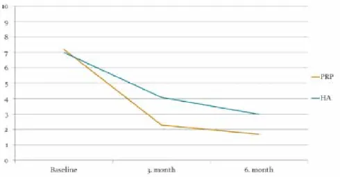
In the PRP group, the mean KOOS score was 76.9 ± 7.5 at the three month follow-up and 84.4 ± 6.2 at the six month follow-up (Fig. 2). The mean VAS score was de-termined as 2.3 ± 1.6 at three months and 1.7 ± 1.4 at six months (Fig. 3). Compared to the pre-treatment scores,
Table 1. Comparison of patients characteristics at baseline
PRP Group
n=45 HA Groupn=45 Statistical analyse
Age (year) 55.2±7.8 56.2±5.1 0.488* Male/Female 5/40 6/39 0.748** BMI (kg/m2) Grade of OA1 1 2 3 32.4±4 1 17 27 32.3±3.3 1 15 29 0.850*** 0.906** KOOS VAS 46±16.27.3±1.6 43.8±8.67±1.3 0.404* 0.234***
1 According to Kellgren-Lawrenceclassification
* Independent sample t-test **Chi-Square test
***Mann-Whitney test
Table 2. Comparison of KOOS and VAS scores of groups at baseline, thirth and sixth month
PRP Group n=45 HA Groupn=45 P value KOOS Baseline 3. month 6. month VAS Baseline 3. month 6. month 46±16.2 76.9±7.5 84.4±6.2 7.3±1.6 2.3±1.6 1.7±1.4 43.8±8.6 68.6±3.7 73.2±4.6 7±1.3 4.1±1.3 3±1 0.404* 0.02* 0.001* 0.234** 0.001** 0.001**
* Independent sample t-test **Mann-Whitney test
281/
ACTA CHIRURGIAE ORTHOPAEDICAEET TRAUMATOLOGIAE ČECHOSL., 80, 2013
ORIGInAL PAPER
PůvODní PRáCE
the difference between thethree and six month scores was statistically signifi-cant.
In the HA group, the mean KOOS score was 68.6 ± 3.7 at the three month follow-up and 73.2 ± 4.6 at the six month fol-low-up (Fig. 2). The mean VAS score was deter-mined as 4.1 ± 1.3 at three months and 3.0 ± 1.0 at six months (Fig. 3). Com-pared to the pre-treatment scores, the difference be-tween the three and six month scores was statisti-cally significant.
When the mean KOOS and VAS scores at the three and six month follow-up of the groups were compared with each other, the clinical scores of the PRP group were determined to be statistically significantly higher (Table 2).
DIScuSSIOn
In the treatment choices for OA, there are both phar-macological and non-pharphar-macological treatment choic-es. The use of these two choices combined together has been recommended in the treatment of OA (2, 16). How-ever, there is as yet no treatment choice which will com-pletely eradicate the damage caused by OA and reverse the course of the disease.
HA is a natural component of cartilage and connec-tive tissue. Although there are some contributory vis-coelastic properties of synovial fluid, it plays a physi-ological role as a trophic factor. It started to be used when it was determined that in OA patients the HA concentration fell and the chain length shortened. De-creased lubrication means inDe-creased weight-bearing on damaged cartilage and in the long-term this leads to impairment in the collagen network and the cartilage surface (6, 9). The mechanism within the joint after ap-plication is not yet known (9). Although it is recom-mended by the European League Against Rheumatism (EULAR) in treatment guidelines, the effect inside the joint is reported to start later than that of steroids which have been applied (19). HA is available in forms with different molecular weights. Negative aspects are that the effect is not long-lasting, it is expensive and the structural modifying effect has not been proven (6). In the current study, three doses of low molecular weight HA were used.
PRP was first used in 1987 in heart surgery to prevent excessive blood transfusion (11). Several studies have reported the use of PRP in orthopaedic joint patholo-gies (1, 8, 12, 13, 15, 21, 22, 26, 30, 31). More than 30 bioactive proteins are found within the alpha granules of platelets (5). Growth factors such as platelet-derived
growth factor, transforming growth factor, vascular en-dothelial growth factor and insulin like growth factor and proteins such as fibrin, fibronectin, vitronectin, and thrombospondin, which are found in PRP, play a role in many stages of tissue healing. The growth factors acti-vate some of the cells which have a function in tissue healing and thus provide soft tissue healing and bone regeneration (1).
The mechanism of the effect of PRP on the degenera-tive knee joint can be listed as inflammation regulation by means of the growth factors and proteins contained in it, correction of angiogenesis, anabolic and cartilage protective effects, cell differentiation and synovial cell modulation. However, as there is not a single mecha-nism of the effects of PRP on joint pathologies, it is more complex than previously thought (3).
Three different methods can be used to obtain PRP; automatic machines and commercial kits with double spin rotation, single spin rotation and manual PRP separation and selective blood filtration (platelet-pheresis). Anitua (5) reported that a platelet count over 300,000 /μl in PRP is effective. In another in vitro study, platelet concentration 2.5 times greater than the basal platelet count was reported to be the most ef-fective (17). The prepared PRP is activated by adding bovine or human thrombin or calcium chloride (27). Growth factors and cytokines are revealed with the formation of platelet gel from the activated PRP (3). In the current study PRP was prepared as single spin rotation and manually. In the analysis of the prepared PRP, concentration was determined as four times greater than the thrombocyte count in the peripheral blood. The prepared PRP was activated by the additon of calcium chloride.
In the evaluation of the results of the current study, the post-treatment thirth month and sixth month knee function scores of both groups were determined to have statistically significantly increased compared to the initial values and the pain scores had statistically significantly decreased compared to the initial values. In the comparison between the groups, the changes in
the functional and pain scores of the PRP group were sta-tistically significantly better than those of the HA group. In a study by Kon et al. (21) in which PRP was adminis-tered to 115 knees, the func-tional scores were reported to be different at the sixth month but the effect reduced by the twelfth month. Filardo et al. (12) reported the median pe-riod of clinical recovery to be nine months in a 24-month follow-up of the same patients. In the current study, although clinical improvement was de-termined in the sixth month of follow-up, it is not known how long this effect will last.
Differences have been shown in literature in the fre-quency of PRP administration at two doses (15), three doses (12, 13, 21, 22, 31) and four doses (8). In a pla-cebo controlled study by Patel et al (26), one group re-ceived a single dose of PRP and the other group rere-ceived two doses and the single dose of PRP was reported to be sufficient. In the current study, clinical benefit to OA patients with a single dose of PRP.
The manual method of obtaining PRP used in the cur-rent study is low-cost and effective. While the cost of automatic devices and kits to obtain PRP is several hun-dreds of dollars, the cost of the manual method used to prepare PRP was approximaely ten dollars (25).
For PRP obtained from autologous blood, there is no risk of immune reaction or disease transfer. There are no studies in literature warning of hyperplasia, carcinogeni-sis or tumour growth of PRP (29). No complications were encountered in any patient in the PRP group of the current study. Pain and mild swelling in eight patients was determined to have been resolved within a few days with ice and paracetamol.
The limitations of this study are that it was not ran-domised, there was no placebo control group, there were no radiological and biological results during follow-up to be compared with the functional and pain scores, the number of patients was low and the follow-up period was short.
In conclusion, the results of this study have shown that the administration of PRP in OA treatment is a cheap and effective method which was more effec-tive than HA injection in reducing pain and providing better functional results at the sixth month of follow--up. This effect was achieved with a single dose of PRP. As PRP alone may not be sufficient in the treat-ment of OA, it can be recommended for use in com-bination therapies. However, prospective, randomised, placebo-controlled, multi-centre studies are required to clarify these results and better understand the ef-fects of PRP.
Acknowledgment
The authors thank Prof. Dr. Nicola Maffulli for his suggestions and also thank Prof. Dr. Yüksel Bek for his help with the statistical analysis.
References
1. ALSOUSOU, J., THOMPSON, M., HULLEY, P., NOBLE, A., WILLETT, K.: The biology of platelet-rich plasma and its appli-cation in trauma and orthopaedic surgery: a review of the litera-ture. J. Bone Jt Surg., 91-B: 987–996, 2009.
2. ALTMAN, R. D.: Practical considerations for the pharmacologic management of osteoarthritis. Am. J. Manag. Care, .15 (8 Suppl): 236–243, 2009.
3. ANDIA, I., SÁNCHEZ, M., MAFFULLI, N.: Joint pathology and platelet-rich plasma therapies. Expert Opin. Biol. Ther., 12: 7–22, 2012.
4. ANITUA, E.: Plasma rich in growth factors: preliminary results of use in the preparation of future sites for implants. Int. J. Oral Maxillofac. Implants, 14: 529–535, 1999.
5. ANITUA, E., ANDIA, I., ARDANZA, B., NURDEN, P., NUR-DEN, A. T.: Autologous platelets as a source of proteins for heal-ing and tissue regeneration. Thromb. Haemost., 91: 4–15, 2004. 6. BROCKMEIER, S. F., SHAFFER, B. S.: Viscosupplementation
therapy for osteoarthritis. Sports Med. Arthrosc., 14: 155–162, 2006.
7. CASTILLO, T. N., POULIOT, M. A., KIM, H. J., DRAGOO, J. L.: Comparison of growth factor and platelet concentration from commercial platelet-rich plasma separation systems. Am. J. Sports Med., 39: 266–271, 2011.
8. CERZA, F., CARNI, S., CARCANGIU, A., DI VAVO, I., SCHI-AVILLA, V., PECORA, A., DE BIASI, G., CIUFFREDA, M.: Comparison between hyaluronic acid and platelet-rich plasma, intra-articular infiltration in the treatment of gonarthrosis. Am. J. Sports Med., 40: 2822–2827, 2012.
9. FAJARDO, M., DI CESARE, P. E.: Disease-modifying therapies for osteoarthritis: current status. Drugs Aging, 22: 141–161, 2005. 10. FELSON, D. T., NAIMARK, A., ANDERSON, J., KAZIS, L.,
CASTELLI, W., MEENAN, R. F.: The prevalence of knee os-teoarthritis in the elderly. the framingham osos-teoarthritis study. Arthritis Rheum., 30: 914–918, 1987.
11. FERRARI, M., ZIA, S., VALBONESI, M., HENRIQUET, F., VENERE, G., SPAGNOLO, S., GRASSO, M. A., PANZANI, I.: A new technique for hemodilution, preparation of autologous platelet-rich plasma and intraoperative blood salvage in cardiac surgery. Int. J. Artif. Organs: 10: 47–50, 1987.
283/
ACTA CHIRURGIAE ORTHOPAEDICAEET TRAUMATOLOGIAE ČECHOSL., 80, 2013
ORIGInAL PAPER
PůvODní PRáCE
12. FILARDO, G., KON, E., BUDA, R., TIMONCINI, A., DIMAR-TINO, A., CENACCHI. A., FORNASARI, P. M., GIANNINI. S., MARCACCI, M.: Platelet-rich plasma intra-articular knee injec-tions for the treatment of degenerative cartilage lesions and os-teoarthritis. Knee Surg. Sports Traumatol. Arthrosc., 19: 528–535, 2011.
13. FILARDO, G., KON, E., DI MARTINO, A., DI MATTEO, B., MERLI, M. L., CENACCHI, A., FORNASARI, P. M., MAR-CACCI, M.. PLATELET-RICH PLASMA VS HYALURONIC ACID TO TREAT KNEE DEGENERATIVE PATHOLOGY: study design and preliminary results of a randomized controlled trial. BMC Musculoskelet. Disord., 13: 229, 2012.
14. GARSTANG, S. V., STITIK, T. P.: Osteoarthritis: epidemiology, risk factors, and pathophysiology. Am. J. Phys. Med. Rehabil., 85 (11Suppl): S2–11; S12–14, 2006.
15. GOBBI, A., KARNATZIKOS, G., MAHAJAN, V., MALCHI-RA, S.: Platelet-rich plasma treatment in symptomatic patients with knee osteoarthritis: preliminary results in a group of active patients. Sports Health, 4: 162–172, 2012.
16. GOLDRING, S. R., GOLDRING, M. B.: linical aspects, pathol-ogy and pathophysiolpathol-ogy of osteoarthritis. J. Musculoskelet Neu-ronal. Interact., 6: 376–378, 2006.
17. GRAZIANI, F., IVANOVSKI, S., CEI, S., DUCCI, F., TONET-TI, M., GABRIELE, M.: The in vitro effect of different PRP concentrations on osteoblasts and fibroblasts. Clin. Oral Implants Res., 17: 212–219, 2006.
18. HUBER, M., TRATTNIG, S., LINTNER, F.: Anatomy, biochem-istry, and physiology of articular cartilage. Invest. Radiol., 35: 573–580, 2000.
19. JORDAN, K. M., ARDEN, N. K., DOHERTY, M., BAN-NWARTH, B., BIJLSMA, J. W., DIEPPE, P., GUNTHER, K., HAUSELMANN, H., HERRERO-BEAUMONT, G., KAK-LAMANIS, P., LOHMANDER, S., LEEB, B., LEQUESNE, M., MAZIERES, B., MARTIN-MOLA, E., PAVELKA, K., PENDLE-TON, A., PUNZI, L., SERNI, U., SWOBODA, B., VERBRUG-GEN, G., ZIMMERMAN-GORSKA, I., DOUGADOS, M.: Standing Committee for International Clinical Studies Including Therapeutic Trials ESCISIT. EULAR Recommendations 2003: an evidence based approach to the management of knee osteo-arthritis: Report of a Task Force of the Standing Committee for International Clinical Studies Including Therapeutic Trials (ES-CISIT). Ann. Rheum. Dis., 62: 1145–1155, 2003.
20. KELLGREN, J. H., LAWRENCE, J. S.: Radiological assessment of osteo-arthrosis. Ann. Rheum. Dis., 16: 494–502, 1957. 21. KON, E., BUDA, R., FILARDO, G., DI MARTINO, A.,
TIMON-CINI, A., CENACCHI, A., FORNASARI, P. M., GIANNINI, S., MARCACCI, M.: Platelet-rich plasma: intra-articular knee injec-tions produced favorable results on degenerative cartilage lesions. Knee Surg. Sports Traumatol. Arthrosc., 18: 472–479, 2010. 22. KON, E., MANDELBAUM, B., BUDA, R., FILARDO, G.,
DELCOGLIANO, M., TIMONCINI, A., FORNASARI, P. M., GIANNINI, S., MARCACCI, M.: Platelet-rich plasma intra-ar-ticular injection versus hyaluronic acid viscosupplementation as treatments for cartilage pathology: from early degeneration to osteoarthritis. Arthroscopy, 27: 1490–1501, 2011.
23. LOPEZ-VIDRIERO, E., GOULDING, K. A., SIMON, D. A., SANCHEZ, M., JOHNSON, D. H.: The use of platelet-rich plas-ma in arthroscopy and sports medicine: optimizing the healing environment. Arthroscopy, 26: 269–278, 2010.
24. MAFFULLI, N., DEL BUONO, A.: Platelet plasma rich prod-ucts in musculoskeletal medicine: any evidence? Surgeon, 10: 148–150, 2012.
25. MEI-DAN, O., MANN, G., MAFFULLI, N.: Platelet-rich plas-ma: any substance into it? Br. J. Sports Med., 44: 618–619, 2010. 26. PATEL, S., DHILLON, M. S., AGGARWAL, S., MARWAHA,
N., JAIN, A.: Treatment with platelet-rich plasma is more effec-tive than placebo for knee osteoarthritis: a prospeceffec-tive, double-blind, randomized trial. Am. J. Sports Med., 41: 356–364, 2013. 27. PIETRZAK, W. S., EPPLEY, B. L.: Platelet rich plasma: biology
and new technology. J. Craniofac. Surg., 16: 1043–1054, 2005. 28. ROOS, E. M., ROOS, H. P., LOHMANDER, L. S., EKDAHL,
C., BEYNNON, B. D.: Knee Injury and Osteoarthritis Outcome Score (KOOS)--development of a self-administered outcome measure. J. Orthop. Sports Phys. Ther., 28: 88–96, 1998. 29. SANCHEZ, M., ANITUA, E., ORIVE, G., MUJIKA, I., ANDIA,
I.: Platelet-rich therapies in the treatment of orthopaedic sport in-juries. Sports Med. 39: 345–354, 2009.
30. SÁNCHEZ, M., GUADILLA, J., FIZ , N., ANDIA, I.: Ultrasound-guided platelet-rich plasma injections for the treatment of osteo-arthritis of the hip. Rheumatology (Oxford), 51: 144–150, 2012. 31. SPAKOVÁ, T., ROSOCHA, J., LACKO, M., HARVANOVÁ,
D., GHARAIBEH, A.: Treatment of knee joint osteoarthritis with autologous platelet-rich plasma in comparison with hyaluronic acid. Am. J. Phys. Med. Rehabil., 91: 411–417, 2012.
Corresponding author:
Ferhat Say, M.D., Assistant Professor
Ondokuz Mayıs Üniversitesi Tıp Fakültesi Hastanesi Ortopedi ve Travmatoloji Anabilim Dalı
Kurupelit, Samsun 55139, Turkey. E-mail: [email protected]
View publication stats View publication stats