Cukurova Medical Journal
Cukurova Med J 2018;43(2):484-486ÇUKUROVA ÜNİVERSİTESİ TIP FAKÜLTESİ DOI: 10.17826/cumj.327703
Yazışma Adresi/Address for Correspondence: Dr. Mehmet Öztürk, Selcuk University Medical Faculty, Department of Radiology, Division of Pediatric Radiology, Konya, Turkey. E-mail: [email protected]
Geliş tarihi/Received: 11.07.2017 Kabul tarihi/Accepted: 30.09.2017
OLGU SUNUMU / CASE REPORT
Spinal cord injury without radiographic abnormality (SCIWORA) in
infant
İnfantta radyolojik bozukluk olmadan spinal kord yaralanması
Semih Sağlık
1, Mehmet Öztürk
2, İbrahim Başar
11Siirt State Hospital, Siirt, Turkey
2Selçuk University, Medical Faculty, Department of Radiology, Division of Pediatric Radiology, Konya, Turkey Cukurova Medical Journal 2018;43(2):484-486
Abstract Öz
SCIWORA (spinal cord injury without radiographic abnormality) is the myelopathy clinic that occurs without radiologically visible damage on direct radiography, computed tomography and myelography after trauma to the vertebral column. This article presents an infant case with SCIWORA to emphasize the importance of early diagnosis and treatment of spinal cord injury.
SCIWORA (radyolojik bozukluk olmadan spinal kord yaralanması) travma sonrası vertebral kolonda direkt radyografide, bilgisayarlı tomografide ve miyelografide radyolojik olarak görünür bir hasar olmaksızın myelopati kliniğinin ortaya çıktığı durumdur. Bu yazıda spinal kord yaralanmasında erken tanı ve tedavinin önemini vurgulamak amacıyla SCIWORA tanısı konan infant olgu sunuldu.
Key words: SCIWORA, Spinal cord, spinal trauma, child Anahtar kelimeler: SCIWORA, spinal kord, spinal
travma, çocuk
INTRODUCTION
SCIWORA is a diagnostic challenge most commonly encountered in pediatric patients. The definition does not include spinal cord injury from electric current, obstetric complications, congenital spinal anomalies or penetrating injury to the spinal canal. Differential diagnosis of non-traumatic degenerative changes versus acute traumatic injuries in adults is difficult, and sometimes even impossible. Early diagnosis and treatment of spinal cord trauma is very important due to the high morbidity and mortality. Spinal cord injuries are more rarely observed in the childhood period compared to adults1. In children, the presence of apophyseal rings
in the vertebral column, anatomic features like hyperelasticity and higher cartilage/bone ratio compared to adults play an important role in low injury rates. The age of the child and level of the lesion affect the severity of spinal cord injury. Spinal deformity linked to trauma and accompanying complications commonly occur in the lower
extremities2. SCIWORA (spinal cord injury without
radiographic abnormality) was first described by Pang and Wilbergen in 1982 as “no identification of any traumatic lesion on direct radiography or computed tomography (CT) tests in spite of neurologic table showing spinal injury after trauma”
3. This article presents an infant case with
SCIWORA diagnosis along with clinical and imaging findings.
CASE
An 18-month old male child was brought to the emergency services after a vehicular traffic accident and physical examination found open consciousness and full cooperation. Thoracic CT identified contusion of the pulmonary parenchyma in the posterior segments. Direct radiography, brain CT investigation, routine biochemistry and full blood counts of the case had no features. Neurological examination did not identify pathologic findings. During monitoring in the 20th hour, rapidly
Sağlık et al. Cukurova Medical Journal
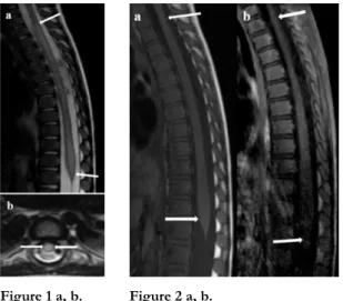
485 advancing sensory defect and motor weakness occurred in the patient’s lower extremities. On first assessment, the case’s sensory examination was normal, abdominal skin reflex and anal reflexes were taken; however deep tendon reflexes (DTR) could not be obtained. Muscle strength examination assessed lower extremity muscle strength as 2/5 on the right and 1/5 on the left. Bladder and sphincter dysfunction were not identified. Thoracolumbar spinal magnetic resonance imaging (MRI) taken nearly 20 hours after trauma identified appearance compatible with myelomalacia between thoracic-2 and lumbar-5 levels of the spinal cord on TA weighted images (Figure 1 a, b). After intravenous contrast material injection, pathologic contrasting was not identified (Figure 2 a, b).
Figure 1 a, b. Figure 2 a, b.
Figure 1 a, b. Thoracolumbar spinal MRI: T2 weighted sagittal (a) and transverse (b) images with thickening of the spinal cord between thoracic-2 and lumbar 5 levels and diffuse signal increase compatible with myelomalacia (between white arrows).
Figure 2 a, b. Thoracolumbar spinal MRI: T1 weighted non-contrast (a) and contrast (b) images with thickening of the spinal cord between thoracic-2 and lumbar 5 levels with no contrasting observed (between white arrows).
With SCIWORA diagnosis, the case was administered intravenous high-dose methylprednisolone treatment. No significant complication was observed related to high-dose steroid treatment. Maintenance treatment was continued for 2 months with oral prednisolone treatment. Included in a physical therapy program, the case had vital DTR in the lower extremities on
the 45th day and muscle strength was observed at 3/5-4/5 levels. At check-up three months later, neurological examination of the case observed lower extremity muscle strength of 4/5-5/5 and vital DTR.
DISCUSSION
A spinal injury specific to the childhood period, the incidence of SCIWORA within all spinal cord injuries varies from 5-67%, with mean accepted as 35%. Linked to the anatomic and biomechanical features of the spinal cord, it is most commonly observed in the first 8 years of life. During trauma, there is flexing and elongation of the vertebral column while the spinal cord does not follow this movement; this situation causes a tendency for SCIWORA (4).
Development of SCIWORA is explained by tension, direct or indirect compression and ischemic injury of the spinal cord as a result of physical effects like flexion, extension or distraction of the vertebral column linked to a variety of trauma like traffic accidents, falls from a height, sporting accidents and childhood fights5, 6. Our case may be explained by a
similar trauma mechanism of a vehicular traffic accident.
SCIWORA most often affects the cervical region rather than the medulla spinalis. It less commonly affects the thoracic and lumbar regions. There are studies reporting that when the thoracic region is affected neurologic prognosis may be bad7.
However, though the thoracolumbar region of the spinal cord was affected in our case, high dose steroid treatment in the early period provided positive functional results. This situation shows the importance of beginning steroid treatment in the shortest possible time and at high doses.
As radiographic imaging methods may be insufficient for diagnosis, detailed neurological examination and monitoring is very important. Though neurological complaints due to trauma may not be present at the time of first application, they may occur suddenly later. As a result, together with other organ injuries linked to trauma, the neurological tableau should be carefully monitored. In our case neurological symptoms occurred while our case was monitored in the ward due to pulmonary contusion, and early intervention prevented worse prognosis. The most beneficial
Cilt/Volume 43 Yıl/Year 2018 Spinal cord injury without radiographic abnormality
486 radiological diagnostic method for SCIWORA diagnosis is MRI. Grabb and Pang were able to show a correlation between injury mechanism and connective and other non-neurological soft tissue injuries on MRI. They stated it could provide information on the form and prognosis of cord injury and cord compression caused by hematoma or disks and reported it should definitely be performed8. Additionally, in their study of MRI
findings, they distinguished 4 groups as severe (haemorrhage involvement in 50% of the cord), mild (haemorrhage involvement in less than 50%), only oedema and no findings. With this classification they stated there was a correlation between MRI findings and prognosis of neurological tableau 8. In our case, only oedema was
observed along the thoracolumbar region of the spinal cord which contributed to a positive prognosis. The other important place for MRI in diagnosis is to reveal extraaxial hematoma that may cause progressive neurological worsening. MRI shows haemorrhagic ligament injury within hours due to methemoglobin forming outside nerve tissue, with hyperintensity on T2 weighted images, while injury in the disk interval may appear hyper or hypointense depending on whether the injury is haemorrhagic or not. Small haemorrhagic soft tissue injury may be masked by surrounding fat and muscle tissue, so fat suppressing methods should be used with T1 weighted sequences. Lesions causing spinal cord compression like hematoma or disk herniation may appear as haemorrhage of spinal parenchyma. If haemorrhage is over 50% of the transverse diameter of the spine it is classified as major, while if it is less than 50% it is classified as minor injury. Edema is observed as hyperintense onT2 weighted sequences, while loss of spinal continuity is hypointense of T1 weighted sequences9.
The first stage of SCIWORA treatment is to ensure stabilisation of the vertebra. Later, especially for patients with severe neurological deficit at time of diagnosis, the National Acute Spinal Cord Injury
Study recommends high dose methylprednisolone administration10.
For early diagnosis of SCIWORA, careful neurological examination and spinal MRI are very effective. Treatment with high-dose steroids begun in the early period may have vital importance in terms of reducing mortality and morbidity.
REFERENCES
1. Cirak B, Ziegfeld S, Knight VM, Chang D, Avellino AM, Paidas CN. Spinal injuries in children. J Pediatr Surg. 2004;39:607-12.
2. Orhun H, Saka G, Berkel T. Injury to the spinal cord without any radiographic abnormality in a child. Acta Orthop Traumatol Turc. 2002;36:268-72.
3. Pollack IF, Pang D, Sclabassi R: Recurrent spinal cord injury without radiographic abnormalities in children. J Neurosurg. 1988;69:177-82.
4. Keleş I. Pediyatrik medulla spinalis yaralanmalarında epidemiyoloji ve patofi zyoloji. Turkiye Fiziksel Tıp ve Rehabilitasyon Dergisi. 2008;54:46–50.
5. Dickman C.A, Rekate H.L, Sonntag V.K.H, Zabramski J.M. Pediatric spinal trauma: vertebral column and spinal cord injuries in children. Pediatr Neurosci. 1989;15:237-56.
6. Hamilton MG, Myles ST: Pediatric spinal injury: review of 174 hospital admissions. J Neurosurg. 1992;77:700-4.
7. Pang D. Spinal cord injury without radiographic abnormality in children, 2 decades later. Neurosurgery. 2004;55:1325-42.
8. Grabb PA, Pang D. Magnetic resonance imaging in the evaluation of spinal cord injury without radiographic abnormality in children. Neurosurgery. 1994;35:406-14.
9. Flanders AE, Schaefer DM, Doan HT, Mishkin MM, Gonzalez CF, Northrup BE. Acute cervical spine trauma: Correlation of MR imaging findings with degree of neurologic deficit. Radiology. 1990;177:25-33.
10. Bracken MB1, Shepard MJ, Collins WF, Holford TR, Young W, Baskin DS, et al. A randomized, controlled trail of methyprednisolone or naloxone in the treatment of acute spinal-cord injury. Results of the Second National Acute Spinal Cord Injury Study. N Eng J Med. 1990;322:1405-11.