Summary
Purpose: Akt, also known as protein kinase B (PKB), is an intracellular signal transduction protein activated by growth hormones. PKB/Akt is frequently activated in a va-riety of cancer types, but its role in the development and progression of lung cancer has not been completely eluci-dated yet. The aim of the present study was to determine the prognostic value of PKB/Akt in non-small cell lung cancer (NSCLC).
Methods: A total of 32 tumor samples from NSCLL pa-tients were examined before treatment. The staining char-acteristics of the cases were evaluated in terms of age, stage (T and N), response to therapy, histological type, tumor size, and ECOG performance status (PS).
Results: No statistical correlation was found between PKB/ Akt expression and gender, ECOG PS and stage (T and N),
while significant correlation between cytoplasmic PKB/akt expression and age was detected (p<0.05). In addition, squa-mous cell carcinoma histology was significantly associated with both nuclear and cytoplasmic staining (p=0.033), and tumor size ( <5 cm) was correlated with nuclear PKB/Akt expression (p=0.03). Both overall survival (OS) and progres-sion-free survival (PFS) were similar in patients with and without both nuclear and cytoplasmic PKB/Akt expression. Conclusion: Our results showed that although PKB/Akt was not associated with survival in NSCLC patients, it may be a potential therapeutic target for NSCLC; more studies with higher numbers of patients are needed to verify this hypothesis.
Key words: intracellular signal transduction, non-small cell lung cancer, phospho-Akt (p-Akt), prognosis, protein kinase B
Prognostic significance of protein kinase B/Akt pathway in
patients with non-small cell lung cancer
Alpaslan Mayadagli1, Sule Karabulut Gul1, Ahmet Bilici2, Ahmet Fatih Oruc1,
Mihriban Kocak1, Alper Ozkan1, Atinc Aksu1, Nagehan Ozdemir Barisik3, Mahmut
Gumus4
1Department of Radiation Oncology, 3Department of Pathology, 4Department of Medical Oncology, Dr.Lutfi Kirdar Kartal Education and Research Hospital, Istanbul; 2Department of Medical Oncology, Istanbul Medipol University, Medical Faculty, Istanbul, Turkey
Correspondence to: Ahmet Bilici, MD. Kemer Mah., Sehit Mustafa Dundar Cad., Kemer Park Evleri, A-8 Blok, Daire 5, Esenler, Istanbul, Turkey. Tel: +90 5325280486, Fax: +90 2164422947, E-mail: [email protected]
Received: 15/06/2013; Accepted: 03/07/2013
Introduction
Lung cancer genesis is based on extremely complex molecular events. The two mechanisms that have been blamed until today for the devel-opment of lung cancer are activation of proto-on-cogenes and inactivation of tumor suppressor genes. Ras and Myc families are among the most blamed oncogenes. Of these, Ras family gener-ally plays a role in NSCLC by means of point mutations, whereas Myc family plays a role in small cell lung cancer (SCLC) by means of am-plification [1,2]. Furthermore, cERB1-2 plays a role, particularly in NSCLC, and c-met, c-src, and
c-raf-1 play a role in SCLC [2,3]. Of tumor sup-pressor genes, variations regarding p53 and RB genes are seen in 75-100% of SCLC and 15-50% of NSCLC. Moreover, apoptosis and cell cycle re-lated genes also play a role in the development of lung cancer [4,5]. Understanding the biology of lung cancer may lead to the identification of novel targets for the treatment of this disease.
PKB/Akt is a serin-threonine kinase that regulates a variety of cellular functions such as cell survival, cell growth, cell differentiation and progression of cell cycle, and participates in a variety of cellular events such as apoptosis and protein synthesis [6,7]. PKB/Akt is activated by
ORIGINAL ARTICLE
phosphoinositide-3 kinase (PI3K) which is ac-tivated by the growth stimulators in the cells, such as epidermal growth factor and insulin-like growth factor-I [8]. This series begins with the activation of Ras, and the kinase cascade pro-gresses in turn by Raf, MEK and Erk proteins. Ras and Raf are proto-oncogenes. Ras proteins are inactive (Ras-GDP) in resting cells [9,10]. Under normal conditions, the efficacy of Ras is minimal in the stimulation of PI-3K pathway by growth factors. On the contrary, oncogenic Ras is a strong activator of PI-3K pathway and sup-presses apoptosis by activating the above men-tioned pathway and forms one of the critical fac-tors of the carcinogenesis process [11,12].
The role of PKB/Akt in the development and progression of lung cancer has not been com-pletely defined. In the present study, the prog-nostic value of PKB/Akt in NSCLC and its cor-relation with clinicopathologic variables was evaluated.
Methods
This study was conducted at the Dr. Lutfi Kirdar Kartal Education and Research Hospital, Department of Radiation Oncology and Medical Oncology, be-tween 2008 and 2009. Thirty-two patients, who had been diagnosed with NSCLC and had not received any anticancer therapy were included. The eligibility cri-teria consisted of measurable disease determined ra-diologically (chest X-ray, thorax CT scan or PET-CT), ECOG PS of 0-2, adequate hematological (absolute neutrophil count > 1500 mm3, platelet count >100000 mm3), hepatic (total serum bilirubin < 1.5 times the upper limit of normal (ULN), ALT and AST < 2.5 times the ULN), and renal function (serum creatinine level < 1.25 mg/dl).
Treatment plan
A weekly chemotherapy schedule including 30 mg/m2 docetaxel and 20 mg/m2 cisplatin was ad-ministered simultaneously with radiotherapy. The treatment continued only with radiotherapy in pa-tients with severe chemotherapy toxicity (grade 3/4 hematological toxicity, liver and renal function im-pairment). Radiotherapy was delivered with 6-15 MV photons in all patients using linear accelerator (GE Saturn 41) as 2 Gy daily fractions 5 days a week for a total of 23 fractions (46 Gy) to the primary tumor and mediastinum, followed by a boost to the primary tu-mor and involved lymph nodes (2 Gy daily fractions, 10 fractions, 26 Gy). The total dose of radiation was 66 Gy in 33 fractions. Response to therapy (complete response (CR), partial response (PR), stable disease (SD), and progressive disease (PD)) were determined
according to the criteria defined by the World Health Organization.
Immunohistochemical staining
Thirty-two NSCLC samples were immunohisto-chemically stained. Three to 5 mµ sections, obtained from paraffin blocks were selected by the pathologists, put on slides and covered with poly-L-lysine. They were kept at 37°C in an incubator overnight. After be-ing kept in xylene for 15 min, they were re-hydrated in 96% alcohol for 15 min, and washed with distilled wa-ter for 5 min. Then, they were put in citrate buffer (pH 6) and boiled for 5 min in a microwave oven 4 times (750W, 350W, 350W, 350W). Then, they were kept at room temperature within the same buffer for 20 min for cooling, and washed with distilled water. In order to eliminate endogenous peroxidase activity, 3% hy-drogen peroxide solution was dripped. After 10 min the sections were washed with PBS (Phosphate Buffer Solution) for 3 times, and protein block (LabVision, Large Volume Ultra V Block, TA-125-UB) was applied for 5 min. The excess amount of block solution was poured and primary (AKT [Ser473] rabbit polyclonal antibody RB-10369-P1 (P&D System, USA) was incu-bated for 40 min without being washed. Afterwards it was washed in PBS 3 times. Secondary antibody (Labvision, Biotinylated Goat Anti Polyvalent TP-125-BN, Thermo Scientific) was applied for 15 min, and washed in PBS 3 times. Tertiary antibody (Labvision, Large Volume Streptavidin Peroxidase, and TS-125-HR, Thermo Scientific) was incubated with the sec-tions for 15 min, and washed in PBS 3 times. After dripping AEC chromogene (Labvision, Large Volume AEC Substrate System, TA-125-HA, Thermo Scientif-ic) for 10 min, the sample was washed with distilled water. The tissues were counterstained with May-er’s Hematoxylin (Bio-Optica, MayMay-er’s hematoxylin 06002L, Milano, Italy) for 1 min. Hematoxylin turned into purple under tap water and the excess amount was washed. The sample was coated with aqueous coating material (Bio-Optica, Mount quick aqueous mounting medium, 05-1740, Milano, Italy) (Figure 1).
Immunohistochemical PKB/Akt expression was examined under light microscope. Samples were evaluated according to the intensity and extensive-ness of nuclear and cytoplasmic staining of tumor cells. Nuclear and cytoplasmic staining percentage of the stained tumor cells was taken into considera-tion for the degree of extensiveness. The nuclear and cytoplasmic staining was analyzed as either present or absent [13,14]. The staining characteristics of the cases were evaluated in terms of age, gender, stage (T and N), response to the therapy, histological type, tumor size and ECOG PS.
Statistics
Statistical analyses were performed using SPSS 16.0 (SPSS Inc., Chicago, IL, USA) software. Chi-square
test and Fisher’s exact test were used to analyze the relationships between the groups and categorical variables. Survival probabilities and curves were ob-tained according to the Kaplan-Meier method and compared by the log-rank test. Univariate analysis was carried out to evaluate the signifi cance of PKB/ Akt expression and other clinicopathological features as prognostic factors, then multivariate analysis with the Cox proportional hazards model was performed in order to further analyze the PKB/Akt expression and all of the signifi cant prognostic factors which were found in the univariate analysis. Multivariate p val-ues were used to characterize the independence of these factors. The 95% confi dence interval (95% CI) was used to quantify the relationship between sur-vival time and each independent factor. All p values were two-sided in the tests and p values < 0.05 were considered as statistically signifi cant.
Results
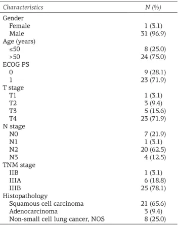
Thirty-one (96.9%) patients were male and one female, with a median age of 63 years (range 18-70). Twenty-four (75%) patients were older than 50 years. The majority of patients (71.9%) had T4 tumors. N stage was as follows: 7 (21.9%) patients had N₀, 1 (3.1%) N₁, 20 (62.5%) N₂ and 4 (12.5%) N₃. One patient (3.1%) was classifi ed as stage IIB, 6 (18.8%) as stage IIIA, and 25 (78.1%) as stage IIIB. ECOG PS was 2 in the majority (65.6%) of the patients. Twenty-nine (90.6%) pa-tients had a history of smoking. Histopathologi-cal subtype was squamous cell carcinoma in 21 (65.6%) patients and adenocarcinoma in 3 (9.4%) patients, while 8 (25%) patients were classifi ed as ‘not otherwise specifi ed’ NSCLC. Demograph-ic, clinical and histopathological characteristics of patients are summarized in Table 1. All
pa-tients completed 66 Gy. Weekly chemotherapy concurrent with radiotherapy could be applied for a median 5 courses (range 3-7).
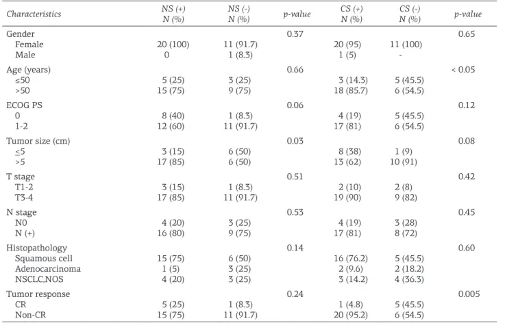
Both nuclear and cytoplasmic staining were observed in 11 of 32 (34.4%) patients. Nuclear staining was positive in 20 (62.5%) of the pa-tients, while cytoplasmic staining was positive in 21 of 32 (65.6%) patients. In 2 (6.3%) patients staining was negative. There was signifi cant cor-relation between cytoplasmic staining and age, when patients with cytoplasmic staining were >50 years compared with patients <50 years (p<0.05). Both nuclear and cytoplasmic staining in patients with T3-T4 stages (17 and 19, respec-tively) was more frequent compared with T1-T2 patients (3 and 2, respectively), but these diff er-ences were not signifi cant (p=0.5 and 0.4, respec-tively). Lymph node involvement was not associ-ated with either nuclear or cytoplasmic staining (p>0.05). In addition, no signifi cant correlation was detected between stage and staining (p=0.67 and p=0.60, respectively).
In patients with squamous cell carcinoma histology both nuclear and cytoplamic staining were signifi cantly more frequent than in pa-tients with non-squamous cell histology (47.6 vs 9.1%, p=0.033). However, there were no
sig-Table 1. Patient and disease characteristics
Characteristics N (%) Gender Female Male Age (years) ≤50 >50 ECOG PS 0 1 T stage T1 T2 T3 T4 N stage N0 N1 N2 N3 TNM stage IIB IIIA IIIB Histopathology
Squamous cell carcinoma Adenocarcinoma
Non-small cell lung cancer, NOS
1 (3.1) 31 (96.9) 8 (25.0) 24 (75.0) 9 (28.1) 23 (71.9) 1 (3.1) 3 (9.4) 5 (15.6) 23 (71.9) 7 (21.9) 1 (3.1) 20 (62.5) 4 (12.5) 1 (3.1) 6 (18.8) 25 (78.1) 21 (65.6) 3 (9.4) 8 (25.0)
NOS: not otherwise specified, ECOG PS: Eastern Cooperative Oncology Group performance status
Figure 1. Immunoperoxidase staining with pAkt (Ser473) antibody (Neomarkers USA). Strong cytoplas-mic staining (×200).
nificant correlations between histopathological subtypes and nuclear or cytoplasmic staining (p>0.05). Nuclear staining in patients with tu-mor size > 5 cm was significantly tu-more frequent (17 of 23 patients, 85%) compared with patients with tumors ≤ 5 cm (15%, p=0.03). The
relation-ships between these parameters and nuclear or cytoplasmic staining are shown in Table 2.
CR was achieved in 6 (18.8%) patients. We observed that there were 5 patients with nuclear and 1 patient with cytoplasmic staining in the CR group. In addition no patient was found with
Table 2. Relationship between patient characteristics and nuclear and cytoplasmic staining
Characteristics NS (+)N (%) NS (-)N (%) p-value CS (+)N (%) N (%)CS (-) p-value Gender Female Male 20 (100)0 11 (91.7)1 (8.3) 0.37 20 (95) 1 (5) 11 (100) -0.65 Age (years) ≤50 >50 15 (75)5 (25) 3 (25)9 (75) 0.66 3 (14.3) 18 (85.7) 5 (45.5)6 (54.5) < 0.05 ECOG PS 0 1-2 12 (60)8 (40) 11 (91.7)1 (8.3) 0.06 4 (19) 17 (81) 5 (45.5)6 (54.5) 0.12 Tumor size (cm) <5 >5 17 (85)3 (15) 6 (50)6 (50) 0.03 8 (38) 13 (62) 10 (91)1 (9) 0.08 T stage T1-2 T3-4 3 (15) 17 (85) 1 (8.3) 11 (91.7) 0.51 2 (10) 19 (90) 2 (8) 9 (82) 0.42 N stage N0 N (+) 4 (20) 16 (80) 3 (25) 9 (75) 0.53 4 (19) 17 (81) 3 (28) 8 (72) 0.45 Histopathology Squamous cell Adenocarcinoma NSCLC,NOS 15 (75) 1 (5) 4 (20) 6 (50) 3 (25) 3 (25) 0.14 16 (76.2) 2 (9.6) 3 (14.2) 5 (45.5) 2 (18.2) 4 (36.3) 0.60 Tumor response CR Non-CR 15 (75)5 (25) 11 (91.7)1 (8.3) 0.24 1 (4.8) 20 (95.2) 5 (45.5)6 (54.5) 0.005
NS: nuclear staining, CS: cytoplasmic staining, CR: complete response, NOS: not otherwise specified, ECOG PS: Eastern Cooperative Oncology Group performance status
Figure 2. Kaplan-Meier estimates for overall survival in the (A) nuclear staining group and (B) the cytoplas-mic staining group.
Figure 3. Kaplan-Meier estimates for progres-sion-free survival in the (A) nuclear staining group and (B) the cytoplasmic staining group.
both nuclear and cytoplasmic staining. Signifi-cant differences were detected in patients with non-CR with respect to cytoplasmic and both nuclear and cytoplasmic staining compared with patients with CR (p=0.005 and 0.049, respective-ly; Table 2).
At a median follow-up of 14.5 months (range 3-51) the median PFS was 13 months (SE: 5; 95% CI: 5-25) and the median OS was 20 months (SE: 5; 95% CI: 11-29) for the entire cohort. Median OS for patients with nuclear staining was worse than that of patients without nuclear staining, but this difference was not statistically signifi-cant (21 vs 16 months, respectively, p=0.270). Fur-thermore, median OS was also similar among patients with or without cytoplasmic staining (20 vs 16 months, respectively, p=0.82). Figure 2 shows OS according to the nuclear or cyto-plasmic staining. Median PFS was similar for patients with and without nuclear and cytoplas-mic staining (p=0.398 and p=0.836, respectively, Figure 3).
Results of univariate survival analyses with respect to the PBK/Akt staining are summarized in Table 3. Prognostic significance could not be proved in multivariate analysis.
Discussion
Akt, or protein kinase B, is a serine/threo-nine kinase that regulates growth factors, such as epidermal growth factor and insulin-like growth factor-I, and through them, cell survival [15]. Akt is cellular homolog product of v-akt and has 3 isoforms: Akt-1, Akt-2 and Akt-3. All 3 iso-forms are expressed in normal tissues, but the level of expression may vary depending on the tissue. Akt-1 and Akt-2 are expressed in brain, thymus and lungs, whereas Akt-3 is expressed in brain and testicles. Akt is activated downstream by various growth factors including insulin,
insulin like growth hormone-I and epidermal growth factor as well as by phosphotidilinositol 3-kinase (PI3K). Complete activation is provid-ed by means of phosphorylation at Thr308/309 location in kinase activation handle and at Ser473/474 location in COOH-terminal tail [16].
The role of Akt in carcinogenesis has been well-documented and Akt is overexpressed in various types of human cancer [16,17]. Akt is associated with initiation of tumorigenesis in pancreatic carcinoma [18] and glioma [19], and seems to correlate with stage and tumor grade in prostate carcinoma [20]. Moreover, although PTEN frequently disappears or is inactivated via mutation [21], PI3K, which is the positive regula-tor of Akt, is frequently upregulated [22]. Finally, it has been shown that activated Akt induces cell transformation [23].
In their study including 61 patients, David et al. put forward the hypothesis that overexpres-sion of Akt might be predictive for short survival [13]. There was a strong staining with pAkt an-tibody in 14 of 61 NSCLC cases and overexpres-sion of PKB/Akt was found to be an independent prognostic factor for survival. In addition, the authors showed that age at the time of diagno-sis was not significant but stage was significant. Overall survival was significantly different with respect to Akt status even after stage has been taken into account and mortality was higher in patients with strong staining as compared with those without staining. There was a tendency for the patients with strong staining to be diag-nosed at lower stages as compared to those with-out staining. Also in their study, patients with strong staining significantly tended to be older on average than those without staining, even if they were at the same stages [13]. In the pres-ent study, cytoplasmic staining alone was seen more frequently in patients aged ≥50 years; no relationship with age and stage could be demon-strated in other groups.
In a study carried out by Balsara et al. in 110 patients, high Akt activity was determined in 23 (21%) patients. In that study, it was demonstrat-ed that there was no relationship between PKB/ Akt expression and histological subtypes or sur-vival. Median OS time was 26 months for the negative group and 23 months for the positive group. The prevalence of PKB/Akt positivity was similar among patients in lower stages (stages I-II) and those with higher stage. Furthermore, PKB/Akt positivity gave similar outcomes both in well-differentiated and poorly-differentiated
Table 3. Results of univariate analysis with respect to the PKB/Akt status
Staining Median OS (months) p-value Median PFS (months) p-value Nuclear staining Absent Present Cytoplasmic staining Absent Present 21 16 20 16 0.270 0.820 15 11.5 14 11 0.398 0.836
tumors. Consequently, this outcome possibly im-lies that Akt activation occurs in the early period of tumor progression [24]. Our results were com-patible with their study with respect to stage, but not with the histological subtypes.
Ninety-six percent of 78 cases that had been evaluated by Shah et al. [14]. were immunoreac-tive for PBB/Akt. PBB/Akt expression is limited only to tumor cells. The percents of tumor cells that were positively stained in each individu-al section ranged between 1-90% (median 15); however, cytoplasmic PBB/Akt staining (CP-Akt) was observed in all positively stained tumor cells. In addition, nuclear staining was observed in 42% of the cases. Nuclear staining was clas-sified as “positive” or “negative”. Membranous staining was observed in only 5% of the cases, and 1-15% of tumor cells stained positively. CP-Akt and NP-Akt had strong correlation with well-differentiated tumors. NP-Akt was corre-lated with the presence of nodal involvement as well as with squamous histology as compared with non-squamous types [14].
In the study performed by Lee et al. [25] only 43 patients were included and no significant cor-relation was observed between CP-Akt and NP-Akt and other pathological factors. PBB/NP-Akt/ alpha-actin and the N stage were the only inde-pendent prognostic factors. Gender, histological subtype, tumor stage and nodal status were not prognostic factors in univariate analysis [25]. In our study both nuclear and cytoplasmic staining in patients with squamous cell carcinoma his-tology were significantly more frequent than in patients with non-squamous cell histology (p=0.033).
The relation between PKB/Akt and distant metastasis has been previously reported in other types of tumors and evidence raises the hypoth-esis that PKB/Akt may have a role in the pro-gression of the disease rather than in its devel-opment [26]. Analyzing the percentage of PKB/ Akt positively stained tumors ignoring subcel-lular localization, Shah et al. obtained similar outcomes [14]; however, it had been surprising to find out a correlation between NK-Akt and lymph node metastasis.
Tsurutani et al. evaluated 252 patients to study for any association between Akt
phos-phorylation with clinical outcomes [27]. Two phosphorylation events are needed for complete activation of Akt. However, a single phospho-rylation site (S473) has been determined up to now in clinical samples of NSCLC, and this led to conflicting results concerning the prognos-tic significance of Akt activation in NSCLC. In the above mentioned study, the authors tried to determine whether Akt phosphorylation en-hances the prognostic accuracy at T308 or not. Phosphorylation of S473 or T308 was positive in most of NSCLC samples, but was rarely de-termined in surrounding tissues. Defining Akt activation using both phosphorylation sites, Akt activation was specific for NSCLC as compared with surrounding tissues (73.4 vs 0%; p<0.05). It was significantly higher in adenocarcinoma as compared to squamous cell carcinoma (78.5 vs 68.5%; p=0.040), and associated with shorter survival in all disease stages (p=0.041). In mul-tivariate analysis, increased phosphorylation of T308 only was a poor prognostic factor in stage I patients or for tumors <5 cm (log-rank p=0.011 and p=0.015, respectively). These results raised the thought that observing Akt phosphorylation at T308 facilitates the evaluation of Akt activa-tion and show that Akt activaactiva-tion is a poor prog-nostic factor for all stages of NSCLC [27]. In the present study we proved that nuclear staining in patients with tumor size > 5 cm was significant-ly more frequentsignificant-ly observed compared with pa-tients with tumors ≤5 cm (85 vs 15%).
In conclusion, no statistical correlation was demonstrated between PKB/Akt expression and gender, stage, ECOG PS, lymph node involve-ment, and T stage. In contrast, significant corre-lation was detected between cytoplasmic PKB/ Akt expression and age. In addition, squamous cell histology was significantly associated with both nuclear and cytoplasmic staining and tumor size ( <5 cm) was related with nuclear PKB/Akt expression. Although PKB/Akt activation may be associated with poor prognosis and chemother-apy and radiotherchemother-apy resistance, no significant difference could be demonstrated between nu-clear or cytoplasmic staining and survival. We maintain that our results need to be confirmed by prospective studies including larger numbers of NSCLC patients.
References
1. Fong KW, Sekido Y, Minna JD. Molecular pathogenesis of lung cancer. J Thorac Cardiovasc Surg 1999;118:1136-1152.
2. Spivack SD, Fasco MJ, Walker VE, Kaminsky LS. The molecular epidemiology of lung cancer. Crit Rev Toxi-col 1997;27:319-365.
3. Mabry M. Activating oncogenes in lung cancer. In : Kane MA, Bunn PA (Eds): Biology of lung cancer. New York, Marcel Dekker Inc., 1998, pp 391-412.
4. Levin WJ, Casey G, Ramos JC et al. Tumor suppressor and immediate early transcription factor genes in nonsmall cell lung cancer. Chest 1994;106 (Suppl) :372S-376S. 5. Hussain SP, Harris CC. Molecular epidemiology and
carcinogenesis: endogenous and exogenous carcino-gens. Mutat Res 2000;462:311-322.
6. Franke TF, Yang SI, Chan TO et al. The protein kinase encoced by the Akt proto-oncogene is a target of the PDGF–activated phosphotidylinositol 3-kinase. Cell 1985;81:727-736.
7. Kitamura T, Ogowa W, Sakaue H et al. Requirement for activation of the serine-threonine kinase Akt (protein kinase B) in insulin stimulation of protein synthesis but not of glucose transport. Mol Cell Biol 1998;18:3708-3717.
8. Coffer PJ, Jin J, Woodgett JR. Protein kinase B (c-Akt): a multifunctional mediator of phosphatidylinositol 3-ki-nase activation. Biochem J 1998;335:1-13.
9. Kolch W. Meaningful relationships: The regulation of the Ras/Raf/MEK/ERK pathway by protein interactions. Biochem J 2000;351:289-305.
10. Lee JT, McCubrey JA. The Raf/MEK/ERK signal trans-duction cascade as a target for chemotherapeutic inter-vention in leukemia. Leukemia 2002;16:486-507. 11. Peggs K, Mackinnon S. Imatinib mesylate-The new gold
standard for treatment of chronic myeloid leukemia. N Engl J Med 2003;348:1048-1050.
12. Blalock WL, Navolanic PM, Steelman LS et al. Require-ment for the PI3K/Akt pathway in MEK1-mediated growth and prevention of apoptosis: Identification of an Achilles heel in leukemia. Leukemia 2003;17:1058-1067.
13. David O, LeBeau H, Brody AR, Friedman M, Jett J. Phos-pho-Akt overexpression in non-small cell lung cancer confers significant stage-independent survival disad-vantage. Chest 2004;125:152S.
14. Shah A, Swain WA, Richardson D et al. Phospho-akt is associated with a favorable outcome in non-small cell lung cancer. Clin Cancer Res 2005;11:2930-2936.
15. Kulik G, Klippel A, Weber MJ. Antiapoptotic signalling by the insulin-like growth factor I receptor,phosphati-dylinostol 3-kinase,and Akt. Mol Cell Biol 1997;17:1595-1606.
16. Coffer PJ, Jin J, Woodgett JR. Protein kinase B(c-Akt): a multifunctional mediator of phosphatidylinositol 3-ki-nase activation. Biochem J 1998;335:1-13.
17. Staal SP. Molecular cloning of the akt oncogene and its human homologues AKT1 and AKT2: amplification of AKT1 in a primary human gastric adenocarcinoma. Proc Natl Acad Sci USA 1987;84:5034-5037.
18. Ruggeri BA, Huang L, Wood M, Cheng JO, Testa JR. Am-plification and overexspression of the AKT2 oncogene in a subset of human pancreatic ductal adenocarcino-mas. Mol Carcinog 1998;21:81-86.
19. Ermonian RP, Furniss CS, Lamborn KR et al. Dysregu-lation of PTEN and protein kinase B is associated with glioma histology and patient survival. Clin Cancer Res 2002;8:1100-1106.
20. Malik SN, Brattain M, Ghosh PM et al. Immuno-histochemical demonstration of phospho-Akt in high Gleason grade prostate cancer. Clin Cancer Res 2002;8:1168-1171.
21. Cantley LC, Neel BG. New insights into tumor suppres-sion:PTEN supresses tumor formation by restraining the phosphoinositide 3-kinase/AKT pathway. Proc Natl Acad Sci USA 1999;96:4240-4245.
22. Shayesteh L, Lu Y, Kuo WL et al. PIK3CA is impli-cated as an oncogene in ovarian cancer. Nat Genet 1999;21:99-102.
23. Mende I, Malstrom S, Tsichlis PN, Vogt PK, Aoki M. Oncogenic transformation induced by membrane-tar-geted Akt2 and Akt3. Oncogene 2001;20:4419-4423. 24. Balsara BR, Pei J, Mitsuuchi Y et al. Frequent activation
of AKT in non-small cell lung carcinomas and preneo-plastic bronchial lesions. Carcinogenesis 2004;25:2053-2059.
25. Lee JH, Koh JT, Shin BA et al. Comparative study of angiostatic and anti-invasive gene expressions as prog-nostic factors in gastric cancer. Int J Oncol 2001;18:355-361.
26. Landreneau RJ, Hazelrigg SR, Mack MJ et al. Thora-coscopic mediastinal lymph node sampling: Useful for mediastinal lymph node stations inaccesible by cervical mediastinoscopy. J Thorac Cardiovasc Surg 1993;106:554-558.
27. Tsurutani J, Fukuoka J, Tsurutani H et al. Evaluation of two phosphorylation sites improves the prognostic sig-nificance of Akt activation in non-small-cell lung cancer tumors. J Clin Oncol 2006;24:306-314.