Postmenopozal Dönemdeki Kadınlarda Stres Üriner
İnkontinans Tanısında Periüretral Doppler Ultrasonografi’nin
Yeri
Periurethral Doppler Ultrasonography in the Evaluation of
the Stress Urinary Incontinence in The Postmenopausal
Women
Geliş Tarihi:09/09/2019 Kabul Tarihi: 11/02/2020
Ege Klin Tıp Derg 2020;58 (1): 13-17
Duygu ADIYAMAN* 0000-0002-5307-8149 A.Akın SİVASLIOĞLU**0000-0003-3711-0118
* Kadın Hastalıkları ve Doğum, Tepecik Eğitim ve
Araştırma Hastanesi, İzmir, Türkiye
**Kadın Hastalıkları ve Doğum, Muğla Sıtkı Koçman
Üniversitesi Tıp Fakültesi, Muğla, Türkiye
Yazışma Adresi: Duygu ADIYAMAN
Kadın Hastalıkları ve Doğum, Tepecik Eğitim ve Araştırma Hastanesi
E-mail: duyguagca@gmail.com.
Öz
Giriş: Stres üriner inkontinans (SUI) psikolojik, hijyenik ve cinsel sorunlara yol açabildiği bilinen bir sağlık sorundur. Peri-üretral vasküleritenin üretra perfüzyonundan sorumlu olduğu ve bu sayede üretral mukozanın normal geriliminin sürdürülebildiği ve kontinansı sağlayabildiği düşünülmektedir. Bu çalışmanın amacı; stres üriner inkontinansı olan postmenopazal kadınlarda kontrol grubuna göre transvajinal ultrasonografi ile peri-üretral damar doppler çalışmasının inkontinans tanısındaki prediktif değerini araştırmaktır.
Materyal ve metod: Bu prospektif çalışmaya 21 kontrol ve 21 postmenopozal SUI’li olmak üzere 42 kadın dahil edildi. Tüm hastalara ve kontrol grubuna endovajinal yolla peri-üretral arteriel doppler ultrasonografi yapılarak elde edilen veriler karşılaştırıldı.
Bulgular: Demografik verileri benzer olan gruplar arasında, stres üriner inkontinansı olanlar ile kontrol grubu arasında periüretral arteriel doppler ultrasonografi ile tespit edilen parametrelerden sistol/diastole (S/D), rezistans indeks (RI) ve pulsatilite indeks (PI) değerlerinde istatistiksel olarak anlamlı fark bulunamadı. Sadece pik sistolik velosite (PSV) değerlerinde iki grup arasında istatistiksel olarak anlamlı ilişki gözlendi. (p=0,04).
Sonuç: İrregüler vaskülarite ve yüksek vasküler rezistansın azalmış üretral basınca neden olarak SUI gelişimine katkıda bulunduğu bilinmektedir. SUI’ nin tedavi edilebilmesi için tanının doğru konulması önemlidir. Endovajinal Doppler ultrasonografi ile periüretral arteriyel PSV’nin değerlendirilmesi kolay ve maliyeti düşük bir alternatif yöntem ortaya çıkmaktadır.
Anahtar Kelimeler: Endovajinal ultrasonografi, periüretral doppler ultrasonografi, stres üriner inkontinans.
Abstract
Objectives: Stress urinary incontinence (SUI) is a major health problem which causes various physiological, hygienic, and sexual problems. It has been claimed that periurethral vascularity is responsible for the perfusion of the urethra so that the normal tension of urethral mucosa can be maintained for preserved urinary continence mechanisms. This study aims to evaluate the predictive value of endovaginal periurethral Doppler ultrasonography in the evaluation of postmenopausal women with SUI.
Methods: Forty-two multiparous women were enrolled in this prospective study (21 SUI vs. 21 control). Endovaginal ultrasonography and periurethral artery Doppler study were performed on both groups.
Results: There were no statistically significant difference between patients with SUI and the control group regarding systole/diastole (S/D), resistance index (RI), and pulsatility index (PI). However, peak systolic velocity (PSV) has been found to be increased in patients with SUI (p=0.04).
Conclusion: Irregular vascularity and high vascular resistance are generally considered to be the main reasons that lead to poor urethral pressure and eventually SUI. Before treating SUI, proper identification by using the correct methods is needed. The increased PSV showed us that among these methods, measuring PSV in endovaginal periurethral Doppler is an easy and cost-effective way.
Keywords: Endovaginal ultrasonography, periurethral Doppler ultrasonography, stress urinary incontinence.
Introduction
Involuntary loss of urine (urinary incontinence) is a major health problem that causes psychological, social, sexual, and hygienic issues (1). Factors that contribute to increased intraurethral pressure are smooth and striated muscles, connective and elastic tissues, and the vascularity of pelvic unit (2). The periurethral vascularity is responsible for 1/3 of urethral closure pressure, and rich vascular plexuses have a key role in urethral closure. Several studies have shown that the mid-urethral blood flow is especially impaired in women with urinary incontinence, and after the application of vaginal estrogen, the pulsatility and the resistance index of the submucosal vessels were shown to be improved (1, 3, 4).
Imaging techniques will be expected to help us clarify the etiology and pathophysiology of pelvic floor dysfunction, therefore improving the outcomes of conservative and surgical treatment while assisting in the development of better therapeutic concepts (5). In pelvic floor assessment, endovaginal Doppler USG is an accessible, cheap, and painless method. Two-dimension endovaginal ultrasound should be the preferred method in the evaluation of urethral vascularity and the anterior compartment of the pelvic floor (6). This study aims to compare periurethral vascular parameters of mid-urethra between multiparous postmenopausal females with stress urinary incontinence and the control group using high-frequency 2D endovaginal color Doppler ultrasound.
Methods
This prospective study was conducted in Izmir Ataturk Training and Research Hospital from January 2016 to December 2016.
The study has been carried out with 21 postmenopausal women, who stress urinary incontinence (SUI) were previously diagnosed with urodynamic tests. Also, 21 postmenopausal continent women were selected as the control group. All patients have had at least a single previous vaginal delivery, and none of them had received any hormone replacement treatment. The control group was selected among women
Patients with known chronic gynecological infections such as pelvic inflammatory disease and cervicitis, urinary tract infections, pelvic organ prolapse grade 1 or greater, and/or history of any kind of gynecologic surgery were excluded from the study. All enrolled subjects were at least 12 months of menopausal state.
This research was approved by the ethics committee of Izmir Kâtip Celebi University. All participants gave their written informed consent. A detailed history of patients and demographic data; age, parity, and body mass index were also included in this study. All 42 patients underwent vaginal examination performed by the same experienced clinician.
The endovaginal ultrasound scan was performed with the transvaginal probe of Medison SonoAce X8 Ultrasound Machine. The patient was lied down in a semi-lithotomy position. The transducer was placed in the vagina and pushed forward by 2-3 cm for the increased visibility of the bladder and urethra.
Tanısında Periüretral Doppler Ultrasonografi’nin Yeri
Urinary Incontinence in The Postmenopausal Women
Figure 1. Anatomy of the urethra
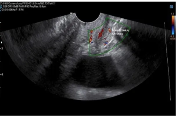
Any pressure on the surrounding structures was avoided, and the examination was conducted with a full bladder. The color Doppler mode was used for assessing the urethral vascularity pattern, both in sagittal and axial plans of the peri-urethral arteries.
Figure 3. Doppler ultrasonography of periurethral arteries
Ultrasound machine calculated Doppler parameters, and they were recorded.
Systole/diastole (S/D), resistance index (RI) pulsatility index (PI), and peak systolic velocity (PSV) were measured in all patients. The IBM SPSS Statistics 22.0 package program (IBM Corp., Armonk, New York, USA) performed all the statistical analyses of this study. Statistically significant differences were accepted as p <0.05.
Results
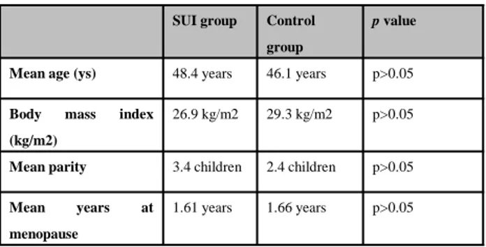
Forty-two patients were included in this prospective clinical study. The characteristics of the study population are shown in table 1.
SUI group Control group
p value
Mean age (ys) 48.4 years 46.1 years p>0.05
Body mass index (kg/m2)
26.9 kg/m2 29.3 kg/m2 p>0.05
Mean parity 3.4 children 2.4 children p>0.05
Mean years at menopause
1.61 years 1.66 years p>0.05
Table 1. Patient demographics
The mean age, body mass index, and the mean parity were 48.4 (±13.2) and 46.1 (±8.6) years, 29.3 (±6.3) and 26.9 (±3.6) kg/m2, 3.4 (±2) and 2.4
(±1.6) children in the SUI and control groups, respectively. There were no significant differences with respect to age, BMI, or parity between the two groups.
Table 2. Doppler differences between SUI and control group
SUI group Control group p value
PSV (cm/s) 9.09±8.94 2.29±4.62 0.04
S/D 5.02±2.70 4.10±2.69 0.27
PI 1.68±0.75 1.55±1,27 0.34
RI 0.76±0.18 1.10±1.70 0.06
Discussion
The arterial blood perfusion of the female urethra derives from the internal pudendal, vaginal, and inferior vesical branches of the vaginal arteries. After the vaginal arteries run along the superior lateral wall of the vagina, they separate into two vessels placed on the dorsal and ventral aspects of the urethra (3). These vessels then deliver arterial blood to the submucosal vascular plexus, which is located next to a few millimeters away from the bladder neck (7). It has clearly been shown with power Doppler imaging that the submucosal vascular plexus comprised two central branches of vessels originating near the proximal or middle urethra and having collateral channels to anterior and posterior superficial vessels overlying the urethra and the middle urethral vessels. However, the anterior branch especially, may not be visible in some postmenopausal women (8).
The increased and stable intraurethral pressure with respect to intra-bladder pressure maintains satisfactory continence. Vascularity within the urethral submucosa and blood supply contribute to the normal tension of the urethral mucosal wall, and peri-urethral vascularity provides 1/3 of this urethral pressure via stiffness of periurethral connective tissue (9-11). Defects in this connective tissue emerge as SUI, cystocele, and urethral hypermobility(12, 13). Management of these cases relies on the proper diagnosis. Wieczorek et al., reported that the color Doppler high-frequency endovaginal ultrasound is a suitable tool in the real-time assessment of urethral vascularity(11, 14).
In color Doppler ultrasound, fluid’s regular flow vibration creates a degree of color saturation. Normal laminary flow creates a homogeneous and constant color. Bifurcations and luminal narrowings bring faster flow velocity and irregular flow patterns. Doppler velocity is the main instrument to define the severity of stenosis. Stenosis can be seen by high velocities, color disturbance, spectral broadening, and post-stenotic waveforms (15). Usually, after stenosis, a mosaic pattern comes out, and turbulent flow occurs. Unlike color persistence, disorganized spread indicates flow resistance (15)
Most Doppler studies mainly include three parameters; systolic/diastolic ratio (S/D), resistive index (RI) and pulsatility index (PI). S/D ratio is simple and can be calculated easily. However, RI and PI are much more complicated to calculate. RI and PI commonly show blood flow resistance. While there is an increased diastolic flow in cases with inflammation or angiogenesis, stenotic, and constricted vessels result in decreased diastolic flow and therefore leading to an inversely increased systolic peak velocity (16). When there is a relatively higher resistance, the diastolic flow would usually be abnormally lower, and RI would be found to be increased up to 1.00 (16). Also, stenotic vessels have higher PI and RI values.
We did not find a statistically significant difference in respect to S/D within the SUI and control group. Additionally, there was no significant difference in PI and RI values between the two groups. However, PSV was increased in SUI group in comparison with the control group, which was statistically significant (p=0,04). Jarmy-Di Bella et al., found that PSV values were lower in incontinent postmenopausal patients than the control group in a study 57 subjects (1). They have measured PSV 8.98 cm/s in continent postmenopausal patients and 6.28 cm/s in incontinent postmenopausal patients respectively, and even though the difference was little, it was statistically significant. However, in that study, the mean age of postmenopausal women was 57 which was much older than our research. The mean menopause age is 45.75 ± 4.7 in Turkey, and our study group was compatible with this data (17). The mean age of the incontinent and continent group in our study were 48,4 and 46,1, respectively. They were relatively early onset of menopause, and their compensatory mechanisms were still active.
Using endovaginal ultrasound and Doppler application, for diagnosis management of SUI is nowadays the interest of studies (18, 19). According to our data increased PSV values can be associated with increased arterial resistance, which leads to poor vascular circulation.
Compensation to this resistant stature, increased systolic velocities for the effort to maintain the flow in resistant periureteral arteries can be reasonable. We resulted that increased PSV values in endovaginal ultrasound could be useful in the diagnosis of SUI as a supporting method.
Indeed, there were some limitations to this study. The low number of patients is one of the limitations of the study. The number could be considered insufficient, although other studies have a similar number of subjects. Also, observer differences did not regard but an intra-observer would be beneficial to increase the reliability of the sonographer.
Quick and easy application of the technique plus its cost-effectiveness is standing out (20). Although we had a limited amount of patients, with the help of future studies, we believe that endovaginal ultrasonography can be a unique method for supporting the diagnosis (21).
In conclusion, endovaginal periurethral Doppler sonography can be an inexpensive alternative for the evaluation of SUI, in early post-menopausal women.
Tanısında Periüretral Doppler Ultrasonografi’nin Yeri
References
1.Jarmy-Di Bella ZI, Girao MJ, Sartori MF, Di Bella Junior V, Lederman HM, Baracat EC, et al. Power Doppler of the urethra in continent or incontinent, pre- and postmenopausal women. Int Urogynecol J Pelvic Floor Dysfunct. 2000;11(3):148-54; discussion 54-5.
2.Petros PE, Ulmsten UI. An integral theory of female urinary incontinence. Experimental and clinical considerations. Acta obstetricia et gynecologica Scandinavica Supplement. 1990;153:7-31.
3.Beco J, Leonard D, Leonard F. Study of the female urethra's submucous vascular plexus by color Doppler. World journal of urology. 1998;16(3):224-8.
4.Girao MJ, Jarmy-Di Bella ZI, Sartori MG, Baracat EC, Lima GR. Doppler velocimetry parameters of periurethral vessels in postmenopausal incontinent women receiving estrogen replacement. International urogynecology journal and pelvic floor dysfunction. 2001;12(4):241-6.
5.Dietz HP. Pelvic floor ultrasound in incontinence: what's in it for the surgeon? International urogynecology journal. 2011;22(9):1085-97.
6.Santoro GA, Wieczorek AP, Dietz HP, Mellgren A, Sultan AH, Shobeiri SA, et al. State of the art: an integrated approach to pelvic floor ultrasonography. Ultrasound in obstetrics & gynecology : the official journal of the International Society of Ultrasound in Obstetrics and Gynecology. 2011;37(4):381-96.
7.Goss CM. Gray’s anatomy of the human body. Philadelphia: Lea & Febiger; 1980. p. 1265–339.
8.Yang JM, Yang SH, Huang WC. Functional correlates of Doppler flow study of the female urethral vasculature. Ultrasound in obstetrics & gynecology : the official journal of the International Society of Ultrasound in Obstetrics and Gynecology. 2006;28(1):96-102.
9.Kobata SA, Girao MJ, Baracat EC, Kajikawa M, Di Bella V, Jr., Sartori MG, et al. Estrogen therapy influence on periurethral vessels in postmenopausal incontinent women using Dopplervelocimetry analysis. Maturitas. 2008;61(3):243-7.
10.Ashton-Miller JA, DeLancey JO. Functional anatomy of the female pelvic floor. Annals of the New York Academy of Sciences. 2007;1101:266-96.
11.Tsai E, Yang C, Chen H, Wu C, Lee J. Bladder neck circulation by Doppler ultrasonography in postmenopausal women with urinary stress incontinence. Obstetrics and gynecology. 2001;98(1):52-6.
12.Rud T, Andersson KE, Asmussen M, Hunting A, Ulmsten U. Factors maintaining the intraurethral pressure in women. Investigative urology. 1980;17(4):343-7.
13.Rada MP, Ciortea R, Malutan AM, Diculescu D, Berceanu C, Mihaela O, et al. Transperineal Ultrasound Assessment of a Cystocele's Impact on the Bladder Neck Mobility in Women with Stress Urinary Incontinence. Medicina (Kaunas, Lithuania). 2019;55(9).
14.Wieczorek AP, Wozniak MM, Stankiewicz A, Santoro GA, Bogusiewicz M, Rechberger T, et al. Quantitative assessment of urethral vascularity in nulliparous females using high-frequency endovaginal ultrasonography. World journal of urology. 2011;29(5):625-32.
15.Donnelly R, Hinwood D, London NJ. ABC of arterial and venous disease. Non-invasive methods of arterial and venous assessment. BMJ (Clinical research ed). 2000;320(7236):698-701.
16.Hall R, Kkhalsa S, Qualls C, Rogers RG. A comparison of periurethral blood flow resistive indices and urethral closure pressure of incontinent women. International urogynecology journal and pelvic floor dysfunction. 2006;17(5):472-7.
17.Yangin HB, Kukulu K, Sozer GA. The perception of menopause among Turkish women. J Women Aging. 2010;22(4):290-305. 18.Shui W, Luo Y, Ying T, Li Q, Dou C, Zhou M. Assessment of female pelvic floor support to the urethra using 3D transperineal ultrasound. International urogynecology journal. 2019.
19.Torella M, De Franciscis P, Russo C, Gallo P, Grimaldi A, Ambrosio D, et al. Stress urinary incontinence: usefulness of perineal ultrasound. La Radiologia medica. 2014;119(3):189-94.
20.Kammerer-Doak D, Svabik K, Bazi T. Variability in practice patterns in stress urinary incontinence and pelvic organ prolapse: results of an IUGA survey. International urogynecology journal. 2017;28(5):735-44.
21.Lone F, Thakar R, Wieczorek AP, Sultan AH, Stankiewicz A. Assessment of urethral vascularity using 2D colour Doppler high-frequency endovaginal ultrasonography in women treated for symptomatic stress urinary incontinence: 1-year prospective follow-up study. International urogynecology journal. 2016;27(1):85-92.

