IDENTIFYING AND TARGETING CODING/NON-CODING
MOLECULAR SWITCHES REGULATING DRUG
RESISTANCE AND METASTASIS IN BREAST CANCER
A DISSERTATION SUBMITTED TO
THE GRADUATE SCHOOL OF ENGINEERING AND SCIENCE OF BILKENT UNIVERSITY
IN PARTIAL FULFILLMENT OF THE REQUIREMENTS FOR THE DEGREE OF
DOCTOR OF PHILOSOPHY IN
MOLECULAR BIOLOGY AND GENETICS
By
Umar Raza
i
IDENTIFYING AND TARGETING CODING/NON-CODING MOLECULAR SWITCHES REGULATING DRUG RESISTANCE AND METASTASIS IN BREAST CANCER
By Umar Raza September, 2017
We certify that we have read this dissertation and that in our opinion it is fully adequate in scope and in quality, as a dissertation for the degree of Doctor of Philosophy.
_________________________ Özgür Şahin (Advisor)
_________________________ Serkan İsmail Göktuna
_________________________ Ayşe Elif Erson Bensan
_________________________ Bala Gür Dedeoğlu
_________________________ Urartu Özgür Şafak Şeker
Approved for Graduate School of Engineering and Science
_________________________ EZHAN KARAŞAN
ii
ABSTRACT
IDENTIFYING AND TARGETING CODING/NON-CODING MOLECULAR SWITCHES REGULATING DRUG RESISTANCE AND
METASTASIS IN BREAST CANCER Umar Raza
Ph.D. in Molecular Biology and Genetics Advisor: Özgür Şahin
September, 2017
Breast cancer is the second most common cancer and the leading cause of cancer associated deaths in women worldwide. Despite the availability of large number and various types of therapy agents which are effective in limiting tumor burden at initial stages, cancer cells still manage to resist to therapy treatment and exhibit re-growth of existing tumor or metastasize to distant organs. Therefore, there is a dire need to identify underlying molecular mechanisms to enhance therapy response and to block metastasis. In addition to coding genome, non-coding RNAs have also play active role in controlling proliferation, apoptosis, invasion and drug resistance in cancer. Therefore, I aimed to identify novel coding/non-coding molecular switches regulating drug resistance and metastasis in breast cancer.
In the first part of this dissertation, I identified miR-644a as a novel tumor suppressor inhibiting both cell survival and epithelial mesenchymal transition (EMT) whereby acting as pleiotropic therapy-sensitizer in breast cancer. Both miR-644a expression and its gene signature are associated with tumor progression and distant metastasis-free survival. Mechanistically, miR-644a directly targets the transcriptional co-repressor C-terminal binding protein 1 (CTBP1) whose knock-outs by the CRISPR-Cas9 system inhibit tumor
iii
growth, metastasis, and drug resistance, mimicking the phenotypes induced by miR-644a. Furthermore, miR-644a/CTBP1-mediated upregulation of wild type- or mutant-p53 acts as a ‘molecular switch’ between G1-arrest and apoptosis by inducing p21 or Noxa, respectively. Interestingly, an increase in mutant-p53 by either overexpression of miR-644a or downregulation of CTBP1 was enough to shift the balance between cell cycle arrest and apoptosis in favor of apoptosis through the upregulation of Noxa. Notably, p53-mutant patients, but not p53-wild type ones, with high CTBP1 level have a shorter survival suggesting that CTBP1 could be a potential prognostic factor for breast cancer patients with p53 mutations. Overall, modulation of the miR-644a/CTBP1/p53 axis may represent a new strategy for overcoming both therapy resistance and metastasis.
In the second part of this dissertation, I performed whole transcriptome sequencing with downstream pathway analysis in the chemoresistant triple negative breast cancer (TNBC) tumors we developed in vivo. This suggested a potential role of integrins and hypoxia in chemoresistance. Mechanistically, we showed that our candidate gene is hypoxia-induced and is overexpressed in resistant tumors, and activates integrin subunit alpha 5 (ITGA5). In the meantime, hypoxia-mediated downregulation of a miRNA targeting our candidate gene, leads to further activation of the ITGA5. This culminates in the activation of FAK/Src signaling thereby mediating resistance. Importantly, higher expression of our candidate gene, or lower expression of miRNA was associated with poorer relapse-free survival only in chemotherapy-treated TNBC patients. Finally, inhibition of candidate gene increased the efficacy of chemotherapy in highly aggressive TNBC models in vivo providing pre-clinical evidence for testing inhibitors against our candidate gene to overcome chemoresistance in TNBC patients.
Keywords: Breast cancer, chemotherapy resistance, EMT, microRNAs, p53 signaling,
iv
ÖZET
MEME KANSERİNDE İLAÇ DİRENCİ VE METASTAZI DÜZENLEYEN KODLANAN/KODLANMAYAN MOLEKÜLER ANAHTARLARIN
TANIMLANMASI VE HEDEFLENMESİ Umar Raza
Moleküler Biyoloji ve Genetik, Doktora Tez Danışmanı: Özgür Şahin
Eylül, 2017
Meme kanseri dünyada en yaygın görülen ikinci kanser olup; kadınlarda kansere bağlı ölümlerin önde gelen nedenidir. Çok sayıda ve farklı çeşitte tedavi ajanlarının varlığına rağmen; kanser hücreleri tedaviye direnç geliştirebilir ve var olan tümör büyümeye devam edebilir ya da uzak organlara metastaz yapabilir. Bu noktada, tedavi yanıtını arttırabilmek ve metastazı engelleyebilmek için altta yatan moleküler mekanizmaları belirlemek önemli bir ihtiyaçtır. Genomun kodlanan bölümünün yanısıra, kodlamayan RNA'lar da kanserde çoğalma, apoptoz, invazyon ve ilaç direncinin kontrolünde aktif rol oynamaktadırlar. Bu nedenle, bu tez çalışmasında meme kanserinde ilaç direncini ve metastazını düzenleyen yeni kodlanan/kodlanmayan moleküler anahtarların belirlenmesi amaçlanmıştır.
Bu tezin ilk bölümünde, hem hücre sağkalımını hem de epiteliyal mezenkimal geçişi (EMG) inhibe eden, meme kanserinde birçok ilaca duyarlılığı artıran ve bir tümör baskılayıcı gibi hareket eden miR-644a’yı tanımladım. Hem miR-644a ifadesi hem de gen imzası, tümör gelişmesi ve uzak metastassız sağkalım ile ilişkilidir. Mekanistik olarak, miR-644a doğrudan transkripsiyonel co-repressor (baskılayıcı) olan C-terminale bağlanan proteini (CTBP1) hedef alır ve bu genin CRISPR-Cas9 sistemi ile baskılanması
v
miR-644a’nın ifadesinin indüklenmesi ile sağlanan fenotipleri taklit ederek tümör büyümesini, metastazı ve ilaç direncini inhibe eder. Ayrıca, miR-644a/CTBP1 aracılığıyla ifadesi artırılan yabanıl tip (wild type) veya mutant-p53 sırasıyla p21 veya Noxa’yı indükleyerek, hücre döngüsünün G1 evresinde tutulması ve apoptoz arasında bir 'moleküler anahtar' olarak işlev görür. İlginç olarak, miR-644a'nın aşırı ifadesi veya CTBP1'in ifadesindeki azalma aracılığı ile oluşan mutant-p53'teki artışın, Noxa'nın ifadesinin artışı ile hücre döngüsü-apoptoz dengesinin apoptoz lehine kayması için yeterli olduğu görülmüştür.Yalnızca p53 mutant ve yüksek CTBP1 ifadesine sahip hastarın daha kısa sağkalıma sahip oldukları tespit edilmiş olup; bu sonuç p53 mutasyonu taşıyan meme kanseri hastaları için CTBP1'in potansiyel prognostik bir biyobelirteç olabileceğini düşündürmektedir. Özetle, miR-644a/CTBP1/p53 ekseninin modülasyonun hedeflenmesi, hem tedaviye direnci hem de metastazı yenmek için yeni bir strateji oluşturabilir.
Bu tezin ikinci bölümünde, in vivo olarak geliştirilen kemoterapi dirençli triple negatif meme kanseri (TNMK) tümörlerde, yeni nesil dizileme ile tüm genom boyutunda gen ifade profili değerlendirilmiştir. Analiz sonuçları, kemoterapi direncinde hipoksinin ve integrinlerin potansiyel bir rolü olduğunu göstermiştir. Seçilen aday genin hipoksi ile indüklendiği ve kemoterapiye dirençli tümörlerde aşırı ifade edildiği; aynı zamanda ITGA5'i aktive ettiği tespit edilmiştir. Diğer taraftan aday genimizi hedefleyen bir miRNA'nın hipoksi aracılığıyla downregülasyonu sağlanarak ITGA5'in daha çok aktifleşmesine yol açtığı gözlenmiştir. Bu olay, dirence aracılık eden FAK/Src yolağının aktivasyonuyla sonuçlanır. Tez çalışmasının bir diğer önemli bulgusu ise aday genin yüksek anlatımının veya miRNA'nın düşük anlatımının, sadece kemoterapi tedavisi uygulanmış TNMK hastalarında kısa nükssüz sağkalımla ilişkili olduğudur. Son olarak, aday genin agresif TNMK in vivo modellerinde inhibisyonunun kemoterapinin etkinliğini
vi
artırması; TNMK hastalarında kemoterapi direncinin üstesinden gelmek amacıyla aday genimize karşı inhibitörlerin test edilmesine ilişkin pre-klinik kanıt sağlamıştır.
Anahtar Kelimeler: Meme kanseri, kemoterapi direnci, EMT, mikroRNA'lar, p53 sinyal yolağı, integrin sinyal yolağı, hipoksi, CTBP1, lisil oksidaz, ITGA5
vii
I dedicate this work to my MOTHER
who sacrificed her present for my future, who supported me at each stage of my life and
who showed an endless patience for me
viii
Acknowledgements
I would like to express my deepest appreciation for Dr. Özgür Şahin for giving me this great opportunity, for directing me with his wise academic experience in my PhD’s study and for providing a healthy helpful research environment.
I would like to acknowledge Özge Saatci for her life-saving efforts to help me for both experiments and data analyses whenever needed. I would like to thank Erol Eyüpoğlu for his friendship and endless support throughout this study. It would have not been possible without any of them. I would also like to thank Pelin Gulizar Ersan for her valuable help in most of the in vivo experiments. I would like to thank Hilal Bal and my intern, Oğuzhan Tarman, for their friendly help and support in the last year.
I would like to thank all present and former members of Şahin Lab: Merve Mutlu, Emre Yurdusev, Ünal Metin Tokat, Özge Akbulut, Selvi Durmuş, Suhail Akhtar Ansari, Rashmi Rekha Mishra, Nevin Belder, Ridho Assidicky and Özlem Şahin for their help and support.
I would like to acknowledge Hayriye Tatlı Doğan for her efforts in immunohistochemistry analyses throughout the study. I would also be grateful to Dr. Jitao David Zhang for his help with analysis of RNA-seq data. I would also like to thank Dr. Yasser Riazalhosseini for his guidance and for sharing inhibitors and shRNA constructs.
I would like to acknowledge Dr. Mehmet Öztürk and Dr. Özlen Konu for recommending me to Dr. Özgür Şahin. I would also like to thank my mates from Dr. Öztürk’s Lab: Çiğdem Özen, Dilek Çevik, Umur Keleş and Gökhan Yıldız for their help and support. I would be thankful to my elder brother, Abdur Rehman Raza Khan, and his family, for always supporting me and pushing me to work hard. I would also be thankful to my dearest and closest friends back in Pakistan, Hassan Ali and Furqan Haider, for being always available to listen to me and support me. I would like to thank Ali Haider, Naveed-ul-Mustafa and Murat Işbilen for all the fun time we spent together at Bilkent. Lastly, I express a deep sense of gratitude to Higher Education Commission, Pakistan for financially supporting me throughout my PhD.
ix
Contents
ABSTRACT ... ii ÖZET ... iv Acknowledgements ... viii Contents ... ixList of Figures ... xiv
List of Tables ... xviii
Abbreviations ... xix
Introduction ... 1
1.1. Breast cancer ... 1
1.2. Subtype-specific first-line treatments for breast cancer ... 2
1.3. Therapy resistance in breast cancer ... 3
1.3.1. Targeted therapy resistance mechanisms in hormone-positive and HER2-amplified breast cancers ... 3
1.3.2. Chemotherapy resistance mechanisms in TNBCs ... 5
1.4. Tumor microenvironment and drug resistance ... 6
1.5. Metastasis in breast cancer ... 7
1.6. MicroRNAs and their dysregulation in cancer ... 9
1.7. MicroRNAs and tumor progression, metastasis and drug resistance ... 11
1.8. Modulation of microRNAs as drug candidates... 14
1.9. The rationale and the aims of the dissertation ... 15
Materials... 16
2.1. Buffers ... 16
2.2. Chemicals and Reagents ... 16
x
2.4. Media and Supplements ... 19
2.5. Kits ... 19
2.6. Equipments ... 20
2.7. Consumables ... 21
Methods ... 23
3.1. Culturing Human Breast Cancer Cell lines... 23
3.2. Transient transfection with miRNA mimics, hairpin inhibitors, siRNAs and expression and reporter constructs ... 23
3.3. Plasmid construction and site-directed mutagenesis ... 24
3.4. Dual luciferase assay ... 27
3.5. In vitro sensitization and cell viability assay ... 27
3.6. Apoptosis assay ... 28
3.7. Poly-hydroxyethylmethacrylate (poly-HEMA) assay ... 28
3.8. 3D matrigel assay ... 29
3.9. Real-Time Cell Analyzer (RTCA) assays ... 29
3.9.1. Real-time cell viability assay ... 29
3.9.2. Real-time migration and invasion assay ... 30
3.10. Migration (wound healing) assay ... 30
3.11. Cell cycle analysis ... 31
3.12. Immunofluorescence ... 31
3.13. Hypoxia assay ... 32
3.14. Quantitative Real-Time Polymerase Chain Reaction (qRT-PCR) ... 32
3.14.1. RNA isolation ... 32
3.14.2. cDNA synthesis ... 33
xi
3.14.4. Reverse transcription for miRNA expression ... 36
3.14.5. qRT-PCR for miRNA expression ... 37
3.15. Protein Biochemistry ... 38
3.15.1. Protein Isolation ... 38
3.15.2. Protein Quantification ... 39
3.15.3. Sodium dodecyl sulfate polyacrylamide gel electrophoresis (SDS-PAGE) ... 40
3.15.4. Western blotting ... 41
3.16. LOX activity assay ... 42
3.17. Lentiviral vector-based stable transfections ... 43
3.18. In vivo animal experiments ... 44
3.18.1. Primary xenografts ... 44
3.18.2. Development of doxorubicin resistance ... 44
3.18.3. Tail-vain metastasis ... 45
3.18.4. IVIS-imaging ... 45
3.19. Immunohistochemistry (IHC) ... 46
3.20. Bioinformatics and statistical analysis ... 47
3.20.1. miRNA target prediction ... 47
3.20.2. Microarray analysis ... 47
3.20.3. Whole transcriptome sequencing (RNA-Seq) and data analysis ... 47
3.20.4. GEO dataset analysis ... 48
3.20.5. DAVID/IPAs ... 48
3.20.6. Generating gene signatures and GSEA ... 48
3.20.7. Statistical analysis ... 50
Results ... 51
The miR-644a/CTBP1/p53 axis suppresses drug resistance by simultaneous inhibition of cell survival and epithelial-mesenchymal transition in breast cancer ... 51
xii
4.1.1. miR-644a inhibits proliferation, promotes apoptosis, and its expression or gene signature correlates with tumor progression in breast cancer ... 51 4.1.2. miR-644a inhibits metastasis and correlates with metastasis-free survival in patients...59 4.1.3. miR-644a is a pleiotropic therapy sensitizer in breast cancer ... 64 4.1.4. CTBP1 is a direct target of miR-644a ... 67 4.1.5. Loss of CTBP1 mimics tumor-suppressive roles of miR-644a in vitro and in
vivo...70
4.1.6. Loss of CTBP1 mimics metastasis-suppressive roles of miR-644a in vitro and in
vivo...75
4.1.7. CTBP1 expression correlates with tumor progression and metastatic spread in
silico...78
4.1.8. CTBP1 is a major functional target of miR-644a mediating drug resistance and EMT...79 4.1.9. miR-644a/CTBP1-mediated wild type or mutant p53 upregulation acts as a switch deciding on G1 arrest or apoptosis ... 82 4.1.10. p53 mutant patients with high CTBP1 level are predicted to have a worse
survival...87 Targeting hypoxia-induced lysyl oxidase overcomes chemotherapy resistance in triple
negative breast cancer ... 91 4.2.1. Whole transcriptome sequencing combined with pathway analyses identifies integrin signaling as a key mediator of chemoresistance in TNBCs ... 91 4.2.2. Hypoxia-induced LOX regulates ITGA5 and is associated with poor RFS in chemotherapy-treated TNBC patients ... 97 4.2.3. LOX hyperactivates ITGA5/FAK/Src axis to confer doxorubicin resistance which is overcome by suppressing LOX ... 103 4.2.4. Targeting LOX overcomes doxorubicin resistance in TNBCs in vivo ... 106 4.2.5. Hypoxia-mediated downregulation of miR-142-3p is a master regulator of
HIF1A/LOX/ITGA5 axis sensitizing TNBCs to doxorubicin, and is associated with worse survival only in chemotherapy-treated TNBC patients... 111 Discussion ... 118 The miR-644a/CTBP1/p53 axis suppresses drug resistance by simultaneous inhibition of cell survival and epithelial-mesenchymal transition in breast cancer ... 118
xiii
5.1.2. CTBP1 and cancer ... 121
5.1.3. p53 mutation status: “gain of pathway” paradigm... 121
5.1.4. miR-644a/CTBP1/p53 axis: Biomarker of drug response in breast cancer ... 123
Targeting hypoxia-induced lysyl oxidase overcomes chemotherapy resistance in triple negative breast cancer ... 124
5.2.1. Tumor microenvironment, hypoxia and therapy resistance in TNBC ... 125
5.2.2. LOX and therapy resistance... 127
5.2.4. miR-142-3p and therapy resistance ... 129
Conclusions and Future Perspectives ... 130
The miR-644a/CTBP1/p53 axis suppresses drug resistance by simultaneous inhibition of cell survival and epithelial-mesenchymal transition in breast cancer ... 130
Targeting hypoxia-induced lysyl oxidase overcomes chemotherapy resistance in triple negative breast cancer ... 133
Bibiliography ... 136
xiv
List of Figures
Figure 1.1. Molecular classification of breast cancer according to gene expression profiling ... 2
Figure 1.2. Most common metastasis sites of breast cancer ... 8
Figure 1.3. Schematic demonstration of miRNA biogenesis ... 10
Figure 4.1. miR-644a inhibits proliferation of breast cancer cells in vitro ... 52
Figure 4.2. miR-644a promotes apoptosis in p53-mut but G1 cell cycle arrest in p53-wt breast cancer cells in vitro ... 53
Figure 4.3. miR-644a inhibits breast cancer tumor progression in vivo... 54
Figure 4.4. miR-644a is downregulated in multiple cancer types ... 55
Figure 4.5. GO Terms associated with miR-644a-GS ... 56
Figure 4.6. Loss of miR-644a correlates with breast cancer progression ... 57
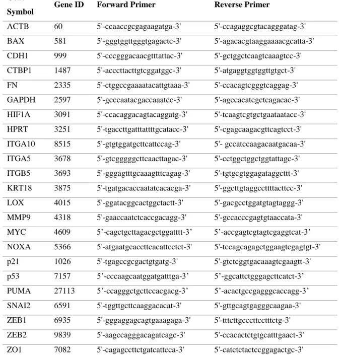
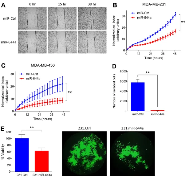
Figure 4.7. miR-644a inhibits migration, invasion and anchorage independent growth in vitro ... 60
Figure 4.8. miR-644a inhibits EMT in vitro ... 61
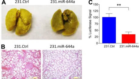
Figure 4.9. miR-644a inhibits metastasis in vivo ... 62
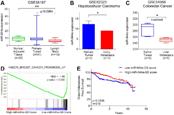
Figure 4.10. miR-644a expression is lower in metastases and correlates with metastasis-free survival ... 63
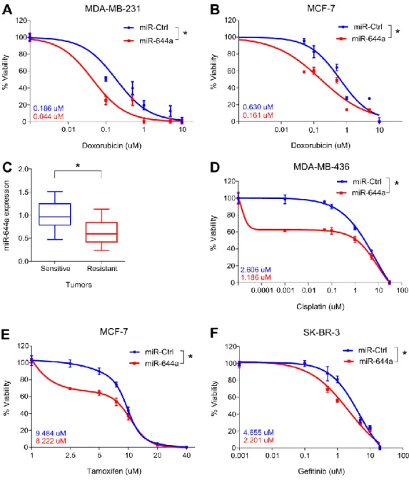
Figure 4.11. miR-644a overexpression acts as a therapy sensitizer in breast cancer cells and its expression correlates with doxorubicin resistance in vivo ... 66
Figure 4.12. Venn diagram for the combinatorial target prediction analysis of miR-644a ... 67
Figure 4.13. Schematic diagram showing miR-644a binding site in CTBP1 3’-UTR (453-460) in different species including human ... 68
Figure 4.14. Modulating miR-644a expression alters CTBP1 expression inversely in vitro ... 69
Figure 4.15. miR-644a directly targets CTBP1 and inversely correlates with CTBP1 expression in breast cancer cell lines...70
Figure 4.16. CTBP1 knockdown inhibits proliferation of breast cancer cells in vitro ... 71
Figure 4.17. CTBP1 knockdown promotes apoptosis in p53-mut but G1 cell cycle arrest in p53-wt breast cancer cells in vitro ... 72
Figure 4.18. Loss of CTBP1 inhibits breast cancer tumor progression in vivo ... 74
Figure 4.19. Loss of CTBP1 inhibits migration, invasion and anchorage independent growth in vitro ... 75
Figure 4.20. Knockdown of CTBP1 inhibits EMT and its upregulation upon miR-644a inhibition promotes EMT in vitro ... 76
xv
Figure 4.22. CTBP1 expression correlates with tumor progression and metastatic spread in
silico... 78
Figure 4.23. CTBP1 expression is associated with poor survival in chemotherapy treated breast cancer patients ... 79 Figure 4.24. CTBP1 knockdown sensitizes MDA-MB-231 cells to doxorubicin... 80 Figure 4.25. CTBP1 rescue overcomes miR-644a associated chemosensitive phenotype ... 80 Figure 4.26. Loss of CTBP1 reverses miR-644a inhibition associated mesenchymal phenotype to epithelial like state ... 81 Figure 4.27. Apoptosis associated genes are enriched in p53-mut tumors as compared to p53-wt ones ... 82 Figure 4.28. miR-644a regulates p53 at post-transcriptional level independent of its mutation status ... 83 Figure 4.29. miR-644a/CTBP1 mediated wild type or mutant p53 regulates p21 or Noxa, respectively at downstream ... 84 Figure 4.30. miR-644a overexpression or CTBP1 knockdown promotes apoptosis in p53-wt cells in the presence of mutant p53 ... 85 Figure 4.31. Ectopic p53 expression does not affect CTBP1 ... 86 Figure 4.32. Modulation of miR-644a/CTBP1/p53 axis upon miR-644a overexpression in different breast cancer cell lines ... 87 Figure 4.33: Mutant p53 is associated with poor survival in breast cancer patients as compared to wt p53 ... 88 Figure 4.34: CTBP1 expression is not associated survival in breast cancer patients ... 88 Figure 4.35. CTBP1 expression is associated with poor survival in p53-mut but not in p53-wt breast cancer patients ... 89 Figure 4.36: CTBP1 expression is associated with poor post-progression survival in p53-mut but not in p53-wt ovarian cancer patients ... 90 Figure 4.37. Schematic representation of developing doxorubicin resistance in mice using MDA-MB-231 TNBC cells ... 92 Figure 4.38. Summary of IPA-based mRNA core analysis showing top deregulated pathways in doxorubicin resistance ... 93 Figure 4.39. DoxoR-GS having enriched integrin/focal adhesion signaling predicts worst survival in chemotherapy-treated TNBC ... 94 Figure 4.40. ITGA5 is upregulated in doxorubicin resistant tumors and is associated with worst RFS specifically in chemotherapy-treated TNBCs ... 95 Figure 4.41. ITGA10 and ITGA5 expression in doxorubicin sensitive and resistant xenografts and association of their expression with survival in different breast cancer subtypes with or without chemotherapy treatment ... 96
xvi
Figure 4.42. ITGA5 expression is associated with worst survival in chemotherapy-treated TNBC
patients. ... 97
Figure 4.43. Hypoxia signaling is associated with chemotherapy resistance and HIF1A signaling is predicted to be activated in doxorubicin resistant xenografts ... 98
Figure 4.44. LOX, a key regulator in hypoxia, is upregulated in doxorubicin resistant xenografts... 100
Figure 4.45. LOX is associated with integrin signaling in chemotherapy treated TNBC tumors... 101
Figure 4.46. HIF1A expression correlates with LOX and ITGA5 expression in silico whereas LOX expression is highly correlated with ITGA5 both in silico and in vivo ... 102
Figure 4.47. LOX is associated with poor RFS in chemotherapy-treated TNBC patients ... 103
Figure 4.48. Hypoxia-induced LOX and ITGA5 hyperactivate integrin signaling ... 104
Figure 4.49. LOX hyperactivates integrin signaling by regulating ITGA5 ... 105
Figure 4.50. Inhibiting LOX activity sensitizes cells to doxorubicin treatment in 3D ... 106
Figure 4.51. Validation of LOX downregulation upon induction of shLOX using doxycycline... 107
Figure 4.52. Knocking down LOX increases tumor suppressive effects of doxorubicin in TNBCs in vivo... 108
Figure 4.53. Combinatorial treatment of xenografts with shLOX and doxorubicin resulted in least proliferation, higher apoptosis and less active integrin signaling ... 109
Figure 4.54. Inhibiting LOX activity increases the efficacy of doxorubicin in syngeneic tumor model of TNBCs ... 110
Figure 4.55. miRNAs predicted to target HIF1A, LOX and ITGA5 ... 111
Figure 4.56. miR-142-3p expression is inversely correlated with HIF1A, LOX and ITGA5 and is associated with better OS in chemotherapy-treated TNBC patients ... 112
Figure 4.57. Correlation analysis of miRNAs (other than miR-142-3p) predicted to target HIF1A, LOX and ITGA5 ... 113
Figure 4.58. Survival analysis of miRNAs (other than miR-142-3p) predicted to target HIF1A, LOX and ITGA5 ... 114
Figure 4.59. miR-142-3p is downregulated in doxorubicin resistant xenografts in vivo and upon hypoxia in vitro ... 115
Figure 4.60: HIF1A, LOX and ITGA5 are downregulated upon miR-142-3p ectopic expression... 115
Figure 4.61. miR-142-3p directly targets HIF1A, LOX and ITGA5 ... 116
xvii
Figure 5.1. Schematic representation of miR-644a/CTBP1/p53 axis-mediated drug resistance by simultaneous modulation of cell survival and EMT in p53-wt (left) and p53-mut (right) cells... 119 Figure 5.2. Schematic representation of hypoxia-induced LOX/ITGA5/FAK/Src axis mediated therapy resistance in TNBCs and targeting approaches for chemosensitization ... 125
xviii
List of Tables
Table 3.1. Sequences of forward and reverse primers used for 3’UTR cloning ... 25
Table 3.2. Protocol for double restriction digestion ... 25
Table 3.3. Protocol for ligation reaction ... 26
Table 3.4. Components of reverse transcription reaction ... 33
Table 3.5. Thermocycler program for cDNA synthesis ... 33
Table 3.6. Sequences of forward and reverse primers used in qRT-PCR analysis ... 34
Table 3.7. Master mix for qRT-PCR reaction ... 35
Table 3.8. qRT-PCR program ... 35
Table 3.9. Thermocycler protocol for Taqman miRNA reverse transcription ... 36
Table 3.10. Components of Taqman miRNA reverse transcription reaction ... 37
Table 3.11. Mastermix for Taqman miRNA PCR amplification ... 37
Table 3.12. qPCR protocol for Taqman miRNA amplification ... 38
Table 3.13. Components of the RIPA buffer ... 39
Table 3.14. Mixture for stacking and resolving gels in different concentrations ... 40
Table 3.15. List of antibodies used in Western blot ... 42
Table 3.16. List of antibodies used in Immunohistochemistry ... 46
Table 4.1. Enrichment of gene sets related to cancer formation and progression of different cancer types in patients with low and high miR-644a-GS scores ... 58
Table 4.2. Enrichment of gene sets related to metastasis in patients with low and high miR-644a-GS scores... 59
Table 4.3. Enrichment of gene sets related to drug resistance in patients with low and high miR-644a-GS scores ... 64
Table 4.4. IPA-based Upstream Regulator Analysis using RNA-Seq data of chemoresistant TNBC xenografts ... 98
xix
Abbreviations
7-AAD 7-Aminoactinomycin D
ACTB Actin beta
AGO Argonaute
AML Acute myeloid leukemia APS Ammonium peroxodisulfate BAPN β-Aminopropionitrile
BAX BCL2 Associated X
BCA Bicinchoninic acid BLI Bioluminescence imaging
BRCA1 Breast cancer type 1 susceptibility protein BrdU 5-bromo-2-deoxyuridine
BSA Bovine Serum Albumin
CAR constitutive androstane receptor Cdc2/CDK1 Cyclin dependent kinase 1 Cdc25C Cell division cycle 25C
CDH1 E-cadherin
CDK2 Cyclin-dependent kinase 2 CDK4 Cyclin dependent kinase 4 CTBP1 C-terminal binding protein 1 CTBP2 C-terminal binding protein 2 DAPI 4',6-diamidino-2-phenylindole
DAVID The database for annotation, visualization and integrated discovery DMEM Dulbecco's modified eagle medium
xx DMFS Distant metstasis free survival DNA Deoxyribonucleic acid dNTP Deoxynucleotide triphosphate ECL Enhanced chemiluminescence ECM Extracellular matrix
EGF Epidermal growth factor
EGFR Epidermal growth factor receptor EMT Epithelial-mesenchymal transition ER-α Estrogen receptor alpha
FAK Focal adhesion kinase FBS Fetal bovine serum
FN Fibronectin
GAPDH Glyceraldehyde 3-phosphate dehydrogenase GEO Gene expression omnibus
GSEA Gene set enrichment analysis H&E Hematoxylin and eosin HCC Hepatocellular carcinoma HCV Hepatitis C virus
HIF1A Hypoxia inducible factor A HIF1B Hypoxia inducible factor B
HPRT Hypoxanthine guanine phosphoribosyl transferase HR+ Hrmone receptor positive
HRP Horseradish peroxidase IDC Invasive ductal carcinoma IHC Immunohistochemistry
xxi ILK Integrin linked kinase
IPA Ingenuity pathway analysis ITGA10 Integrin subunit alpha 10 ITGA5 Integrin subunit alpha 5 ITGB5 Integrin subunit beta 5
kDa Kilo dalton
KRT18 Keratin 18
LNA Locked nucliec acid
LOX Lysyl oxidase
MDR Multi drug resistance
METABRIC Molecular taxonomy of breast cancer international consortium
MFP Mammary fat pad
miRNA microRNA
MMP1 Matrix metallopeptidase 1 MMP9 Matrix metallopeptidase 9
MTDH Metadherin
mut Mutant
MYC MYC proto-oncogene
NOXA Phorbol-12-myristate-13-acetate-induced protein 1 (PMAIP1) NSCLC Non-small cell lung carcinoma
ORF Open reading frame
OS Overall survival
P/S Penicilllin/Streptomycin
p21 Cyclin-dependent kinase inhibitor 1A (CDKN1A) PBS Phosphate buffered saline
xxii pCR Pathological complete response PDX Patient derived xenograft Poly-HEMA Poly-hydroxyethylmethacrylate PR Progesterone receptor
pre-miRNA Precursor miRNA pri-miRNA Primary miRNA
PTEN Phosphatase and tensin homolog PUMA P53-upregulated modulator of apoptosis
qRT-PCR Quantitative real time polymerase chain reaction Rb Retinoblastoma protein
RDX Radixin
RFS Relapse free survival
RHOA Ras homolog gene family, member A RTCA Real time cell analyzer
RTK Receptor tyrosine kinase
SDS-PAGE Sodium dodecyl sulfate polyacrylamide gel electrophoresis SERD Selective ER downregulator
SNAI2 Snail family transcriptional repressor 2
Src Src proto-oncogene, non-receptor tyrosine kinase TAE Tris-acetate EDTA
TBST Tris buffer saline Tween20 TCGA The cancer genome atlas TNBC Triple negative breast cancer TP53 Tumor protein 53
xxiii
v/v Volume/volume
w/v Weight/volume
wt Wild type
XPO5 Exportin 5
ZEB1 Zinc finger e-box binding homeobox 1 ZEB2 Zinc finger e-box binding homeobox 2
1
Chapter 1
Introduction
1.1. Breast cancer
Although last decades have witnessed a great success in prevention, diagnosis and therapy, cancer is still one of the leading causes of death worldwide with nearly 1.6 million new cases reported only in the United States in 2016 [1]. Breast cancer is the most common malignancy and the second leading cause of cancer deaths among women [2] whose incidence rate is drastically increasing worldwide as compared to other cancer types[3]. It has been estimated that approximately 60,000 women will be diagnosed with non-invasive whereas nearly 250,000 women will be diagnosed with invasive breast cancer only in the United States, of which around 40,000 will die from breast cancer in 2017 [1].
Being a highly heterogeneous disease, breast cancer represents very distinct molecular, morphologic and clinical features among different subtypes. Clinically, it has been classified into 3 major subtypes based on the expression status of specific receptors, namely, hormone receptors (estrogen receptor alpha (ER-α) and progesterone receptor (PR)) and human epidermal growth factor receptor 2 (HER2/ErbB2). These subtypes are 1) hormone receptor positive (HR+) expressing higher levels of ER and PR; 2) HER2-amplified; and 3) triple-negative breast cancer (TNBC) which lacks the expression of all three receptors[4]. At the molecular level, breast cancer has been classified into 5 major subtypes largely overlapping with the clinical subtypes (Figure 1.1) [5] where Luminal subtype A and B are hormone positive, ErbB2 positive is HER2-amplified, and the most of Basal subtype is TNBCs.
2
Figure 1.1. Molecular classification of breast cancer according to gene expression profiling [5].
1.2. Subtype-specific first-line treatments for breast cancer
Most of the breast cancer patients (around 70%) are HR+ subtype. Due to the overexpression of ER-α, patients in this subtype are mainly treated with hormone-targeted therapies such as tamoxifen and aromatase inhibitors in clinics [6]. On the other hand, HER2+ group, in which HER2 gene is amplified/overexpressed, encompasses around 20% of all the breast cancer cases. HER2 targeting monoclonal antibodies such as trastuzumab (Herceptin) or pertuzumab (Perjeta) are in clinical use to treat HER2+ breast cancers. These antibodies have shown efficiency towards blocking tumor growth as well as to enhance chemotherapy response[7, 8]. In addition, a dual inhibitor of EGFR and HER2 called lapatinib has been approved since 2007 to be used in combination with chemotherapy in clinics to treat HER2 overexpressing metastatic breast cancers [9]. Lastly, TNBCs are the most aggressive subtype of breast cancer which accounts for nearly 10-15% breast cancer diagnosed worldwide. Targeted-therapies are not available for TNBCs due to lack of druggable receptors e.g. ER-α or HER2; therefore, chemotherapy is the only treatment option for patients diagnosed with both early-stage and advanced-stage TNBCs [10]. Among chemotherapy agents, anthracycline and taxane-based chemotherapy agents are mainly used in both neo-adjuvant and adjuvant settings to treat TNBC [11].
3 1.3. Therapy resistance in breast cancer
Despite the availability of large number and various types of therapy agents (both chemo and targeted therapy) which are effective in limiting tumor burden at initial stages, cancer cells still manage to evade and exhibit re-growth of existing tumor or metastasize to distant organs. Currently, one of the major obstacles in achieving an effective cancer treatment is the emergence of therapy resistance: both “de novo” and “acquired”[12]. De novo resistance occur at the very beginning of treatment where tumor mass does not respond to treatment due to pre-existing molecular signature which interferes with the anti-cancer mechanisms of given treatment. In contrary, acquired resistance occurs in response to long-term treatments where cancer cells manage to survive under given anti-cancer treatment after initial response and become resistant. This kind of resistance is mostly associated with genomic, transcriptomic and epigenetic changes in tumor in response to prolonged treatments.
1.3.1. Targeted therapy resistance mechanisms in hormone-positive and HER2-amplified breast cancers
Cross-talk between ER-α and HER2 is one of the well-studied mechanisms of tamoxifen resistance. In the presence of HER2, tamoxifen can serve agonistically to ER-α by bringing transcription co-activators rather than repressors to ER-α transcription complex; thus, promoting transcription of ER-α target genes [13-15]. In addition to HER2 signaling, other growth factor receptors (i.e., insulin-like growth factor receptor or fibroblast growth factor receptor) can confer tamoxifen resistance by activating MAPK and PI3K/AKT/mTOR signaling cascades[16, 17]. Other resistance mechanisms include lack of ER-α expression due to hypermethylation of ER-α promoter[18] and dysregulated drug metabolisms [19, 20]. Constitutively active ligand-independent mutant ER-α can
4
confer resistance to aromatase inhibitors by altered transcriptional regulation at downstream [21, 22]. Other mechanisms of resistance to aromatase inhibitors include hyperactive interferon signaling [23] and disturbance in the balance of pro- and anti-apoptotic genes [24]. In order to overcome resistance, mTOR and CDK4/6 inhibitors have shown promising results in combination with other therapy agents in clinical trials and have been commercialized. For instance, everolimus, an allosteric inhibitor of mTOR, has been approved for use in post-menopausal woman with HR+ breast cancer as well as in other cancer types [25]. Recently, FDA has approved two different CDK4/6 inhibitors, palbociclib and ribociclib, to treat HR+ and HER2-negative advanced therapy resistant or metastatic tumors based on results of clinical trials where significant improvement in clinical outcome was observed when these inhibitors were combined with chemotherapy agents, letrozole or fulvestrant[26].
Multiple mechanisms responsible for trastuzumab resistance have been identified. In order to inhibit binding of antibody to HER2, cells often adapt to cleavage of the extracellular domain of the HER2 receptor [27-29]. Overexpression of other members of ErbB family such as HER3 or other tyrosine kinase receptors like IGF1R to promote downstream signaling have also been reported in the context of trastuzumab resistance [30, 31]. Other well-studied resistance mechanisms include loss of phosphatase and tensin homolog (PTEN) tumor suppressor or activation of oncogenes like SRC to activate HER2 signaling pathway even in the presence of trastuzumab [32-35]. Due to identification of proteins responsible for developing resistance, combination therapies have been tested to treat advanced stage therapy resistant HER2-amplified breast cancer [36]. For instance, combining trastuzumab with small molecule tyrosine kinase inhibitor, lapatinib, followed by PI3K/mTOR inhibition has shown to improve the survival outcome [37]. Furthermore, combination of mTOR inhibitor, evorolimus, with
5
trastuzmab has benefitted the patients in clinical trials [38]. In addition, antibody drug conjugate TDM1 (trastuzumab + emtansin) has been approved for metastatic HER2-amplified breast cancer patients after superior effect of T-DM1 over lapatinib and capecitabine combination in patients pretreated with trastuzumab and taxane therapies [39].
1.3.2. Chemotherapy resistance mechanisms in TNBCs
The extent of pathological response to neo-adjuvant treatment at surgery has been established as a prognostic marker for disease recurrence in breast cancer [40]. As compared to other subtypes, TNBC patients show low risk of recurrence if pathological complete response (pCR) is achieved [41]. While only 30-40% of TNBC patients show pCR towards treatment, others have less than 60% 5-year survival due to aggressive relapse as a response of chemotherapy resistance [42]. Recently, PTEN deletions/mutations and copy number amplifications of proto-oncogenes MYC, JAK2, PIM1 have been frequently observed in residual disease after neo-adjuvant chemotherapy treatment of TNBCs, thus, suggesting high rate of chromosomal instability as a driving force to develop chemotherapy resistance in TNBCs. However, due to subclonal nature of driver mutations in TNBCs, single agent targeted therapy is unlikely to be successful [43]. Aberrant NFKB signaling has also been associated with chemotherapy resistance in cancer, in general, but found difficult to be targeted because of the off- target effects and toxicities as a large spectrum of cellular processes revolve around NFKB [44]. Bortezomib, a ubiquitin-proteasome inhibitor known to suppress NFKB signaling by inhibiting IKB degradation [45] showed promising results in myeloma and lymphoma, but failed to prove its efficacy in combination with chemotherapy agents in solid tumors [46]. Interestingly, loss of proto-oncogene MYC has been shown to confer chemotherapy
6
resistance in breast cancer by misbalancing the expression of genes regulating apoptosis [47]. Other reported mechanisms of chemotherapy resistance in breast cancer include mutations in DNA repair enzymes [48], altered expression of drug transporter proteins [49], greater drug detoxification [50].
1.4. Tumor microenvironment and drug resistance
Other than malignant cells, tumor microenvironment comprises of immune cells, fibroblasts, tumor vasculature, signaling molecules and extracellular matrix (ECM) which also play pivitol roles in tumor cell maintainence and survival. Although, ECM has been proposed as safegaurd preventing cancer initiation in early life stages, it has been shown playing active roles in pathological incidences like tumorigenesis [51]. Tumor microenvironment associated ECM largely differs from that of the normal tissue and has been shown to serve as a basic scaffold for chemotaxis driven cancer cell invasion and metastasis [52]. Interplay between cancer cells and ECM elements (ECM modulating enzymes, collagens, laminin etc) is dynamic. Attachment to ECM has shown to alter polarization of malignant cells and cause resistance to etoposide-induced apoptosis in breast cancer [53]. Specifically, cell adhesion molecules-mediated drug resistance depends on association of integrin to ECM components including fibronectin, collagen and laminin [54]. Accumulating evidence has shown that changes in composition and topography of ECM upon therapy are directly sensed by multiple cell adhesion molecules including integrins which activate downstream pro-survival signaling to resist the given therapy [15]. ECM remodelling and integrin signaling mediated resistance has also been implicated to radiation and targeted therapies by attenuating activities of receptor tyrosine kinases (RTKs) such as EGFR[55, 56]. Desmoplastic stroma in tumor microenvironment
7
also poses a physical barrier for efficient delivery of therapy agents, thus rendering treatment options ineffecient and/or unsucessful[57].
As solid tumors grow in size, the inner core of the tumor mass gets deprived of oxygen. Tumor cells usually counteract this by adapting to hypoxia via upregulating transcription factors known as hypoxia-inducible factors (HIFs) [16]. Although human genome express three HIF-α isoforms, hypoxia-driven phenotypes and mechanisms have been mainly associated with HIF1-α which is widely expressed in tissues as compared to other family members [17] and transactivates distinct set of genes [18]. Patches of hypoxic tumor microenvironment has been well-established as significant contributor to chemotherapy failure and drug resistance due to fact that hypoxia is being causally involved in regulating multiple aspects of tumor biology such as resistance to apoptosis, metabolic reprogramming, angiogenesis and pH homeostasis [19, 20]. Therefore, studying drug resistance in settings where the hypoxic tumor microenvironment taken into account is critical towards the identification of true driving factors with high potential for successful translation in the clinic.
1.5. Metastasis in breast cancer
The majority of cancer deaths are not caused by primary tumors, but rather by the dissemination of the disease, i.e. the development of distant metastases. Metastasis is accomplished in two major steps: dissemination and colonization. Dissemination phase includes local invasion, intravasation into the systemic circulation, survival in the circulatory system and extravasation. Colonization phase includes the adaptation of these cells to a foreign microenvironment where the microscopic cells turn into macroscopic tumors. The whole process is outcome of the interplay between genetic and epigenetic modifications in tumor as well as in the tumor microenvironment [58].
8
Nearly 15% of all breast cancer patients experience an agressive disease and develop distant metastasis within 3 years of diagnosis of primary tumors, but it can also take 10 or more years after the intial detection of primary tumor to establish metastatic growth at distant organs [59]. Once disseminated, breast carcinoma can metastasize to various organs. The most preferential sites of breast cancer metastasis include bone, lungs and liver (Figure 1.2). Analysis of advanced stage breast cancer patients has shown subtype-specific tendency of breast cancer to metastasize. While HR+ patients show more bone metastasis, HER2-amplified patients exhibit increased incidence rate of liver metastasis, and TNBCs preferentially metastasized to brain and lungs [60]. In addition, breast cancer recurrence and mortality rate have also been associted with its subtypes. ER negative patients, in general, and TNBCs, in particular, usually develop metastasis within first 5 year of diagnosis. On the other hand, more than 50% of recurrence incidences in ER positive patients appear after 5 years of first diagnosis[61].
Figure 1.2: Most common metastasis sites of breast cancer [59]. See Appendix for the copyright permission.
9
Established prognostic markers of breast cancer metastasis include presence of lymph-node metastasis, larger-sized primary tumors and loss of histopathological differentiation. However, these traditional markers are only able to predict the course of disease confidently in very small population of patients. Therefore, additional prognostic markers, especially molecular markers, are needed to better predict the course of disease and metastasis incidence [59]. In this regard, there have been few studies which determined a set of protein-coding genes mediating organ-tropic metastasis in breast cancer. It has been shown that there are two sets of genes, one of which regulates both primary breast tumor growth (in case of ID1, CXCL1, PTGS2 and MMP1) and lung metastasis capacity while the other set involves gene e.g. SPARC and MMP2 which regulate lung metastatic virulence specifically [62]. Gene expression analysis of brain-tropic cancer cells and of clinical samples, coupled with functional analysis, identified the cyclooxygenase COX2, the epidermal growth factor receptor (EGFR) ligand HB-EGF, and the alpha2,6-sialyltransferase ST6GALNAC5 as mediators of cancer cell passage through the blood-brain barrier [63]. Set of genes including VCAM-1, NF-kB, JAGGED1, Src, matrix metallopeptidase 1 (MMP1), lysyl oxidase (LOX) and certain cytokines (CXCR4, CXCL12, TGFβ) have been associated with metastatic spread of breast cancer to bones [61].
1.6. MicroRNAs and their dysregulation in cancer
MicroRNAs (miRNAs) are a large family of small regulatory RNAs, acting in post-transcriptional gene regulation. They are 20-22 nucleotides long and recognize their target mRNAs by complementary base pairing. They control gene expression by mRNA cleavage, mRNA destabilization or inhibition of translation [64]. Almost half of miRNAs reside in clusters and transcribed as polycistronic precursor miRNAs [65]. Other
10
miRNAs, located in intergenic regions, are transcribed by their own promoters while those present in intronic regions are likely under the control of the host genes’ promoters [66]. Currently, it has been reported that there are more than 2600 unique mature miRNAs in human (miRBase version 21) [67]. Most miRNAs are transcribed by RNA polymerase II as primary transcripts (pri-miRNAs), usually several kilobases long, which fold into hairpin structures containing imperfectly base-paired stem-loop structures [68]. RNase III endonuclease Drosha then cleaves primary miRNAs (pri-miRNAs) into ~70 nt long precursor miRNAs (pre-miRNAs) which are later transported to cytoplasm by RanGTP-dependent dsRNA-binding protein exportin-5 (XPO5) [69]. In cytoplasm, RNase III endonuclease Dicer cleaves pre-miRNAs into mature miRNAs which are loaded to RNA-induced silencing complexes. Along with Argonaute (Ago) proteins (mainly Ago1 and Ago2 in mammals) of the complex, miRNAs downregulate gene expression by binding to target mRNAs (Figure 1.2).
Figure 1.3: Schematic demonstration of miRNA biogenesis [70]. See Appendix for the
11
Although miRNA binding sites have also been found in 5’-UTR [71] and open reading frames (ORFs) [72] of the genes, they preferentially interact with seed-matching sequences in the 3’-UTR of mRNA. One miRNA can downregulate multiple genes due to the short sequence required for mRNA recognition, which is known as the ‘seed region’ spanning between the 2nd and the 7th (or 8th) nucleotide of mature miRNAs. Taking both direct and indirect regulations together, it is not rare that a single miRNA can regulate the expression of tens or hundreds of genes. Out of all the identified human miRNAs, almost 50% are located at fragile sites on chromosomes known for having common alterations (i.e. amplification, deletion and rearrangements) in cancer [73]. Roles of miRNAs in cellular processes like cell cycle progression, proliferation, metabolism, apoptosis, and stress resistance [74] cannot be overlooked as more than 60% of human protein coding genes are predicted to be under selective pressure to be regulated by miRNAs [75].
1.7. MicroRNAs and tumor progression, metastasis and drug resistance
Considering the enormous regulatory potential of miRNAs, it is not surprising that they play crucial roles in cancer development, progression, metastasis and drug resistance [76]. For instance, elevated expression of oncogenic miR-17∽92 cluster (comprising of 6 miRNAs) has been reported during lymphomagenesis which allows continuous activation of oncogenic PI3K and NF-kB signaling by suppressing negative regulators of these pathways [77]. Another oncogenic miRNA, miR-21, is upregulated in lung, prostate, breast and pancreatic cancers compared to normal tissues [78]. In line with this, knockdown of miR-21 in breast cancer inhibited tumor growth and enhanced apoptosis by downregulating anti-apoptotic protein Bcl-2 [79]. miR-155 is overexpressed in pancreatic cancer where it promotes tumor development by repressing the expression of tumor suppressor Tp53INP1, and oligonucleotide-mediated inhibition of miR-155
12
restored Tp53INP1 levels along with significant increase in apoptotic cell death [80]. Tumor suppressor let-7 family miRNAs have shown to target a network of cell cycle-associated genes including E2F5, CCNA2, CDK8, hence playing important roles in regulating multiple proliferation pathways and controlling tumor growth [81]. miR-34 is another well-studied tumor-suppressor miRNA which is directly regulated by p53 and controls p53-mediated cell death. Low miR-34 expression attenuates p53-mediated apoptosis and contributes to tumor development [82]. Taken together, their function as negative regulators of multiple targets in biological networks and their common dysregulation in cancer make miRNAs attractive targets for cancer therapy.
miRNAs have been associated with drug resistance to both chemo- and targeted-therapies. Inhibition of miR-21 has been reported to increase gemcitabine sensitivity in cholangiocarcinoma and to inhibit the growth of topotecan-treated MCF7 cells [83]. In medulloblastoma, miR-34a has been demonstrated to sensitize cancer cells to mitomycin C and cisplatin by directly targeting the oncogenic gene MAGE-A, and to induce apoptotic cell death by modulating tumor suppressor p53 levels in a positive feedback loop [84]. In another study, miR-137 has been shown to target constitutive androstane receptor (CAR), which is an important regulator of multi-drug resistance (MDR), and its overexpression sensitized neuroblastoma, hepatocellular carcinoma (HCC) and colon cancer cells to doxorubicin [85]. Similarly, miRNAs have also been shown to regulate resistance to targeted therapies. Two independent studies have associated miR-221/222 with resistance to tamoxifen, by establishing miR-221/222 downregulating the expression of ER alpha [86] and p27/Kip1 [87]. Furthermore, it has been shown that miR-221/222 also confers resistance to fulvestrant, a selective ER downregulator (SERD), by modulating both Wnt/β-catenin and TGF-β pathways [88]. We have recently demonstrated that miR-375 is downregulated in tamoxifen resistant MCF-7 cells
13
compared with parental ones and re-expression of miR-375 sensitized resistant cells to tamoxifen partially by downregulating the oncogene metadherin (MTDH) [89]. Altogether, these reports clearly indicate the involvement of miRNAs in resistance to both chemotherapy and targeted therapy, and these miRNAs may be therapeutically modulated to sensitize tumor cells again to the drugs.
miRNAs have also been implicated in regulation of metastatic cascade. For instance, the miR-200 family has been found downregulated in metastases compared to primary tumors [48, 49] and plays a central role in the inhibition of EMT by forming a double-negative feedback loop with ZEB1 and ZEB2, both of which are the transcriptional repressors of cell-cell contact protein E-Cadherin [50, 51]. miR-31 has been demonstrated to regulate several post-intravasation steps including intraluminal viability, extravasation and survival at distal tissue in addition to the invasion and metastatic colonization steps by simultaneous targeting of three key genes: integrin α5 (ITGA5), radixin (RDX) and Ras homolog gene family , member A (RHOA) [90]. Recently, it was shown that miR-520/373 family inhibits both in vitro cell invasion and in vivo intravasation of highly invasive ER (-) breast cancer cells. Decreased expression of miR-520c was found to be correlated with the lymph node metastasis of ER (-) breast cancer patients [91]. miR-200 has been shown to promote the colonization of breast cancer cells by directly targeting the Sec23a gene which is involved in the secretion of metastasis-suppressive proteins [92]. Recently, miR-612 is suggested to suppress the colonization of HCC cells to the lungs [93]. Altogether, these findings suggest that miRNAs regulate metastasis at multiple steps by modulating different components of the cellular networks.
14
1.8. Modulation of microRNAs as drug candidates
With the discovery of microRNAs as crucial regulators of biological processes and their dysregulation in various human diseases, in general, but in cancer, in particular, therapeutic targeting of miRNAs is very attractive for scientists for novel therapy development in human pathologies[94]. There are several strategies being evaluated to target miRNAs or use them as targeting agents in cancer, such as i) inhibition of oncogenic miRNAs by antisense DNA oligonucleotides, antagomirs, locked nucleic acids (LNAs), RNA sponges or miR-masking; ii) exogenous expression of tumor suppressor miRNAs; and iii) targeting miRNAs by using small molecules [95]. Recently, an antisense nucleotide, Miravirsen, has been introduced as a potential drug to treat hepatitis C virus (HCV) infection. In principle, HCV requires liver specific miR-122 expression for replication. Miravirsen binds to miR-122 and inhibits its biogenesis in liver; thus, inhibiting HCV to replicate and multiply. After showing its success in treating HCV in chimpanzees, Miravirsen is now being tested in human clinical trials and has been found safe in term of toxicity[96]. MRX34, a liposomal encapsulated nanoparticle formulation, is the first miRNA-based cancer therapy agent. It is a double-stranded mimic of miR-34, a well-established tumor suppressor miRNA known for inhibiting oncogenicity and for inducing cancer cell death [97]. MRX34 has shown promising results in the Phase 1 clinical trial of different cancer types including renal cell carcinoma, melanoma and hepatocellular carcinoma [98]. Combination of miRNA delivery system with therapy agents has also shown promising results. For instance, delivery of LNAs encapsulated miR-10b in combination with a low dose of doxorubicin was enough to achieve a significantly greater decrease in tumor burden compared with doxorubicin monotherapy without any evidence of damage to normal tissues[99]. These studies clearly suggest that miRNAs have high potential to be used as the therapeutic drugs without toxicity in future.
15
1.9. The rationale and the aims of the dissertation
Therapy resistance and metastasis are two major hallmarks of cancer. Despite the huge advances in the field of drug design, cancer cells still manage to evade therapy, exhibit regrowth of tumor and/or metastasize to distant organs. Not only coding, but also non-coding genome (long non-non-coding RNAs, miRNAs, circular RNAs) is being extensively studied to understand the underlying mechanisms of therapy resistance and metastasis. Notably, treatment options are restricted to chemotherapy in TNBC due to lack of available druggable targets. This dissertation starts from the view to utilize unbiased genome-wide approaches to identify novel hubs of therapy resistance and metastasis in breast cancer, then to apply appropriate in vitro approaches to explore molecular mechanisms regulated by these hubs, and to validate findings using in vivo models in combination with in silico cross validation using online available patient data.
In this dissertation, the aims are as following:
- To identify novel coding/non-coding molecular switches regulating tumor progression, metastasis and chemo-response in breast cancer
- To identify novel drug targets overcoming chemotherapy resistance specifically in TNBCs
16
Chapter 2
Materials
2.1. Buffers
1X Anode Buffer I 300 mM Tris, 20% (v/v) methanol
1X Anode Buffer II 25 mM Tris, 20% (v/v) methanol
1X Cathode Buffer 40 mM 6-aminocaparoic acid, 20% (v/v) methanol
1X PBS 137 mM NaCl, 2.7 mM KCl, 10 mM Na2HPO4,
2 mM KH2PO4 (pH: 7.4)
1X SDS-PAGE Running Buffer 25 mM Tris, 14.41 g/l glycine, 1% (v/v) SDS
1X TAE 40 mM Tris, 20 mM acetic acid, 1 mM EDTA
1X TBST 20 nM Tris, 137 mM NaCl, 0.2% (v/v)
Tween20
RIPA Lysis Buffer 150 mM NaCl, 1% (v/v) NP-40, 0.5% (v/v) Sodium DOC, 50 mM Tris-HCl (pH:8.0), 50 mM NAF, 1 mM NaVO4, 4% (v/v) Protease inhibitor, 4% (v/v) Phospatase inhibitor
2.2. Chemicals and Reagents
4X Protein Loading Dye 250 mM Tris HCl (pH: 6.8), 10% (w/v) SDS, 0.1% (w/v) Bromophenol blue, 50% Glycerol (v/v), 25% (v/v) β-mercaptoethanol
6-aminocaparoic acid Sigma Aldrich, St Louis, MO, USA
17
7-AAD BD Biosciences, San Diego, CA, USA
Acetic acid Sigma Aldrich, St Louis, MO, USA
Acrylamide/bisacrylamide Applichem, Darmstadt, Germany
Agar Sigma Aldrich, St Louis, MO, USA
Agarose Promega, Madison, WI, USA
Ammonium peroxisulfate Carlo Erba, Cornaredu, Italy
Bouin’s solution Sigma Aldrich, St Louis, MO, USA
Bovine Serum Albumin Santa Cruz, Dallas, TX, USA
Protease inhibitor cocktail Roche Applied Science, Mannheim, Germany D-Luciferin Potassium salt Sigma Aldrich, St Louis, MO, USA
DAPI Sigma Aldrich, St Louis, MO, USA
dNTPs Thermo Fisher Scientific, Waltham, MA, USA
ECL Amersham Pharmacia Biotech, Amersham, UK
Thermo Fisher Scientific, Waltham, MA, USA
Ethanol Sigma Aldrich, St Louis, MO, USA
Isopronapol Sigma Aldrich, St Louis, MO, USA
Ethidium Bromide Thermo Fisher Scientific, Waltham, MA, USA Gene Ruler 100bp DNA Ladder New England Biolabs, Ipswich, MA, USA Gene Ruler 1kb DNA Ladder New England Biolabs, Ipswich, MA, USA
Isopronapol Sigma Aldrich, St Louis, MO, USA
LightCycler 480 SYBR Green I Master
Roche Applied Science, Mannheim, Germany
Lipofectamine 2000 Invitrogen, Carlsbad, CA, USA
18
Milk powder Sigma Aldrich, St Louis, MO, USA
Nuclease free water Applied Biosystems/Ambion, Austin, TX, USA Page Ruler Protein Ladder Thermo Fisher Scientific, Waltham, MA, USA
Phosstop Roche Applied Science, Mannheim, Germany
Ponceu S Sigma Aldrich, St Louis, MO, USA
Shandon Immu-mount Thermo Fisher Scientific, Waltham, MA, USA
Sodium Chloride Merck, Darmstandt, Germany
Sodium Dodecyl Sulfate (SDS) Merck, Darmstandt, Germany
TEMED Serva, Heidelberg, Germany
TRIsure Bioline, Luckenwalde, Germany
Triton X-100 Sigma Aldrich, St Louis, MO, USA
Trizma base Sigma Aldrich, St Louis, MO, USA
Trypton Sigma Aldrich, St Louis, MO, USA
Tween-20 VWR, Radnor, PA, USA
WST-1 Roche Applied Science, Mannheim, Germany
Yeast Extract Sigma Aldrich, St Louis, MO, USA
2.3. Enzymes and Enzyme Buffers
10X Cut Smart Buffer New England Biolabs, Ipswich, MA, USA 10X T4 DNA ligase Buffer Thermo Fisher Scientific, Waltham, MA, USA BsmBI Restriction enzyme New England Biolabs, Ipswich, MA, USA Notl Restriction enzyme New England Biolabs, Ipswich, MA, USA Phusion Polymerase New England Biolabs, Ipswich, MA, USA
19
T4 DNA Ligase Thermo Fisher Scientific, Waltham, MA, USA
T4 Kinase Thermo Fisher Scientific, Waltham, MA, USA
Taqman universal mix Thermo Fisher Scientific, Waltham, MA, USA XhoI Restriction enzyme New England Biolabs, Ipswich, MA, USA
2.4. Media and Supplements
DMEM Lonza, Basel, Switzerland
Fetal Bovine Serum (FBS) Biowest, Nuaille, France
LB Agar 1.5% (w/v) agar, 10 g/l trypton, 5 g/l yeast extract, 10 g/l NaCl
LB Broth 10 g/l trypton, 5 g/l yeast extract, 10 g/l NaCl
Matrigel BD, Flanklin Lakes, NJ, USA
Non-essential amino acids Lonza, Basel, Switzerland
optiMEM Invitrogen, Carlsbad, CA, USA
Penicillin/Streptomycin Lonza, Basel, Switzerland
2.5. Kits
BCA Protein Assay kit Pierce, Rockford, IL, USA
BrdU FITC Flow kit BD, Flanklin Lakes, NJ, USA
Caspase-Glo® 3/7 Assay kit Promega, Madison, WI, USA Cell Titer-Glo and 3D cell Titer-Glo
Luminescent Cell Viability Assay kit
20
Trans-lentiviral Packaging kit Dharmacon, Lafayette, Colorado, United States Dual Luciferase Reporter kit Promega, Madison, WI, USA
First strand cDNA synthesis kit Fermantas, St. Leon-Roth, Germany Gel and PCR clean up kit MN, Duren, Germany
Lysyl Oxidase (LOX) Activity Assay kit (Fluorometric)
Abcam, Cambridge, United Kingdom
MycoAlert detection kit Lonza, Basel, Switzerland
Plasmid isolation kit MN, Duren, Germany
Plasmid Maxi kit Qiagen, Hilden, Germany
Taqman miRNA Assays Applied Biosystems, Foster City, CA, USA
2.6. Equipments
Accuri FACS BD Biosciences, San Diego, CA, USA
Axiovision 4.3 microscopy Carl Zeiss, Munich, Germany
Centrifuges Thermo Fisher Scientific, Waltham, MA, USA
Beckman, Pasadena, CA, USA
Cell culture hood Nüve, Ankara, Turkey
Cell culture incubator Nüve, Ankara, Turkey
Counting chamber Marienfeld, Königshofen, Germany
Freezer (-80oC) Hettich, Geldermansen, Holland
Freezer (-20oC) and Fridge (4oC) Bosch, Stuttgart, Germany
Horizontal Shakers Bellco, Vineland, NJ, USA
21
LightCycler 96 Roche Applied Science, Mannheim, Germany
Mini-PROTEAN Gel casting module Biorad, Hercules, CA, USA Mini-PROTEAN Tetra Cell Biorad, Hercules, CA, USA
Multichannel Pipette Thermo Fisher Scientific, Waltham, MA, USA
Nanodrop 1000 Thermo Fisher Scientific, Waltham, MA, USA
Nikon TS300 Inverted microscope Nikon, Tokyo, Japan
Thermocycler Thermo Fisher Scientific, Waltham, MA, USA
Power supplies for electrophoresis Biorad, Hercules, CA, USA Semidry Western Blot transfer unit Biorad, Hercules, CA, USA Synergy HT microplate reader Biotek, Winooski, VT, USA
UV-Reader Vilber Lourmat, Eberhardzell, Germany
Vortex Isolab, Wertheim, Germany
Water bath Nüve, Ankara, Turkey
X-ray cassette Amersham Pharmacia Biotech, Amersham, UK
X-ray hyper processor Amersham Pharmacia Biotech, Amersham, UK
2.7. Consumables
100 mm dishes Greiner bio-one, Frickenhausen, Germany
145 mm dishes Greiner bio-one, Frickenhausen, Germany
96-well plates Greiner bio-one, Frickenhausen, Germany
6-well plates Greiner bio-one, Frickenhausen, Germany
Filtered pipette tips (10 ul, 20 ul, 200 ul, 1000 ul)
22
Cell scrapers Greiner bio-one, Frickenhausen, Germany
Coverslips Marienfeld, Königshofen, Germany
Cryovials Greiner bio-one, Frickenhausen, Germany
Microscope slides Marienfeld, Königshofen, Germany
Parafilm VWR, Radnor, PA, USA
PCR tubes Axygen, Corning, NY, USA
Plastic pipettes (10 ml, 25 ml) Corning Incorporated, Corning, NY, USA
PVDF Membrane Biorad, Hercules, CA, USA
Reaction tubes (500 ul, 1.5 ml, 2 ml) Axygen, Corning, NY, USA
Storage bottles (250 ml, 500 ml, 1 L) Corning Incorporated, Corning, NY, USA
Whatmann paper GE Healthcare, Little Chalfont, UK
X-ray films Kodak, Rochester, NY, USA
Cuvettes VWR, Radnor,PA, USA
qPCR Plates Roche Applied Science, Mannheim, Germany
23
Chapter 3
Methods
3.1. Culturing Human Breast Cancer Cell lines
Human breast cancer cell lines MDA-MB-157, MDA-MB-231, MCF-7, BT474, SK-BR-3, ZR-75-1 together with normal breast epithelial cell lines MCF-10A and MCF-12A and mouse mammary cancer cell line 4T1 were obtained from ATCC (Manassas, VA, USA). 231.Luc.GFP was a kind gift from Dr. Dihua Yu (MD Anderson Cancer Center, Houston, TX, United States). MDA-MB-231 and SK-BR-3 cell lines were cultured with Dulbecco Modified Eagle Medium while MCF-7, BT474 and ZR-75-1 cell lines were cultured with DMEM supplemented with 0.1% insulin. MCF10A and MCF-12A cell line was cultured with DMEM supplemented with 0.1% insulin (0.01 mg/ml), 0.002% EGF (20 ng/ml). All media were supplemented with 50 U/ml penicillin/streptomycin, 1% non-essential amino acids and 10% fetal bovine serum. All cell lines were tested for mycoplasma contamination regularly using MycoAlert mycoplasma detection kit (Lonza, Basel, Switzerland).
3.2. Transient transfection with miRNA mimics, hairpin inhibitors, siRNAs and expression and reporter constructs
Transient transfection was performed 24 hours after cell seeding using Lipofectamine 2000 (Invitrogen, Carlsbad, CA, USA) and OptiMEM. Briefly, 4000-6000 cells/well of 96-well plate or 150,000-200,000 cells/well of 6-well plate were seeded in 100 μl or 1.5 ml Penicillin/Streptomycin free (P/S-free) media, respectively. For one well of 6-well transfection, 2-3 μl of lipofectamine (2 μl in case of miRNA mimics, siRNAs, hairpin inhibitors whereas 3 μl in case of expression or reporter constructs) was diluted in 250 μl
24
optiMEM (mix A) and vortexed for 20 seconds. Simultaneously, miRNA mimics, siRNAs, hairpin inhibitors, expression or reporter constructs were separately diluted in 250 μl optiMEM (mix B) and vortexed for 20 seconds. Both vials (mix A and mix B) were incubated at room temperature for 5 minutes, then mixed in 1:1 (v/v) ratio and vortexed for 20 seconds followed by incubation at room temperature for 20 minutes. Media was aspirated from cells and replaced with 1 ml fresh P/S-free media. 500 μl of transfection mixture was added on top of the cells resulting final volume of 1.5ml transfection medium. Cells were incubated at 37oC, 5% CO2 for further experiments. For one well of 96-well format, transfection mixture was prepared 10 times less as compared to that used for transfection in one well of 6-well plate.
For miRNA mimic viability screen, miRNA mimics were transfected at a concentration of 20nM for 48 hours. For other experiments, miR-644a mimic and siCTBP1s were transfected at a concentration of 40 nM whereas miR-142-3p and siLOX were transfected at a concentration of 20 nM for either 48 or 72 hours. Hairpin inhibitors were transfected at a concentration of 100nM for either 48 hours or 72 hours. Transfection of GFP-tagged CTBP1 human ORF clone (NM_001012614; Cat. No. RG208594), and Myc-DDK tagged TP53 human mutant ORF clone (NM_000546; Cat. No. RC200003, expressing transcript variant 1 of Homo sapiens protein p53 having an R175H mutation) and reported constructs carrying 3’-UTRs of human CTBP1, HIF1A, LOX and ITGA5 were carried out at 50 ng (for 96-well plate) or 500 ng (for 6-well plate) per well.
3.3. Plasmid construction and site-directed mutagenesis
The 3’-UTR sequence of CTBP1 containing binding site for miR-644a and the 3′-UTRs of HIF1A, LOX and ITGA5 containing binding sites for miR-142-3p were amplified from genomic DNA of MDA-MB-231 cells using primers listed in Table 3.1.
![Figure 1.1. Molecular classification of breast cancer according to gene expression profiling [5]](https://thumb-eu.123doks.com/thumbv2/9libnet/5789561.117744/26.892.183.772.110.293/figure-molecular-classification-breast-cancer-according-expression-profiling.webp)
![Figure 1.2: Most common metastasis sites of breast cancer [59]. See Appendix for the copyright permission](https://thumb-eu.123doks.com/thumbv2/9libnet/5789561.117744/32.892.198.749.690.1004/figure-common-metastasis-breast-cancer-appendix-copyright-permission.webp)
![Figure 1.3: Schematic demonstration of miRNA biogenesis [70]. See Appendix for the copyright permission](https://thumb-eu.123doks.com/thumbv2/9libnet/5789561.117744/34.892.290.664.657.1035/figure-schematic-demonstration-mirna-biogenesis-appendix-copyright-permission.webp)