http://journals.tubitak.gov.tr/biology/ © TÜBİTAK
doi:10.3906/biy-1502-37
Amplification of GC-rich ADAMTS-2 and URG4/URGCP promoter regions with
optimized combination of PCR enhancers
Meltem ALPER1, Esra TOKAY2, Feray KÖÇKAR2,*
1Aksaray Vocational School of Technical Sciences, Aksaray University, Aksaray, Turkey
2Department of Biology, Faculty of Science and Literature, Balıkesir University, Balıkesir, Turkey
1. Introduction
Polymerase chain reaction (PCR) is a conventional method for the amplification of target DNA regions. A large variety of parameters can influence the outcome of a PCR amplification. These variables include cycling parameters as well as the concentrations of Mg+2, H+, dNTP, primers, templates, and added enhancers (Innis and Gelfand, 1990; Hecker and Roux, 1996). Most DNA does not require special modifications or additives for amplification. However, the templates that have tandem repeats, high guanine-cytosine (GC) content, and strong secondary structures are difficult to amplify with basic PCR conditions. Increased hydrogen bonding with neighboring cytosine and guanine generates complex hairpins and loops in the GC-rich regions (McDowell et al., 1998; Musso et al., 2006). In the human genome there are often GC-rich sequences including important regulatory domains such as promoters, enhancers, and control elements (Wilson, 1997; Mamedov et al., 2008). The PCR amplification of the GC-rich DNA is often problematic because of stable secondary structures in the DNA that are resistant to melting. These secondary structures cause DNA polymerases to stall, resulting in incomplete and nonspecific amplification (Sahdev et al., 2007).
To amplify these problematic GC-rich DNA fragments, several approaches have been developed. Organic molecules such as dimethyl sulfoxide (DMSO), glycerol, polyethylene glycol, formamide, betaine, 7-deaza GTP, and dUTP have been included in the reaction mixture and have been shown to improve the amplification of GC-rich DNA sequences (Pamp and Medrano, 1991; Sun et al., 1993; Mutter and Baynton, 1995; Turner and Jenkins, 1995; Baskaran et al., 1996; Henke et al., 1997; Chakrabarti and Schutt, 2001; Kang et al., 2005; Musso et al., 2006). Moreover, different PCR strategies were improved, such as hot-start PCR, step-down PCR, and a hot-start method combined with touch-down to increase PCR yield (Korbie and Mattek, 2008).
ADAMTS-2 (a disintegrin and metalloproteinase with
thrombospondin type I, motif II) is a metalloproteinase playing a key role in the processing of fibrillar collagen precursors into mature collagen molecules by cleaving of the amino terminus of the type I, II, III, and V procollagens. This maturation process allows the correct fibril formation (Colige et al., 2005). The functional analysis of the human ADAMTS-2 protein has been widely studied and mutations in the ADAMTS-2 gene have been illustrated with Ehlers– Danlos syndrome type VIIC in humans (Colige et al.,
Abstract: PCR is an effective and widely used method for the amplification of target DNA fragments in vitro. The templates that have
high guanine-cytosine (GC) content, complex secondary structures, or short tandem repeats are difficult to amplify by the conventional PCR methods. Human genome regulatory regions such as promoters are rich in terms of GC base compositions. To amplify these regions some special modifications or additives are required. Using different PCR strategies such as hot-start/touchdown PCR or the utilization of various additives into the reaction mixture such as organic molecules can improve PCR yield. In the present study we optimized the PCR conditions for the amplification of the human ADAMTS-2 gene promoter region featuring an extremely high GC content and a secondary structure. We show that a combination of three additives, betaine, dimethyl sulfoxide, and 7-deaza GTP, is essential to obtain a correct PCR amplicon. To demonstrate whether the optimized PCR condition was applicable to other GC-enriched regions, a similar procedure was repeated for the amplification of the human URG4/URGCP gene promoter.
Key words: GC-rich PCR, PCR enhancers, promoter amplifications, robust PCR
Received: 12.02.2015 Accepted/Published Online: 21.06.2015 Final Version: 05.01.2016
2004). Antiangiogenic and antitumor activities of this gene have also been identified in recent studies (Dubail et al., 2010).
The human upregulator of cell proliferation (URG4/
URGCP; GenBank NM_017920) encodes 922 amino
acids in cytoplasm. Overexpression of URG4/URGCP in HepG2 cells promotes hepatocellular growth and survival in tissue culture and in soft agar, and accelerated tumor development in nude mice. URG4/URGCP is strongly expressed in hepatocellular carcinoma, gastric cancer, and osteosarcoma. It may be a putative oncogene that contributes importantly to multistep carcinogenesis and cell cycle regulation (Tufan et al., 2002; Dodurga et al., 2012).
To find out the molecular mechanisms involved in the regulation of the ADAMTS-2 and URG4/URGCP genes it is necessary to amplify their promoter regions. Due to the high GC content, multiple CpG island regions, short tandem repeats, and perfect hairpin structures, the
ADAMTS-2 and URG4/URGCP promoter regions are
extremely difficult to amplify by conventional PCR. In the present study 658 bp of the putative human
ADAMTS-2 gene promoter were amplified using a
PCR-based strategy with conventional Taq DNA polymerase. Different PCR enhancers, Taq DNA polymerase enzymes, and PCR methods were used to obtain the correct amplicon. Because of the failure of these approaches, optimized concentrations of some PCR enhancers such as DMSO, 7-deaza GTP, and betaine were introduced into the reaction mixture as enhancers. In this case a specific PCR product yield was obtained. The PCR fragment was cloned into a pGEMT Easy Vector system and confirmed by automated DNA sequencing.
Afterwards, to demonstrate whether the optimized PCR condition was applicable for other similar GC-enriched regions, the same procedure was repeated for the amplification of the putative human URG4/
URGCP promoter. We obtained a high yield of specific
PCR product including the combinations of the PCR enhancers as mentioned above. The PCR fragment was also cloned into the pGEMT Easy Vector and confirmed by automated DNA sequencing. In both cases, we obtained a specific PCR amplification, demonstrating that these low-cost additives can be used for the amplification of a variety of DNA templates with extremely high G and C deoxyribonucleotide content and complex secondary structures.
2. Materials and methods
2.1. Genomic DNA isolation from cultured cells
Saos-2 cells were kindly provided by Dr Kenneth Wann (Cardiff School of Biosciences, Cardiff, UK). Monolayers of the cells were maintained in Dulbecco’s modified Eagle
medium (Sigma) containing 10% heat inactivated (56 °C for 1 h) fetal calf serum (Invitrogen) and 2 mM L-glutamine (HyClone). The cultures were maintained at 37 °C in a humid incubator with a 5% (v/v) CO2 atmosphere. Genomic DNA was prepared from Saos-2 cells using a Blood & Cell Culture DNA Mini Kit (QIAGEN) according to the manufacturer’s instructions.
2.2. Bioinformatic analysis
To determine the putative human ADAMTS-2 and URG4/
URGCP promoter regions we obtained the sequences
approximately 1.5 kb upstream of the translation start site of the ADAMTS-2 gene (GenBank accession no. NM_014244) and the human URG4/URGCP gene (GenBank accession no. NM_017920) using the genome browser feature of the National Center for Biotechnology Information. Potential promoter sequences of these genes were submitted to Genomatix (https://www.genomatix. de/) to predict potential core promoter elements, transcription factors, and putative islands. GC content and CpG nucleotide composition of the ADAMTS-2 and URG4/URGCP promoters were identified by using the bioinformatic tool EMBOSS CpGPlot /CpGReport/ Isochore (http://www.ebi.ac.uk/Tools/emboss/cpgplot/). Secondary structure analysis was also performed to display hairpin and loop structures using the web-based mfold program (http://mfold.rna.albany.edu/?q=mfold/ DNA-Folding-Form).
2.3. Genomic PCR
The ADAMTS-2 and URG4/URGCP putative promoter regions were amplified using Saos-2 genomic DNA as the template. The primers were designed 760 bp and 545 bp upstream of the translational start sites of the ADAMTS-2 and URG4/URGCP genes based on the genomic sequence mentioned above. The sequences of both forward and reverse primers and the size of the amplified gene products are listed in the Table. The basic reaction mixture consisted of 100 ng/µL of each primer set, 10 mM of each dNTP, 1X PCR buffer with KCl, and 1 U of Taq DNA polymerase. As for cycling conditions, the initial denaturation was performed at 94 °C for 3 min, followed by 35 cycles of denaturation at 94 °C for 30 s, gradient annealing at 60 °C/62 °C/65 °C/68 °C for 30 s, and an extension at 72 °C for 60 s, with a final extension at 72 °C for 10 min. To amplify these GC-rich promoters, some enhancers such as ethylene glycol, formamide, 7-deaza GTP, and DMSO were added to the PCR mix. A hot-start PCR was also performed with a combination of touch-down PCR (TD-PCR). Cycling conditions consisted of two separate phases. After 10 min of initial denaturation at 95 °C (Taq DNA polymerase was added to the reaction mixture after this
step or another hot-start DNA polymerase was added to the reaction mixture at the beginning of the reaction), the first cycling phase began with an annealing temperature of Tm 10 °C and then the annealing temperature was decreased by 1 °C per cycle until the Tm of the primers was reached for a total of 10–15 cycles. Phase 2 of the TD-PCR program is a generic amplification stage of 20 or 25 cycles using the final annealing temperature reached in Phase 1. PCR products were size-fractioned on 0.8% agarose gel and visualized in the gel imaging system. The verification of the PCR conditions were controlled using GAPDH primers as positive controls (Table). A negative control was also included in all PCR reactions to control for genomic DNA contamination.
2.4. Cloning and sequence analysis of the PCR fragments
PCR products were purified using a gel extraction kit (Fermentas) and cloned into the pGEMT Easy Vector system with a T:A cloning strategy. Then 3 U/µL T4 DNA ligase, 1X ligation buffer, 50 ng of the pGEM-T Easy plasmid vector, and purified PCR product were added into tubes at a final volume of 20 µL. The ligation reaction was incubated at 4 °C overnight and then transformed into the
E. coli DH5α strain. Recombinant colonies were screened
with blue-white screening and then automated DNA sequencing was performed by RefGen (Ankara).
3. Results
3.1. Putative promoter prediction and amplification of the human ADAMTS-2 gene
A putative ADAMTS-2 promoter region at position –1400/+110 relative to the transcription start site (TSS) was chosen for the amplification. Figure 1A indicates 1400 bp upstream of the putative promoter region of the
ADAMTS-2 gene. The GC content and CpG nucleotide
composition of the putative promoter region were determined and are presented in Figure 1B using the EMBOSS CpGPlot/CpGReport/Isochore bioinformatic tool. The –658/+110 promoter region was chosen for further cloning studies to eliminate the severe secondary structure present in the 1500-bp promoter region. However, the –658/+110 region also included potential GC islands such as the region spanning 500 bp (–450/+50) from the translation start site, with an observed-to-expected ratio of CpG 1.4 (Figure 1B). The secondary structure prediction of these regions indicated the formation of 11 independent structures owing to its high internal energy in the range of –158.86 to –165.67 kcal/mol, as indicated in Figure 1C.
Because of the high GC content and complex secondary structures, an amplification of 768 bp of the 5’ flanking region of the ADAMTS-2 gene was carried out using different PCR strategies and Taq DNA polymerase enzymes to obtain a proper amplicon. Saos-2 genomic
Table. Forward and reverse primer sets for PCR amplification of URG4/URGCP and ADAMTS-2 promoters.
Gene Primer sequence (5’ – 3’) Product length (bp)
ADAMTS-2
(–658/+110) Frw: TTCGAAGATCCATGGCAGCCGGACT Rev: CTC GAG GACTTCAGA GGAAGAGGAACTGG 760
ADAMTS-2
(–805/+110) Rev: CTCGAGGACAGAATGCATACGTG 915
ADAMTS-2
(–1257/+110) Rev: CTCGAGCTGCTGTGTAAGTCGTGTTTCCC 1367
URG4/URGCP
(–482/+63) Frw: AAGCTTCGACGCCATGAGCGCAGCGAGGTCRev: CTCGAGGTGCTTCGCATTGCCTAGAGCT 548
URG4/URGCP (–1500/+63) Rev :ATCTCCTTCATTTCATTACTCCCTCGAG 1563 URG4/URGCP (–736/+63) Rev: CTCGAGACAATGGTTTACATTGTCCA 1101 URG4/URGCP (–1038/+63) Rev: CTCGAGTAATTGGTGGAGCCCAGATT 799
H-β2 Frw: TTTCTGGCCTGGAGGCTATCRev: CATGTCTCGATCCCACTTAACT 294
Figure 1. (A) Putative human ADAMTS-2 promoter region spanning –1400/+110 bp; (B) GC contents and CpG nucleotide composition
graphs of the ADAMTS-2 promoter. First, 500 bp of the 5’ UTR and promoter region showed extremely rich G+C content (80%). The
ADAMTS-2 promoter contains a CpG island region spanning 500 bp (–450/+50) from the translation start site, with an
observed-to-expected ratio of CpG of 1.4. (C) Predicted graphs of the ADAMTS-2 secondary structure with the highest and lowest internal
energy. (D) Agarose gel electrophoresis of the PCR amplification of the human ADAMTS-2 promoter (768-bp region). Effects of MgCl2
concentration and DMSO on PCR amplification with Taq DNA polymerase: 1–4: different MgCl2 concentrations (2–4 mM) with
or without 4% DMSO, 5: nontemplate control, 6: GAPDH as positive control. (E) PCR amplification with Dream Taq Green DNA
Polymerase: 1: 2 mM MgCl2 with 4% DMSO, 2: 2 mM MgCl2 without DMSO. (F) PCR amplification with QIAGEN LongRange PCR
Enzyme Mix (907-bp region): 1: nontemplate control, 2: GAPDH as positive control, 3: 2.5 mM MgCl2 with 20% Q Solution. (G) PCR
amplification with Platinum Pfx DNA Polymerase (1360-bp region): 1: 1 mM MgSO4 without DMSO, 2: 1 mM MgSO4 with 2% DMSO,
3: 2 mM MgSO4 with 4% DMSO. (H) PCR amplification with Expand High Fidelity Taq DNA Polymerase (1360-bp region): 1: 2 mM
MgCl2 with 10% DMSO, 2: 2 mM MgCl2 with 4% DMSO, 3: 2 mM MgCl2 with 2% DMSO, 4: 2 mM MgCl2 with 10% Q Solution, 5: 2 mM
MgCl2 with 4% Q Solution, 6: 2 mM MgCl2 with 2% Q Solution. (I) Hot-start combined with touch-down PCR: 1–4: Different MgCl2
concentrations (2 mM–4 mM) with or without 10% DMSO, 5: GAPDH as positive control. (J) PCR amplification using a combination
of 2 mM MgCl2, 50 µM/L 7-deaza GTP, 10% DMSO, and 20% betaine (0.26 M) in the reaction mixture with conventional Taq DNA
Polymerase, M: 1-kb DNA ladder (Thermo SM0311).
DNA was used as an initial template for the amplification of 768 bp of the putative human ADAMTS-2 promoter with a GC content of 80%. PCR was carried out as described in Section 2. First, cycling conditions and MgCl2 concentrations were optimized. Figure 1D shows the PCR amplification results of the human ADAMTS-2 promoter for varying MgCl2 concentrations within the range of 2–4 mM and 4% DMSO. As can be seen in Figure 1D, neither the optimization of MgCl2 concentration nor the addition of 4% DMSO to the reaction mixture could provide a successful amplification with the conventional Taq DNA polymerase enzyme, but the positive control worked. Thus, different GC-rich targeting DNA polymerase enzyme systems were used in the reactions.
Dream Taq Green DNA polymerase (Thermo Scientific) was used in combination with 10X Dream Taq Green Buffer containing KCl, MgCl2, and (NH4)2SO4 at a ratio optimized for a robust performance in PCR. As shown in Figure 1E, the amplification resulted in products derived from regions other than the target DNA location, leading to multiple bands on the gel electrophoresis. Among the high-fidelity DNA polymerase enzyme and buffer systems, the QIAGEN LongRange provides a highly specific long-range PCR amplification, even from GC-rich and complex genomic DNA sequences. It was attempted to amplify 907 bp of the ADAMTS-2 promoter region using 2 U of a high-fidelity enzyme mix (QIAGEN), 2.5 mM MgCl2, and 1X Q solution as an enhancer. As shown in Figure 1F,
the ADAMTS-2 promoter region could not be amplified even when the positive control worked. Another high-fidelity DNA polymerase enzyme, Platinum Pfx (Life Technologies), provides a hot-start method for problematic and/or GC-rich sequences and has 26 times higher fidelity than conventional Taq DNA polymerase. PCR was carried out to amplify 1360 bp of the ADAMTS-2 promoter region using 1 U of Platinum Pfx DNA Polymerase, 1–2 mM MgSO4, DMSO(2%–4%),and a hot-start PCR cycling condition performed as described before Section 2. As indicated in Figure 1G, the correct amplicon could not be obtained. We tried to amplify 1360 bp of the ADAMTS-2 promoter region according to the Expand High Fidelity Enzyme System (Roche) instructions. Briefly, we used 2.6 U of DNA polymerase and 2 mM MgCl2,DMSO(2%, 4%, 10%) or PCRx Enhancer solutions (2%, 4%, 10%) (Life Technologies). As indicated in Figure 1H, this high-fidelity enzyme and enhancer system could not solve the amplification problem.
A hot-start PCR method combined with TD-PCR was also performed with Taq DNA polymerase to amplify the target DNA fragment as described in Section 2. As indicated in Figure 1I, with varying MgCl2 concentrations (2–4 mM) and with or without 10% DMSO, nonspecific PCR products were obtained in the reactions.
Finally, to overcome the problems associated with the amplification of the GC-rich ADAMTS-2 promoter region, 50 µM 7-deaza GTP, 5% DMSO, and 20% betaine (0.26 M) were included in the reaction tubes at given concentrations. As presented in Figure 1J, by combining these additives, the region at position –658/+110 bp could be amplified. The resulting fragment was cloned into a pGEM-T Easy Vector system (Promega). The amplified fragment was confirmed by automated DNA sequence analysis.
3.2. PCR amplification of the putative URG4/URGCP promoter region
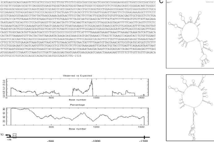
The putative URG4/URGCP promoter region was determined with in silico analysis, showing an extremely high GC content, with 80% in the first 300 bp as shown in Figure 2A. This region also contains two probable CpG island regions spanning 400 bp from the translation start site, with an observed-to-expected ratio ranging from 0.6 to 1 as shown in Figure 2B. In addition, a potential CpG island is present at around 900 bp of the URG4/URGCP promoter with an observed-to-expected ratio ranging from 1 to 2. Secondary structure prediction analysis indicated four independent secondary structures owing to its highest and lowest internal energy in the range of –78.02 to –79.6 kcal/mol as shown in Figure 2C. First we tried to amplify the region at position –1516/+63 of the putative human URG4/URGCP promoter by the conventional PCR method, as described in Section 2, with
the specific primer pairs shown in the Table. Figure 2D shows the PCR amplification results for 1500 bp of the human URG4/URGCP promoter for varying MgCl2 and DMSOconcentrations within the range of 1–2 mM and 2%–10%, respectively. The target DNA fragment could not be amplified with the addition of varying concentrations of DMSO in the PCR amplifications as an enhancer. Furthermore, nonspecific amplifications were observed in agarose gel electrophoresis. In order to facilitate the amplification, the target region was shortened to 548 bp and we also tried to amplify the –485/+63 region of the
URG4/URGCP promoter by PCR using specific primers
(Table) and introducing different PCR enhancers into the buffer composition. Formamide was used in reactions with a 2.5% concentration to amplify 1500 bp and 548 bp of the promoter regions. Higher concentrations of formamide were not used in the reactions because of inhibitory effects on Taq DNA polymerase and presumably other DNA polymerases (Varadaraj and Skinner, 1994). As shown in Figure 2E, the target DNA fragment could not be amplified with this composition. Another PCR additive used to enhance the amplification of GC-rich sequences is ethylene glycol; it was also introduced into the reaction mixture at different concentrations (2% 6%, 8%, and 10%) to improve PCR yields. As shown in Figure 2F, ethylene glycol could not provide a successful amplification. We also tried to amplify the URG4/URGCP promoter region with the Phusion High Fidelity DNA Polymerase Enzyme (Life Technologies). Briefly, to amplify 736-bp and 1038-bp promoter regions, 1X Phusion Master Mix with 1.5% DMSOand50µM 7-deaza GTP was used in the reactions. Unlike the standard PCR cycling conditions, denaturation steps were performed at 98 °C. As can be seen in Figure 2G, the target DNA region could not be amplified.
To demonstrate whether the optimized PCR conditions for the putative human ADAMTS-2 promoter are applicable to other GC-rich URG4/URGCP promoter regions, a similar procedure was repeated. Primer sets were used specifically in the –485/+63 region of the URG4/
URGCP promoter. Similarly, the 548-bp region of the
putative human URG4/URGCP promoter was successfully amplified introducing the same concentrations of the PCR additives into the reaction mixture as shown in Figure 2H. The resulting fragment was purified and cloned into a pGEM-T Easy Vector system and confirmed by automated DNA sequence analysis.
4. Discussion
The expression of eukaryotic protein-coding genes can be regulated at several levels. Regulation at the level of transcription initiation is the most critical one. Promoter regions, proximal regulator elements, enhancers, silencers, insulators, or locus control regions are cis-acting
transcriptional regulatory DNA elements (Köçkar et al., 2001). In humans, approximately 60% of all promoters fall near a CpG island (Venter et al., 2001; Maston et al., 2006). The presence of a CpG island is the most reliable indicator for predicting the presence of a gene (Ioshikhes and Zhang, 2000; Maston et al., 2006). CpG island promoters may often lack TATA boxes and display heterogeneous TSSs. According to the in silico analysis, the ADAMTS-2 gene has a TATA-less and probably CpG island promoter having 80% G+C content and a CpG island located in the
–450/+50 region. Furthermore, 1250 bp of the putative human ADAMTS-2 promoter region contains perfect hairpin structures and the GC percentages of these hairpins are on average 75%. This region also includes the repeated segment GAGG, constituting 34% of the 150-bp region starting at +98. It is well known that templates that have very rich AT or GC base compositions or long short tandem repeats are difficult to amplify by PCR. Among these, templates with GC content greater than 65% frequently give very weak signals when amplified under
Figure 2. (A) Putative human URG4/URGCP promoter region spanning –1516/+63 bp. (B) Bioinformatic analysis revealed a promoter
region around –1500 bp including TSS at the +58 bp position for the URG4/URGCP gene, and 300 bp of the promoter region showed extremely high G+C content (80%). The URG4/URGCP promoter contains CpG island regions of minimum length of 100 bp spanning 150 bp (–100/+50) from the translation start site, with an observed-to-expected ratio of CpG of 0.6. (C) Two predicted secondary structure graphs of the URG4/URGCP promoter site. The internal energy required for formation of these constructs is indicated as kcal/ mol and the lowest and the highest forms of the secondary structure are shown. (D) Agarose gel electrophoresis of the PCR amplification of the human URG4/URGCP promoter (–1516/+63) region: 1: Positive control with GAPDH primers, 2–3: 2% DMSO with 1 mM and
2 mM MgCl2 concentrations, 4–5: 10% DMSO with 1 mM and 2 mM MgCl2 concentrations, 6: nontemplate control. (E) Agarose gel
electrophoresis of 1500 bp and 548 bp of human URG4/URGCP promoter amplifications with formamide: 1: Positive control with h-β2
primers, 2: 2.5% formamide with 1 mM MgCl2 (1500-bp region), 3: 2.5% formamide with 1 mM MgCl2 concentrations (548-bp region).
(F) Agarose gel electrophoresis of the PCR amplification of the human URG4/URGCP promoter (548-bp region): 1: Positive control with
GAPDH primers, 2–3: 6% ethylene glycol with 1–2 mM MgCl2, 4–5: 8% ethylene glycol with 1–2 mM MgCl2, 6–7: 10% ethylene glycol
with 1–2 mM MgCl2, 8–9: 2% ethylene glycol with 1–2 mM MgCl2 concentration, 10: PCR without ethylene glycol with 2 mM MgCl2.
(G) Agarose gel electrophoresis of the PCR amplification of the human URG4/URGCP promoter (736- and 1038-bp regions) with Phusion High Fidelity PCR Master Mix: 1–2: 1X master mix with 1.5% DMSO and 50 µM 7-deaza GTP (736-bp region and 1038-bp, respectively). (H) PCR amplification with conventional Taq DNA Polymerase using 50 µM 7-deaza GTP, 10% DMSO, and 20% betaine (0.26 M) in the reaction mixture, M: 1-kb DNA ladder (Thermo SM0311).
standard conditions. Furthermore, the amplification may result in products derived from regions other than the target DNA location (Sahdev et al., 2007). Because of the high GC content and complex hairpin structures, it was hard to amplify the ADAMTS-2 and URG4/URGCP promoter regions with standard PCR conditions. Despite the optimization of the MgCl2 concentration, buffer pH, and cycling conditions, the target regions could not be amplified. Different additives were introduced into the reaction buffers. In addition to KCl- and MgCl2 -containing buffers,buffers with (NH4)2SO4 and MgSO4 were also used in PCR. Ammonium ions can make an amplification reaction more tolerant of nonoptimal conditions and MgSO4 helps to destabilize secondary structures in DNA (Seifi et al., 2012). Previous studies have indicated that formamide and ethylene glycol can be used as PCR enhancers. These compounds were used in varying concentrations in the reactions. Formamide interferes with the formation of hydrogen bonds between the two DNA strands (Varadaraj and Skinner, 1994). Ethylene glycol is able to decrease the melting temperatures of DNA and modulates the annealing process of GC-rich templates (Zhang et al., 2009). These additives and enhancers could not provide a successful amplification.
We tried to amplify these problematic promoter fragments with GC-rich targeted high-fidelity DNA polymerases as well as the conventional Taq DNA polymerase. A hot-start PCR approach was used in combination with a TD-PCR; however, that did not help in getting the correct amplification. In previous studies betaine and DMSO or betaine, DMSO, and 7-deaza GTP combinations were found to enhance amplification by resolving the complex secondary structure formation of the GC-rich regions (Henke et al., 1997; Kang et. al.,
2005; Musso et al., 2006; Ralser et al., 2006). In the attempt to amplify 760 bp of the putative human ADAMTS-2 promoter region, a 5% DMSO, 20% betaine (0.26 M), and 50 µM 7-deaza GTP combination was included in the reaction mixture. Specific and exclusive amplification of the target sequence was obtained with the addition of these organic molecules, as expected.
In order to test whether the optimized concentrations of these three additives could be useful to amplify another GC-rich region, the same procedure was used to amplify the URG4/URGCP promoter region containing 80% GC base composition and perfect hairpin structures. When approximately 1500 bp upstream of the URGCP gene was analyzed bioinformatically, a perfect hairpin structure between the sequences at positions 203 bp and 903 bp was observed with a GC percentage of 100%. A repeated segment GGGG was also present along the 150-bp region starting at 61 bp. Because of the high GC content and complex hairpin structures, the 1500 bp upstream of the URGCP gene region could not be amplified by conventional PCR or by the addition of the different additives into the reactions. Only the region at +63/–485, about 548 bp long, could it be amplified using the same additives at the same concentrations. In both cases, we could obtain specific amplification, demonstrating that the optimized concentrations of these additives can be used for the amplification of GC-rich DNA fragments.
Acknowledgments
This work was supported mainly by the Scientific and Technological Research Council of Turkey (TÜBİTAK, 212T200) and partially by the Balıkesir University Scientific Research Projects Unit (BAP, 2010/39).
References
Baskaran N, Kandpal RP, Bhargava AK, Glynn MW, Bale A, Weissman SM (1996). Uniform amplification of a mixture of deoxyribonucleic acids with varying GC content. Genome Res 6: 633–638.
Chakrabarti R, Schutt CE (2001). The enhancement of PCR amplification by low molecular-weight sulfones. Gene 274: 293–298.
Colige A, Nuytinck L, Hausser I, van Essen AJ, Thiry M, Herens C, Adès LC, Malfait F, Paepe AD, Franck P et al. (2004). Novel types of mutation responsible for the dermatosparactic type of Ehlers-Danlos syndrome (type VIIC) and common polymorphisms in the ADAMTS2 gene. J Invest Dermatol 123: 656–663
Colige A, Ruggiero F, Vandenberghe I, Dubail J, Kesteloot F, Van Beeumen J, Beschin A, Brys L, Lapière CM, Nusgens B (2005). Domains and maturation processes that regulate the activity of ADAMTS-2, a metalloproteinase cleaving the aminopropeptide of fibrillar procollagens types I-III and V. J Biol Chem 2005; 280: 34397–34408.
Dodurga Y, Avcı CB, Susluer SY, Satıroğlu Tufan NL, Gündüz C (2012). The expression of URGCP gene in prostate cancer cell lines: correlation with rapamycin. Mol Biol Rep 39: 10173– 10177.
Dubail J, Kesteloot F, Deroanne C, Motte P, Lambert V, Rakic JM, Lapière C, Nusgens B, Colige A (2010). ADAMTS-2 functions as anti-angiogenic and anti-tumoral molecule independently of its catalytic activity. Cell Mol Life Sci 67: 4213–4232.
Hecker KH, Roux HR (1996). High and low annealing temperatures increase both specificity and yield in touchdown and stepdown PCR. Biotechniques 20: 478–485.
Henke W, Herdel K, Jung K, Schnorr D, Loening SA (1997). Betaine improves the PCR amplification of GC-rich DNA sequences. Nucleic Acids Res 25: 3957–3958.
Innis MA, Gelfand DH (1990). PCR Protocols: A Guide to Methods and Applications. San Diego, CA, USA: Academic Press.
Ioshikhes IP, Zhang MQ (2000). Large-scale human promoter mapping using CpG islands. Nat Genet 26: 61–63.
Kang J, Lee MS, Gorenstein DG (2005). The enhancement of PCR amplification of a random sequence DNA library by DMSO and betaine: application to in vitro combinatorial selection of aptamers. J Biochem Biophys Methods 64: 147–151.
Köçkar FT, Foka P, Hughes TR, Kousteni S, Ramji DP (2001). Analysis of the Xenopus laevis CCAAT-enhancer binding protein alpha gene promoter demonstrates species-specific differences in the mechanisms for both auto-activation and regulation by Sp1. Nucleic Acids Res 29: 362–372.
Korbie DJ, Mattick JS (2008). Touchdown PCR for increased specificity and sensitivity in PCR amplification. Nat Protoc 3: 1452–1456.
Mamedov TG, Pienaar E, Whitney SE, TerMaat JR, Carvill G, Goliath R, Subramanian A, Viljoen HJ (2008). A fundamental study of the PCR amplification of GC-rich DNA templates. Comput Biol Chem 32: 452–457.
Maston GA, Evans SK, Green MR (2006). Transcriptional regulatory elements in the human genome. Annu Rev Genomics Hum Genet 7: 29–59.
McDowell DG, Burns NA, Parkes HC (1998). Localised sequence regions possessing high melting temperatures prevent the amplification of a DNA mimic in competitive PCR. Nucleic Acids Res 26: 3340–3347.
Musso M, Bocciardi R, Parodi S, Ravazzolo R, Ceccherini I (2006). Betaine, dimethyl sulfoxide, and 7-deaza-dGTP, a powerful mixture for amplification of GC-rich DNA sequences. J Mol Diagn 8: 544–550.
Mutter GL, Boynton KA (1995). PCR bias in amplification of androgen receptor alleles, a trinucleotide repeat marker used in clonality studies. Nucleic Acids Res 23: 1411–1418.
Pomp D, Medrano JF (1991). Organic solvents as facilitators of polymerase chain reaction. Biotechniques 10: 58–59.
Ralser M, Querfurth R, Warnatz HJ, Lehrach H, Yaspo ML, Krobitsch S (2006). An efficient and economic enhancer mix for PCR. Biochem Biophys Res Commun 347: 747–751.
Sahdev S, Saini S, Tiwari P, Saxena S, Singh SK (2007). Amplification of GC-rich genes by following a combination strategy of primer design, enhancers, and modified PCR cycle conditions. Mol Cell Probes 21: 303–307.
Seifi T, Ghaedi K, Salamian A, Tanhaei S, Safari F, Hojati Z, Tavassoli M, Baharvand H, Nasr Esfahani MH (2012). Amplification of GC-rich putative mouse pep promoter using betaine and DMSO in ammonium sulfate polymerase chain reaction buffer. Avicenna J Med Biotech 4: 206–209.
Sun YG, Hegamyer G, Colburn NH (1993). PCR-direct sequencing of a GC-rich region by inclusion of 10% DMSO: application to mouse c-jun. Biotechniques 15: 372–374.
Tufan NL, Lian Z, Liu J, Pan J, Arbuthnot P, Kew M, Clayton MM, Zhu M, Feitelson MA (2002). Hepatitis Bx antigen stimulates expression of a novel cellular gene, URG4, that promotes hepatocellular growth and survival. Neoplasia 4: 355–368.
Turner SL, Jenkins FJ (1995). Use of deoxyinosine in PCR to improve amplification of GC-rich DNA. Biotechniques 19: 48–52. Varadaraj K, Skinner DM (1994). Denaturants or cosolvents improve
the specificity of PCR amplification of a G + C-rich DNA using genetically engineered DNA polymerases. Gene 11: 1–5.
Venter JC, Adams MD, Myers EW, Li PW, Mural RJ, Sutton GG, Smith HO, Yandell M, Evans CA, Holt RA et al. (2001). The sequence of the human genome. Science 291: 1304–1351.
Wilson IG (1997). Inhibition and facilitation of nucleic acid amplification. Appl Environ Microbiol 63: 3741–3751.
Zhang Z, YangX, MengL, LiuF, ShenC, YangW (2009). Enhanced amplification of GC-rich DNA with two organic reagents. BioTechniques 47: 775–779.