PROGRAMMING MICROENVIRONMENTAL SIGNALS WITH BIOACTIVE PEPTIDE AMPHIPHILES FOR SKELETAL AND CARDIAC MYOGENESIS
A THESIS
SUBMITTED TO THE MATERIALS SCIENCE AND NANOTECHNOLOGY PROGRAM
OF THE GRADUATE SCHOOL OF ENGINEERING AND SCIENCE OF BILKENT UNIVERSITY
IN PARTIAL FULFILLMENT OF THE REQUIREMENTS FOR THE DEGREE OF
MASTER OF SCIENCE
By
İmmihan Ceren Garip
I certify that I have read this thesis and that in my opinion it is fully adequate, in scope and in quality, as a thesis of the degree of Master of Science.
...
Assist. Prof. Dr. Ayşe Begüm Tekinay (Advisor)
I certify that I have read this thesis and that in my opinion it is fully adequate, in scope and in quality, as a thesis of the degree of Master of Science.
... Assoc. Prof. Dr. Mustafa Özgür Güler
I certify that I have read this thesis and that in my opinion it is fully adequate, in scope and in quality, as a thesis of the degree of Master of Science.
... Assoc. Prof. Dr. Çağdaş Devrim Son
Approved for the Graduate School of Engineering and Science: ...
Prof. Dr. Levent Onural
i ABSTRACT
PROGRAMMING MICROENVIRONMENTAL SIGNALS WITH BIOACTIVE PEPTIDE AMPHIPHILES FOR SKELETAL AND CARDIAC MYOGENESIS
İmmihan Ceren Garip
M.S. in Materials Science and Nanotechnology Supervisor: Assist. Prof. Dr. Ayşe Begüm Tekinay
August, 2014
The extracellular matrix (ECM) is crucial for the coordination and regulation of various cellular processes, including cell adhesion, recruitment, differentiation and death. ECM components structurally support tissue function and regeneration by acting as a substrate for cell migration and differentiation. In addition, by facilitating the fine localization of signals within their structural framework, these components activate receptors on the cell membrane for the initiation of signal transduction cascades. As such, cell-matrix interactions and matrix-associated signals are important for the normal functioning of cells, as well as for natural or artificially assisted tissue regeneration. In keeping with this ECM-centric approach, we designed and synthesized peptide amphiphiles with bioactive epitopes to resemble the native microenvironment of muscle tissue and to examined their potential in the induction of progenitor cell differentiation into skeletal myotubes and cardiac myocytes. The formation of skeletal myotubes was promoted through the use of basal lamina-mimetic peptide nanofibers inspired by the chemical structures of laminin and fibronectin, two proteins strongly represented in the skeletal muscle extracellular matrix. We demonstrated that our basal lamina mimetic peptide nanofiber system actively interacts with the cells it contains and enhances their differentiation within 3 days. Morphological analysis and immunocytochemical stainings indicated the formation of differentiated myotubes.
ii
We also designed glycosaminoglycan-mimetic peptide amphiphiles to mimic the glycosaminoglycans found in the myocardium. Glycosaminoglycans have been reported to play substantial roles in growth factor binding and the induction of angiogenesis, and their mimicry through peptide amphiphile nanofibers is promising as a combined approach for generating multifunctional cardiovascular tissue engineering scaffolds. We demonstrated that peptide nanofibers enhance the adhesion of cells to the surface and induce cardiac myoblast cells to differentiate into cardiomyocytes through both gene expression analysis and immunostainings.
In summary, myogenic platforms were developed by programming signal rich environment from self-assembled peptide nanofibers inspired from the components of the ECM to induce the differentiation of cells. These bioactive nanofiber systems serve as promising platforms for muscle tissue engineering applications.
Keywords: Peptide nanofibers, extracellular matrix, biomimetic, basal lamina, skeletal muscle tissue, laminin, fibronectin, glycosaminoglycan, myocardial regeneration.
iii ÖZET
BİYOAKTİF PEPTİT AMFİFİLLER İLE ÇİZGİLİ KAS VE KALP KASI OLUŞUMU İÇİN MİKROÇEVRESEL SİNYALLERİ PROGRAMLAMA
İmmihan Ceren Garip
Malzeme Bilimi ve Nanoteknoloji Programı, Yüksek Lisans Tez Yöneticisi: Yrd. Doç. Dr. Ayşe Begüm Tekinay
Ağustos, 2014
Hücrelerarası ekstrasellüler matris, hücre yapışması, hareketi, farklılaşması ve ölümü gibi hücresel olaylarda önemli düzenleyici ve yönlendirici roller oynamaktadır. Dokunun görevini yerine getirebilmesi ve yenilenebilmesi için, matris elemanları, hücrelere yapısal olarak destek olmakla birlikte, hücrelerin hareket edip farklılaşmalarını sağlayacak yüzey görevi de görmektedirler. Ayrıca, hücrelerde tepki oluşturacak sinyallerin düzgün bir şekilde hücrelere sunulmasını sağlayarak, reseptörlerin aktive edilip sinyal iletiminin sağlanmasında rol alırlar. Bu nedenle hücre-matris etkileşimi ve matrise bağlı sinyaller hücrelerin normal fonksiyonlarını yerine getirmelerinin yanı sıra dokunun yenilenmesinde de önemlidir. Bu tezde, kas dokusunun doğal çevresini taklit eden biyoaktif peptit nanofiberler tasarlanarak, öncül hücrelerin çizgili kas ve kalp kası hücrelerine dönüşmelerindeki etkileri araştırılmıştır. İlk olarak, çizgili kas oluşumunu indüklemek için, çizgili kas hücrelerarası matrisinde bulunan laminin ve fibronectin proteinlerinin kimyasal yapılarından esinlenerek, bazal laminayı taklit eden peptit nanofiberler tasarlanmıştır. Bu peptit nanofiber sisteminin hücrelerle etkileştiği ve üç gün içerisinde farklılaşmayı arttırdığı gösterilmiştir. Morfolojik ve immunojenik boyamalar farklılaşmış myotüplerin oluştuğunu göstermiştir.
Ayrıca, kalp dokusunun hücrelerarası iskele yapısında bulunan glikozaminoglikanların kimyasal yapıları kullanılarak glikozaminoglikanları taklit edebilen peptit nanofiberler tasarlanmıştır. Büyüme faktörlerine bağlandığı ve damarlanmayı indüklediği bilinen glikozaminoglikan taklidi peptit nanofiberler tümleşik kardiyovasküler doku mühendisliği iskeleleri için umut verici yapılardır. Bu
iv
çalışmada, peptit nanofiberlerin hücre yapışmasını arttırdığı ve kardiyak miyoblast hücrelerinin kalp kası hücrelerine farklılaştığı gösterilmiştir. Bu farklılaşma gen ekspresyonu analizi ve immunoboyamalarla kanıtlanmıştır.
Özet olarak, kas oluşumunu indükleyen, hücrelerarası iskelenin yapısal elemanlarından esinlenilerek tasarlanan, kendiliğinden toplanan peptit nanofiber platformlar hücrelerin farklılaşmasını desteklemişlerdir. Bu biyoaktif nanofiber sistemlerin kas doku mühendisliği uygulamalarında kullanılması umut vaadetmektedir.
Anahtar Kelimeler: Peptit Nanofiberler, Hücrelerarası İskele, Biyomimetik, Basal Lamina, Çizgili Kas dokusu, Laminin, Fibronektin, Glikozaminoglikan, Kalp kası Rejenerasyonu.
v
ACKNOWLEDGEMENTS
First of all, I would like to thank and express my gratitude to my advisor Dr. Ayşe Begüm Tekinay for her support and guidance at all times. She not only supported my scientific career but also contributed to my personal development. I also would like to thank Dr. Mustafa Özgür Güler for his guidance and support. This work could not be accomplished without Dr.Güler.
I would like to thank Murat Kılınç, Nuray Gündüz and Gülcihan Gülseren for their collobration and friendship in different projects which could not be possible without their sincere efforts.
I was lucky to work with Hakan Ceylan from whom I learned many techniques. I am greatfull for his kindness and contributions to my technical skills and view of science. Also, all NBT and BML lab members were there when I needed them. It was great to know and work with them. I would like to thank Elif Arslan, Yasin Tümtaş, Didem Mumcuoğlu, Berna Şentürk, Melis Göktaş, Seher Yaylacı, Gülistan Tansık, Gözde Uzunallı, Melike Sever, Mevhibe Geçer, Büşra Mammadov and Seda Koyuncu for their support and friendship.
I would like to thank UNAM (National Nanotechnology Research Center) for providing facilities and equipments for my research and to thank TUBITAK (The Scientific and Technological Research Council of Turkey) for financial support, provided in the form of a BIDEB 2210-E MSc fellowship.
I have also special thanks to Öncay Yaşa, who was with me either in good and bad times with endless support.
Lastly, I always feel the love and support of my family with me. I owe the greatest thanks to them.
vi TABLE OF CONTENTS ABSTRACT ... I ÖZET... III ACKNOWLEDGEMENTS ... V TABLE OF CONTENT ... VI LIST OF ABBREVIATIONS ... X LIST OF FIGURES AND TABLES ... XI
CHAPTER 1. ... 1
INTRODUCTION ... 1
1.1Overview of Skeletal Muscle Tissue ... 2
1.2 Skeletal Muscle Extracellular Matrix and Regeneration ... 3
1.2.1 Function and composition of the basal lamina ... 4
1.2.2 Course of Differentiation ... 5
1.2Muscle Injuries and Tissue Engineering Strategies for Repair ... 7
1.4 Heart And Cardiac Muscle Tissue ... 9
1.4.1 Myocardial extracellular matrix ... 10
1.5 Cardiovascular Diseases and Cardiac Regeneration ... 11
1.6 Cardiovascular Tissue Engineering Strategies ... 13
1.7 Motivation and Goals ... 17
CHAPTER 2. ... 19
BASAL LAMINA MIMETIC PEPTIDE NANOFIBERS FOR SKELETAL MUSCLE DIFFERENTIATION ... 19
vii
2.2 EXPERIMENTAL ... 22
2.2.1 Chemicals and Solutions ... 22
2.2.2 Synthesis of Peptide Amphiphiles ... 22
2.2.3 Preparation of Peptide Nanofiber Gels ... 23
2.2.4 Characterizations of Self-Assembled Peptide Nanostructures ... 24
2.2.4.1 Secondary Structure Analaysis by Circular Dichroism: ... 24
2.2.4.1 Scanning Electron Microscopy: ... 24
2.2.4.3 Transmission Electron Microscopy: ... 24
2.2.4.4 Mechanical Characterization by Rheology: ... 25
2.2.5 Cell Culture ... 25
2.2.5.1 Cell Adhesion and Viability ... 25
2.2.5.2 Cell Proliferation ... 26
2.2.6 Cell Differentiation ... 26
2.2.6.1 Morphology ... 27
2.2.6.2 Myogenin and Myosin Heavy Chain Gene Expression ... 27
2.2.6.3 Myosin Heavy Chain Immunocytochemical Staining ... 28
2.3 RESULTS AND DISCUSSION ... 29
2.3.1 Sythesis and Characterization of Peptide Amphiphiles ... 29
2.3.1.1 Mass Spectrometry and HPLC Purification ... 29
2.3.1.2 Circular Dichroism ... 29
2.3.1.3 Scanning Electron Microscopy ... 33
2.3.1.4 Transmission Electron Microscopy... 33
2.3.1.5 Oscillatory Rheology ... 33
2.3.2 Cell Culture on Peptide Nanofibers ... 36
2.3.2.1 Design of 2D Cell Culture Experiments ... 36
2.3.2.2 Biocompatibility of Peptide Nanonetworks ... 36
viii
2.3.2.4 Morphology of Differentiated Cells ... 41
2.3.2.5 Gene Expression analysis ... 41
2.3.2.6 Immunocytochemical Staining ... 43
2.4 Conclusion ... 47
CHAPTER 3. ANGIOGENIC PEPTIDE NANOFIBERS FOR CARDIOMYOCYTE DIFFERENTIATION ... 49
3.1 INTRODUCTION ... 50
3.2 EXPERIMENTAL ... 52
3.2.1 Chemicals and Solutions ... 52
3.2.2 Synthesis of Glycosaminoglycan-mimetic Peptide Amphiphiles ... 52
3.2.3 Purification of Peptide Amphiphiles ... 53
3.2.4 Characterizations of Self-Assembled Peptide Nanostructures ... 53
3.2.4.1 Circular Dichroism ... 53
3.2.4.2 Scanning Electron Microscopy ... 54
3.2.4.3 Transmission Electron Microscopy... 54
3.2.4.4 Oscillatory Rheology ... 54
3.2.5 Cell Culture ... 54
3.2.5.1 Cell Viability and Adhesion ... 55
3.2.5.2 Cell Proliferation ... 56
3.2.6 Gene Expression Analysis ... 56
3.2.7 Immunocytochemical Staining... 57
3.3 RESULTS AND DISCUSSION ... 57
3.3.1 Self-assembly and Characterization of Peptide Amphiphiles ... 58
3.3.1.1 Circular Dichroism ... 58
3.3.1.2 Zeta Potential Measurement ... 58
ix
3.3.1.4 TEM ... 62
3.3.1.5 Oscillatory Rheology ... 62
3.3.2 Cell Culture on Peptide Nanofibers ... 62
3.3.2.1 Safety of Peptide Nanonetworks ... 65
3.3.2.2 Adhesion of cells ... 65
3.3.2.3 Gene Expression analysis ... 65
3.3.2.4 Immunocytochemical Staining ... 68
3.4 CONCLUSION ... 72
CHAPTER 4. CONCLUSION AND FUTURE PERSPECTIVES ... 73
x LIST OF ABBREVIATIONS BrdU : Bromodeoxyuridine CD : Circular Dichroism CS : Chondroitin Sulfate cTnT : Cardiac Troponin T
C2C12 : Mouse Myoblast Cells
DCM Dichloromethane
DMEM : Dulbecco’s Modified Eagle Medium
DMF Dimethlyformamide
FGF : Fibroblast Growth Factor
GAG : Glycosaminoglycan
GAPDH Glyceraldehyde 3-phosphate dehydrogenase
GF : Growth Factor
HM-PA : Heparin Mimetic Peptide Amphiphile
HS : Heparan Sulfate
HSPG : Heparan Sulfate Proteoglycan
H9C2 : Ventricular Rat Myoblast Cells
Mlc-2v Ventricular Myosin Light Chain
MHC : Myosin Heavy Chain
PA : Peptide Amphiphile
SEM : Scanning Electron Microscope
TCP : Tissue Culture Plate
TEM : Transmission Electron Microscope VEGF : Vascular Endothelial Growth Factor
xi
LIST OF FIGURES AND TABLES
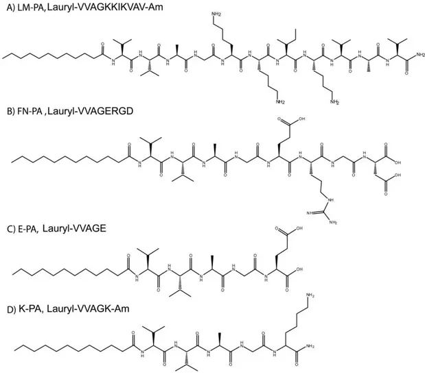
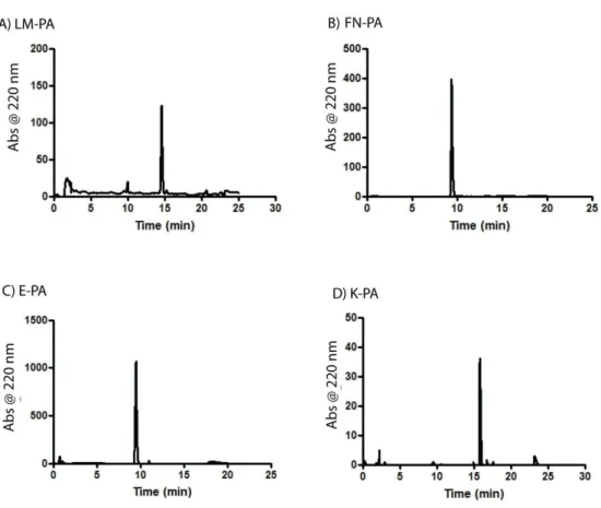
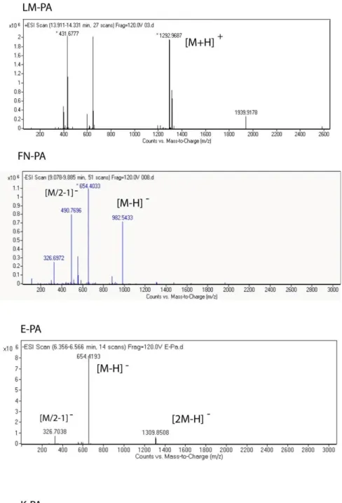
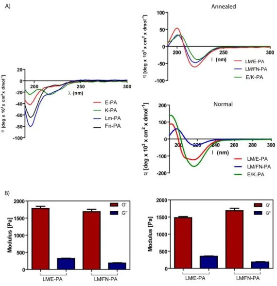
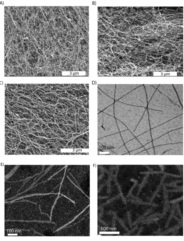
Figure 1.1 Hierarchical organization of skeletal muscle tissue. Epimysium is the outermost layer that surrounds whole muscle tissue, perimysium is the connective tissue around fascicles and endomysium surrounds muscle fiber. Image acquired from wadeyoder.com... 6 Figure 1.2 Scheme of the present major strategies for cardiac tissue engineering using (a) collagen strings, (b) biodegradable gels or (c) cardiac cell sheets (Adapted with permission from Zammaretti et al. Current Opinion in Biotechnology 2004, 15:430–434.) ... 16 Figure 2.1 Chemical structures of peptide amphiphile molecules, A) LM-PA, B) FN-PA, C) E-PA and D) K-PA ... 30 Figure 2.2 Liquid Chromatography of A) LM-PA, B) FN-PA, C) E-PA and D) K-PA ... 31 Figure 2.3. Electrospray ionization mass spectra of A) LM-PA, B) FN-PA, C) E-PA and D) K-P ... 32 Figure 2.4 Characterization of peptide amphiphile molecules by using circular dichoism and oscillatory rheology. A) Secondary structure characterization of annealed and normal PA molecules with circular dichoism, and B) storage and loss moduli of PA mixtures prepared through annealing and without annealing procedure, to compare mechanical differences between standard and annealing protocol. ... 34 Figure 2.5 Characterization of peptide amphiphile molecules by using scanning electron microscopy and transmission electron microscopy. A) SEM images of LM/FN-PA and B) LM/E-PA and C) E/K-PA gels that reveal the ECM mimicking morphology (Scale bars = 3 µm). Representative TEM images of individual
xii
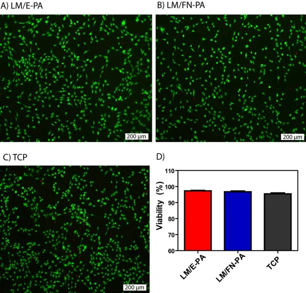
nanofibers of (D) LM/E-PA and E) STEM image of LM/FN-PA, and F) E/K-PA (Scale bars = 100 nm). ... 35 Figure 2.6 Viability of C2C12 cells on peptide nanofiber networks. Live-dead assay of C2C12 cells at 24 h. Dead cells were stained red and live cells were stained green. The scale bar is 200 µm in all images. (A) LM/E-PA, (B) LM/FN-PA and (C) TCP. (D) Relative viability of C2C12 cells on LM/FN-PA and LM/E-PA coated surfaces compared to tissue culture plate surface at 24 h ... 38 Figure 2.7 Cellular viability and proliferation of C2C12 cells on control peptide group. A) Optical microscope image of C2C12 cells on E/K-PA coating, 10x objective, B) live (green) and dead (red) cells stained with live/dead assay, C) quantitative analysis of viability assay and D) proliferation of cells cultured in E/K-PA. ... 39 Figure 2.8 Adhesion and proliferation of C2C12 cells on peptide nanofiber networks. Fluorescence images of 2 h adhesion assay, A) C2C12 cells on LM/E-PA, B) LM/FN-PA and C) TCP and D) Relative adhesion of C2C12 cells normalized to TCP. E) Relative proliferation of cells normalized to TCP at 48 h. Error bars represent mean ± SEM (*p<0.05). ... 40 Figure 2.9 Bright-field images of C2C12 cells cultured on peptide nanofiber networks after 3 days and 4 days of myogenic induction. (Scale bars = 200 µm) .... 42 Figure 2.10 The relative myotube number, average length and diameter quantified with Image J. Error bars represent mean ± SEM. ... 44 Figure 2.11 Gene expression analyses of C2C12 cells cultured on nanofiber networks after myogenic induction. A) Myogenin expression at day 3, B) Myogenin expression at day 4, C) Myosin Heavy Chain (MHC) expression at day 3, D) Myogenin expression at day 4. The expression level of each gene was normalized
xiii
against TCP and GAPDH was used as the internal control. Error bars represent mean ± SEM, (*p<0.05). ... 45 Figure 2.12 Representative immunofluorescent staining of Myosin Heavy Chain (MHC) and nuclei of myotubes 4 days after myogenic induction on the peptide nanofibers A) LM/E-PA, B) LM/FN-PA, and C) TCP. Green: MHC, Red: Nucleus. (Scale bar = 20 µm) ... 46 Figure 2.12 The relative fusion index and maturation index were calculated from images as reported in materials and methods. Error bars represent mean ± SEM. .... 48 Figure 3.1 Chemical structures of peptide amphiphile nanofibers used in this study. Chemical structure of A) GAG-PA and B) K-PA ... 59 Figure 3.2 LC-MS chromatograms and electrospray ionization mass spectra of K-PA and GAG-PA. Liquid chromatography and Mass spectroscopy analysis of A, C) K-PA and B, D) GAG-K-PA. ... 60 Figure 3.3 Characterization of PA molecules, A) circular dichroism spectra of PA molecules showing the β-sheet structure and B) zeta potentials of individual and mixed PA molecules. ... 61 Figure 3.4 Morphological properties of PA molecules analyzed with electron microscopy. A) SEM and B) STEM images revealed the nanofibrous network that mimic the native matrix architecture... 63 Figure 3.5 Mechanical characterization of peptide amphiphile network at 10 mM and 1 mM concentration by oscillatory rheology. A) Storage and loss moduli of PA system and frequency and strain sweep of 10 mM PA network showing the behavior of gels under varying strain/stress amplitudes and strain, respectively. B) Storage and loss moduli of 1 mM PA mixture showing the gel characteristic of the system and frequency and amplitude sweep tests results. ... 64
xiv
Figure 3.6 A) Viability of H9C2 cells cultured on GAG/K-PA coated surfaces and tissue culture plates at 24 h compared to TCP analyzed by Alamar Blue assay, B) relative proliferation of H9C2 cells normalized to TCP at 72 h, analyzed with BrdU incorporation. ... 66 Figure 3.7 Adhesion of H9C2 cells cultured on GAG/K-PA coated surfaces and tissue culture plates. A) Representative Calcein-Am stained fluorescent images of adhered H9C2 cells on GAG/K-PA and TCP at 2 h, B) Relative adhesion of H9C2 cells to PA nanofibers at 2 h with respect to TCP... 67 Figure 3.8 Gene expression analysis of Mlc-2v and myogenin. A) qRT-PCR analysis of cardiac differentiation marker, Mlc-2v in H9C2 cells incubated for 7 and 10 days in the presence of 10 nM RA, results were normalized to GAPDH and compared to TCP. B) Expression of Mlc-2v when the results were normalized to GAPDH and compared with Mlc-2v expression in growth medium. PA nanofiber system along with RA increases gene expression ca. 15 folds greater than uncoated TCP incubated with GM at day 7 and 20 fold higher at day 10. C) Expression analysis of skeletal muscle marker myogenin compared to TCP in the differentiation medium. p<0.05 indicates statistical significance. ... 69 Figure 3.9 Immunocytochemistry staining of cardiac specific protein, cTnT, and cytoskeleton. Confocal images of cTnT staining in H9C2 cells cultured on GAG/K-PA for 10 days, D) Confocal images of cTnT staining in control TCP group. Green: cTnT, red: Actin, blue: nucleus. (Scale bar = 20 µm) ... 70 Figure 3.10 Characterization of cellular morphology with SEM imaging and confocal microscopy. A) SEM image of H9C2 cells incubated in 2% FBS supplemented with 10 nM RA for 10 days, B) H9C2 cells incubated in growth medium for 10 days kept
xv
their undifferentiated phenotype. Red=Phalloidin, Blue=ToPro staining. (Scale bar = 20 μm) ... 71
Table 2.1 Mass and molecular weights of synthesized peptide amphiphiles ... 23 Table 2.2 Primer sequences of myogenin and myosin heavy chain for C2C12 cells. 27 Table 3.1 Primer sequences and annealing temperatures with efficiencies ... 57
1 CHAPTER 1.
2 1.1 Overview of Skeletal Muscle Tissue
Skeletal muscle is a highly complex, hierarchically organized and heterogeneous tissue that is attached to, and allows the movement of, the various bones of the skeleton. It serves a multitude of functions, the most important of which is to generate longitudinal forces. This is established by the contraction of multiple myofibrils inside uniaxially directed bundles of myofibers 1, which are bundled together and attached to the skeleton by tendons.
Skeletal muscle is composed of thousands of muscle fibers (myofibers) that are formed during development by the fusion of mononucleated myoblasts of mesodermal origin. Myofibers are multinucleated cellular syncytia and, since the generation of force requires substantial energy, must be supported constantly through an extensive network of blood vessels. As their constituent cells are terminally differentiated, mature muscle fibers are not capable of self-renewal. However, skeletal muscle is a dynamic tissue and can either increase or decrease its mass in response to a variety of environmental signals, such as starvation, exercise and nutrients 2. This self-renewal capacity is derived from a subpopulation of cells called satellite cells or myoblasts, which reside beneath the basal lamina. They constitute up to 1-5% of total skeletal muscle nuclei, depending on age and muscle fiber composition 3. These cells, first described in 1961, are found in healthy muscle fibers, remain in the mitotic quiescent state G0 and express CD 34, M-cadherin, PAX 7, syndecan 3 and c-met 4. Satellite cells are unipotent adult stem cells and possess an exceptional self-renewal capacity. This ensures their persistence within the muscle while preserving the muscle’s ability to regenerate and repair after injury. These cells are activated in response to severe muscle damage, which triggers their asymetrical division and proliferation. Through asymmetrical division, some of these myocytes repopulate the stem cell niche, while others differentiate into myoblasts and rebuild the muscle by fusing with one another or with residual myofibers 5. As such, large numbers of new myotubes are formed only a few days after acute muscle damage, 6 which serves as a testament to the remarkable regenerative capacity of skeletal muscle. This regeneration is regulated by locally produced growth factors that control cellular proliferation and differentiation 7.
3 Characteristics of the skeletal muscle fibers
Skeletal muscle cells are postmitotic, multinucleated myotubes formed during the embryonic development by the fusion of myoblasts. The myotube matures into the long muscle cell, which possess diameter of 10 to 100 μm and lengths of up to several centimeters. Their total lengths can vary from several millimeters to more than 30 cm 8.
Skeletal muscle fibers are characterized by their striated myofibrils, which are precisely organized contractile and regulatory proteins composed of repeating units arranged in series known as sarcomeres 9. Myofibrils are the smallest functional unit of a myofiber and a muscle fiber is composed of hundreds of myofibrils, which occupy about 80% of the sarcoplasm and are surrounded by mitochondria (also called sarcosomes). Myofibrils are principally composed of two major filaments formed by contractile proteins: thin filaments contain actin, while thick filaments incorporate myosin as well as many other cytoskeletal proteins 10. The capacity of the myofiber to produce force is directly related to the number of parallelly arranged sarcomeres within a muscle fibe
1.2 Skeletal Muscle Extracellular Matrix and Regeneration
The extracellular matrix (ECM) is the non-cellular component present within all tissues and organs, and plays an important role not only in providing a structural framework for the cellular constituents but also by regulating cell behavior by initiating crucial biochemical and biomechanical cues that are required for tissue morphogenesis, differentiation and homeostasis 11. The ECM serves as a substrate for cell migration, modulates growth factor activity and transmits instructive and permissive signals from the microenvironment to cells to support their functional integrity 12. In muscle development, the primary role of the ECM is to function as a structural support for cell attachment, resulting in cell-to-extracellular matrix interactions that are required for cell migration. Depending on the tissue and development stage of the organism, ECM has a diverse nature and composition, containing several distinct families of molecules with disparate evolutionary origins. Although all ECMs are principally composed of water, proteins and polysaccharides, each tissue has an ECM with a unique composition and topology. ECM
4
glycoproteins include both collagens and a diverse array of non-collagenous proteins such as laminins, tenascins and fibronectin 13.
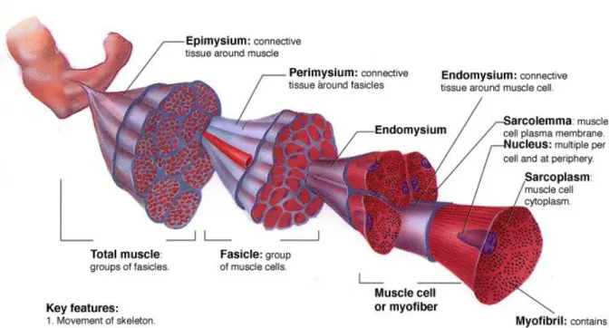
Skeletal muscle is a hierarchically organized tissue and its ECM is ordered in several layers around whole muscle, mucle fascicles and muscle fibers. A layer of connective tissue, called the epimysium, surrounds each muscle, and bundles or fascicles of muscle cells are grouped together by perimysia, which are derived from the epimysial layer and contain the nerves and blood vessels that run through the muscle. Lastly, each muscle fiber is surrounded by a basal lamina and a mesh-like sheath of connective tissue called the endomysium. The basal lamina serves as a scaffold for muscle fiber formation and recovery from injury 14. Figure 1.1 demonstrates the structure of skeletal muscle tissue and connective layers.
1.2.1 Function and composition of the basal lamina
The basement membrane that surrounds muscle fibers is closely associated with the sarcolemma, and provides a protective niche in which muscle regenerative cells (satellite cells) reside 14. It is a specialized ECM sheath composed of both an internal basal lamina and an external reticular lamina. The basement membrane is now known to be critical in muscle fiber structure and function. The actin cytoskeletons of muscle fibers are linked to their basal laminae, which support muscle function and protect against contraction-induced damage. Genetic studies of muscular dystrophy patients and animal models of muscular dystrophy have demonstrated the importance of the basement membrane in the maintenance of muscle integrity. In addition to the maintenance of muscle integrity, it is also essential in the promotion of myogenesis and muscle development. 15.
The muscle ECM is formed by numerous components, including collagen, perlecan, laminin, entactin, fibronectin and several glycoproteins and proteoglycans. These proteins function as a scaffold that provides mechanical support for the cells and facilitates the formation of parallel myofibers by coordinating their attachment and alignment.
Collagens are the most abundant proteins in the animal kingdom and, as a general feature, function to limit the distensibility of tissues, which are effectively 'kept in place' by the enormous tensile strengths of collagen fibrils 16. The muscle basement
5
membrane consists primarily of a type IV collagen network; however, types VI, XV, and XVIII are also present within the matrix 17. Fibronectin in particular is important for the maintenance of skeletal muscle, because it can bind to both the integrin receptors on cells and other molecules in the ECM.
At the sarcolemma, laminins are bound by integrins and α-dystroglycan 18
. Laminins are among the best studied of basement membrane components and can bind directly to type IV collagen, and may also be indirectly linked by fibronectin 19. Laminins are composed of three chains (α, β and γ) that exist in different combinations and form at least 16 different types of laminins. Laminins 211 and laminin 221 in the mature muscle bind to each other and to matrix proteins and serve to connect the muscle networks. Laminin-211 in the basement membrane is extremely important for the maintenance and stabilization of differentiated muscle 20. Interactions between matrix glycoproteins provide potential mechanisms for lateral force transmission from the myofiber 21. Together with the branched network structure of type IV collagen, these glycoproteins form the basis for basement membrane architecture. 1.2.2 Course of Differentiation
During embryonic development, skeletal muscle arises from mesodermal precursor cells 22.Adult skeletal muscles have a robust system of repair. It has been found that a large number of new myotubes arise within a couple days of acute muscle damage. Normally, satellite cells are quiescent and remain in the G0 phase of the cell cycle until they receive certain stimuli 6. Satellite cells express various proteins, including Pax7, CD34, c-met, Mcadherin, syndecan-3 and syndecan-4 23-24, that are important for their activation and proliferation in response to muscle damage. Several common stimuli that trigger the activation of satellite cells include exercise, injury from myotoxins, disease, denervation or other muscle damage. Following damage to the skeletal muscle, growth factors (GFs) and cytokines, such as hepatocyte growth factor, epidermal growth factor; platelet-derived growth factor BB; and members of the insulin-like growth factor (IGF) and fibroblast growth factor family 25 are released from the ECM 26, myofibers, endothelial cells, interstitial cells 27 and leukocytes 28. Interactions among these GFs and receptors on
6
Figure 1.1 Hierarchical organization of skeletal muscle tissue. Epimysium is the outermost layer that surrounds whole muscle tissue, perimysium is the connective tissue around fascicles and endomysium surrounds muscle fiber. Image acquired from wadeyoder.com.
7
quiescent satellite cells trigger satellite cell activation and result in their asymmetric proliferation. Activated satellite cells downregulate Pax7 expression and upregulate MyoD and Myf5 transcriptional activators, which direct these precursor cells to the myogenic linage 29-30. MyoD/Myf5 positive cells first become myoblasts and then stop proliferating and terminally differentiate into cells that begin expressing myogenin, myosin heavy chain (MHC), and muscle creatine kinase (MCK), which are muscle-specific genes. These myoblasts continue proliferating until they reach a sufficient number to begin fusing with existing fibers or with other myoblasts to form new multinucleated myotubes, which later on mature into muscle fibers 31. These sequential events are regulated through ECM components, growth factors and cytokines and occur within a dynamic microenvironment. Therefore, programming the environmental signals to induce the differentiation of progenitor cells for regenerative purposes is a challenging yet promising approach for functional restoration of damaged tissue. As the focus of the present work is differentiation of skeletal muscle cells with bioactive scaffolds using advanced nanoscale systems, the next section will be devoted to an in-depth discussion of muscle injuries and tissue engineering strategies for the regeneration of muscle tissue.
1.2 Muscle Injuries and Tissue Engineering Strategies for Repair
Damage to skeletal muscle may occur throughout life due to exercise and contraction, abnormalities in the immune system, acute physical and chemical injury, or muscle degenerative diseases. Skeletal muscle injuries are a common cause of severe long-term pain and physical disability. Acute damage to skeletal muscle can occur by physical or chemical trauma. For example, excessively hot or cold temperatures and sharp or blunt traumas induce rapid myofiber necrosis. Likewise, ischemia/reperfusion also causes acute muscle damage. Also, during organ transplantation surgery, stroke and hypovolemic shock, or prolonged periods of zero blood flow induce muscle dysfunction through the loss of cellular energy supplies and results in the accumulation of potentially toxic tissue metabolites. Restoration of blood flow is essential for the rescue of ischemic muscle. Therefore, there is a strong need to improve the short- and long-term management of skeletal muscle injuries. According to the principle of conservative management, muscle injuries heal conservatively. It follows the RICE protocol (rest, ice, compression, and elevation). Other therapies also exist, including the local application of heat and passive motion
8
exercises or drug therapy, which typically consists of nonsteroidal anti-inflammatory drugs (NSAIDs) and intramuscular corticosteroids 32. However, current treatment methods do not seem to obtain complete restoration of normal functionality, and new biological therapies could represent new and more effective strategies for the restoration of muscle tissue. Current biological strategies for the management of muscle injuries are growth factor therapy 33-36, cell therapy 37, platelet-rich plasma (PRP) therapy 38-39 and tissue engineering scaffolds.
In skeletal muscle injuries, tissue engineering represents a biological alternative for the replacement of large tissue loss after severe damage. Tissue engineering could allow skeletal muscle fibers to be grown in culture and then transplanted into a patient to improve their physical and psychological symptoms. A comprehensive understanding of tissue structure and the ability to imitate its molecular composition are important to effectively grow tissues for therapeutic use. Biomaterials that emulate the natural tissue structure can be designed and used as controlled microenvironments for regeneration and tissue development.
Efficient skeletal muscle regeneration is strongly correlated with the properties of the biomaterials used for scaffold generation and with the regenerative potential of the cells used for scaffold seeding. Biomaterials with different physiochemical features and compositions, as well as different biological characteristics, have been used to fabricate efficient tissue engineering scaffolds. Both natural and synthetic materials are used to mimic the structural and biomechanical properties of the native tissue. Materials commonly used as scaffolds are hydrogels, polymer scaffolds (PLA, PLLA, PDMS) 40-42 and natural products such as collagen 43, fibrin 44-45 hyaluronan 41, chitosan 46 and matrigel. Kroehne et al. have used collagen scaffolds with parallel oriented pores to reproduce the three-dimensional organization of skeletal muscle and showed that this scaffold has the ability to induce skeletal muscle-like tissue regeneration 43. In addition, Ma et al. reported the ability of 3-D collagen scaffolds seeded with myoblasts to improve muscle healing with an increased quantity of innervated and vascularized regenerated muscle fibers 47. Fibrin is another natural material used as a scaffold and Page et al. demonstrated that fibrin microthread scaffolds provide an efficient delivery system for cell-based therapies and improve the regeneration of a large defect in the tibialis anterior of the mouse 48.
9
Another approach is the use of decellularized tissues as scaffolds, which readily provide the native extracellular microenvironment experienced by cells. Borschel et al. used acellular muscles with injected myoblasts and showed the production of longitudinal contractile force upon electrical stimulation after differentiation 49. In another study, Sicari et al. implanted porcine urinary bladder ECM scaffold to mice and demonstrated new skeletal muscle formation. Moreover, in a parallel human clinical study, scaffold-treated patients with volumetric muscle loss showed similar outcomes with mice and improved limb strength during physical therapy 50.
In addition to its composition, the structural features of biomaterial are important for the alignment of cells, since skeletal muscle tissue is ordered into aligned bundles and tissue constructs should be aligned to function after engraftment. Therefore, biodegradable materials with parallel oriented pores and aligned or grooved morphologies have been used to reproduce the three-dimensional organization of skeletal muscle. Ricotti et al. fabricated electrospun nanofibrous PHB scaffolds and demonstrated that the aligned nanofibrous mesh decreases the proliferation activity and provides a higher differentiative stimulus 51. In another study, grooved polystyrene substrates were conjugated with RGD and were shown to enhance adhesion, growth and differentiation of C2C12 cells 52.
1.4 Heart And Cardiac Muscle Tissue
Heart is an organ that operates as an engine for the circulatory system. It accomplishes this task with a tissue structure that is specially adapted to meet the demands of constant activity during times of rest and exercise. It has three layers; epicardium, myocardium and endocardium, where myocardium is the muscular middle layer of the heart wall. Myocardium is composed of cardiac muscle fibers, which occupy most of the tissue volume but constitute only 30% of the total number of cells in a normal adult heart. Remaining cell populations are non-cardiomyocyte cell types, among which cardiac fibroblasts represent the vast majority 53.
Heart contraction is an autonomic function of the peripheral nervous system, and the myocardium stimulates contractions and relaxations to pump blood from the ventricles and to allow the atria to receive blood, respectively.
10
Cardiac muscle is involuntary, so functionally, it is smilar to smooth muscle. However, anatomically cardiac muscle more closely resembles skeletal muscle due to being striated as a result of the parallel arrangement of actin and myosin filaments. Cardiac muscle fibres are long, cylindrical cells with one or sometimes two nuclei which are centrally located within the cell. Also, the cardiac muscle has several unique features such as intercalated discs, which are connections between two adjacent cardiac cells and help muscle cells to contract rapidly as a single unit. Intercalated discs are irregular transverse thickenings of the sarcolemma that contain desmosomes and gap junctions and ensure proper functioning of the cardiac muscle as an effective pump 54-55.
1.4.1 Myocardial extracellular matrix
There is a connective tissue layer between the muscle fibers, analogous to the endomysium of skeletal muscle, and it supports capillary network necessary to meet the high metabolic demand of strong continuous activity. The ECM not only facilitates the exchange of mechanical, electrical and chemical signals during homeostasis, but also plays essential roles in development, remodeling and signaling in the cardiovascular system 56. This three-dimensional structure acts as a framework for myocytes, fibroblasts and endothelial cells contained in the myocardium and the vasculature to align and build a network. ECM components are also important in determining the mechanics of myocardium, pericardium, blood vessels and valves 57. The extracellular space can be divided into solid and fluid components, of which organization, composition and density are dynamic both in normal and pathological conditions. The fluid components of the matrix contribute to the distribution of soluble factors and other molecules related to different processes. Also, these components act as binding sites for soluble factors and effect their distribution and availability. Other components play roles in accumulating water due to their hydrophilic nature and therefore providing resistance to compression. The extracellular space and its components also serve to connect the cellular components of the myocardium, effectively linking each individual cell and coordinating the contraction of the whole tissue. In addition, cellular and acellular signals and ECM components involved in cell-ECM interactions during post-myocardial infarction (MI) or hypertensive remodeling may contribute to the progression toward recovery or heart failure by playing agonistic or antagonistic roles 56.
11
The ECM is a collection of macromolecular proteoglycans, glycoproteins, proteases, collagens, growth factors and cytokines. Fibroblasts and vascular smooth muscle cells (VSMC) primarily contribute to the secretion of fibronectin and collagens type I and III, while VSMCs, myocytes and endothelial cells produce type IV collagen and laminins 58.
Proteoglycans have recently begun to receive increased attention for their potential signalling roles. During the synthesis of proteoglycans; glycosaminoglycans, which are polysaccharide chains consisting of repeating amino sugar and uronic acid disaccharide units, are covalently linked to the core protein, sulphated and then secreted into the microenvironment. Glycosaminoglycans (GAGs), and especially sulphated GAGs, have a strongly negative charge, which attracts water and results in large hydrodynamic volumes. When the muscle is placed under pressure, the water is removed and GAGs return to their original volume, which allows proteoglycans to act as a lubricant and makes them essential in tissues undergoing continuous cycles of pressure-relief, such as cartilage or cardiac muscle 59. Furthermore, glycosaminoglycans also play major roles in cell signalling, angiogenesis 60, axonal growth 61, tumor progression 62, and anti-coagulation 63.
Heparan sulphate is a common glycosaminoglycan and can bind to multiple growth factors and cytokines through polysaccharide chains. These factors include vascular endothelial growth factor (VEGF), BMPs, and fibroblast growth factors (FGFs), and TGF-βs, which have been demonstrated to be important for cardiac development 64. Numerous studies have revealed that ECM composition differs during different physiological states and differences in the microenvironment are largely driven by cell-cell and cell-matrix interactions within the developing and adult heart. Alterations in the signaling pathways that regulate the production of ECM components, or perturbations in their expression patterns, can lead to congenital heart diseases and cardiac malfunctions 64.
1.5 Cardiovascular Diseases and Cardiac Regeneration
One of the leading causes of death worldwide, ischemic heart disease is characterized by reduced blood supply to the heart muscle, which leads to a deficit in the production of the energy required for cardiac contraction. It is a multi-factorial
12
condition, resulting from a combination of genetics, life style and environment factors. Risk factors for ischaemic heart disease include family history, LDL cholesterol, high blood pressure and obesity.
Acute myocardial infarction (AMI) is a subcategory of ischaemic heart disease and occurs when the blood supply is completely cut at the level of a coronary artery 65, leading to a permanent tissue ischemia. While death does not always occur, the disease nonetheless results in the large-scale loss of cardiac muscle, diminishes the contraction capability of the heart 66 and frequently causes heart failure.
Current therapies for AMI include medical treatment, heart transplantation and implantation of mechanical ventricular assist devices. With the exception of transplantation and mechanical assistance devices, current pharmacological, interventional or operative therapies for the disease suffer from an inability to compensate the decreased pumping capacity of heart following the irreversible loss of functional cardiomyocytes 67. Thus, new solutions are required to regenerate the damaged myocardium, to overcome the bad prognosis of patients with heart failure, and to address the shortage of heart donors.
Cellular repair strategies for heart disease include direct transplantation of cells into the damaged tissue, tissue engineering techniques for the ex vivo development of replacement tissue and therapies that prompt the heart to regenerate damaged tissues. Cell Therapy
The basis of cell therapy is the repopulation of injured myocardium by transplantation of healthy cells. Although heart was once thought to be a terminally differentiated organ and therefore incapable of replenishing lost myocytes, Beltrami et al. have demonstrated that mammalian cardiac myocytes retain some capacity for division 68 and identified endogenous cardiac progenitor cells, which retain some potential for differentiation into endothelial cells, smooth muscle cells and cardiac myocytes 69.Following the discovery that native progenitor cells exist in the adult heart, the delivery of these progenitor cells have been studied as a method to facilitate the generation of a functional myocardium, and this has eventually been translated into the clinic as a cell-based therapy to treat heart disease 70. However, a key concern for stem cell transplantation is the selection of cell source, which
13
determines the safety and efficacy after engraftment. In addition, clinical trials with bone marrow progenitors have shown modest improvements in ventricular function 70-72
.
Various cell types have been considered as candidates for therapeutic delivery in humans, including BMCs, myocardium or adipose tissue derived cells (which were already used in clinical trials), endothelial progenitors, hematopoietic stem cells and pluripotent stem cells 65,67.
Currently, bone marrow is the most frequent source of cells used for cell therapy. Jackson et al. have reported that the transplantation of bone marrow with labelled haematopoietic stem cells followed by myocardial infarction results in a low rate of cardiomyocyte differentiation from the transplanted cells 73. However, many other studies in animals could not demonstrate cardiomyocyte differentiation from haematopoietic progenitor cells, 74-75 nor did they find any improvement in cardiac function following haematopoietic stem cell transplantation 76.On the other hand, bone marrow-derived cells have been used in small clinical trials with apparent success, 77-79 yet most studies demonstrated modest cell therapy-mediated improvements in ventricular function 80.
Another source for cell therapy is endogenous cardiac progenitor cells, since the initial discovery had demonstrated that there existed an endogenous population of progenitor cells resident in the mammalian heart. The advantage of this approach is the feasibility of autologous transfer, although the ex vivo expansion of these cells is necessary. However, a principal problem with cardiac progenitors is the lack of definitive markers 65.Small populations of stem cells in the mammalian myocardium express the cell-surface markers Kit12 or Sca1 81. Animal studies reported that these cells differentiated into cardiomyocytes, smooth muscle cells and vascular endothelium, replacing the majority of the infarcted tissue and improving ventricular function significantly when engrafted to heart 82-83.
1.6 Cardiovascular Tissue Engineering Strategies
As an extension of cell transplantation, tissue engineering with biocompatible scaffolds can provide an appropriate structural and three-dimensional
14
microenvironment to integrate myocytes with the host tissue and develop the vasculature necessary to support blood flow.
Design of the biomaterial scaffold is important to mimic the native cardiac tissue in terms of mechanical properties, structure of the underlying matrix, and presence of vasculature. As such, tissue engineering constructs should be mechanically robust yet flexible, electrophysiologically stable, vascularized or capable of vascularization after implantation, non-immunogenic and contractile 84.
Recent studies have explored the possibility of using decellularized hearts as scaffolds; however, although decellularized scaffolds are suitable for replacing native tissue with a new tissue layer of the same thickness, injectible hydrogels are less invasive in acute MI 85.
A common approach is to seed cardiomyocytes onto porous scaffolds, typically from a biodegradable polymer such as poly(lactic-coglycolicacid) or natural polymers such as collagen and fibrin 86. Zimmermann et al. developed collagen gels into which cardiomyocytes are embedded and mechanically conditioned in a cyclic stretching device, and studies with this gel suggest that some of the cardiomyocytes survive after transplantation into uninjured hearts and undergo further proliferation 87.
Similarly, Zhang et al. used fibrin gels and compared the maturation of human embryonic stem cell-derived cardiomyocytes in 2D monolayers and 3D patch cultures. As a result, they observed that cells in 3D fibrin-based patches exhibited longer sarcomeres and increased expression of genes associated with contractile function 86.
Another study has shown that porous alginate scaffolds with seeded cardiomyocytes yield 3D high-density cardiac constructs and optimized the cell seeding requirements for a uniform distribution in a 3-dimensional structure 88.
The ability to support vascularization is one of the most important requirements for biomaterial or tissue engineering constructs to correctly facilitate the regeneration of damaged myocardial tissue 89. 3D cell constructs generally lack the vascular network that exists in normal tissues. However, a bioengineered myocardial tissue graft requires persistent neovascularization, or angiogenesis, for its growth and survival after implantation. This factor is essential because cardiomyocytes are very sensitive
15
to prolonged ischemia and may die by necrosis and apoptosis if deprived of oxygen, and the engineered heart muscle should survive the ischemic period to maintain viability and function 84. Strategies to promote vessel formation include cell tri-culture, use of growth factors and peptides and the engineering of novel proangiogenic scaffolds 90.
Xiang et al. reported succesful neovascularization in the infarct area following implantation of collagen-GAG scaffolds in combination with bone marrow-derived mesenchymal stem cells 91.
Leor et al. also used porous sodium alginate scaffolds to grow fetal rat cardiac cells within a 3D biograft, and transplanted this scaffold into cardiac tissue. Intensive neovascularization from the neighbouring coronary network was observed through histological analysis 92. In another study, Chiu et al. developed angiogenic and cardioprotective peptide-encapsulated collagen–chitosan hydrogels and observed increased formation of mature blood vessels after the injection of the hydrogel into rat myocardium. The increase in mature blood vessel numbers in turn enhanced the presence of cardiomyocytes while reducing tissue loss 3 weeks post-MI 93.
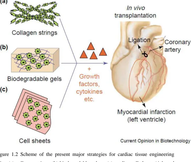
Another approach for cardiac tissue regeneration is the site-specific delivery of angiogenic growth factors from tissue-engineered devices to stimulate localized vessel recruitment to the cell transplant and wound area. For example, Perets et al. constructed a porous alginate scaffold that incorporates tiny poly (lactic-co-glycolic acid) microspheres capable of controlling the release of angiogenic factors such as bFGF, which was released from the scaffolds in a controlled manner in vitro and accelerated the vascularization of the mesenteric membrane in the rat peritoneum 94. Figure 1.2 demonstrates the current myocardial tissue engineering strategies.
16
Figure 1.2 Scheme of the present major strategies for cardiac tissue engineering using (a) collagen strings, (b) biodegradable gels or (c) cardiac cell sheets (Adapted with permission from Zammaretti et al. Current Opinion in Biotechnology 2004, 15:430–434.)
Angiogenesis can also be promoted under certain structure geometries. Madden et al. created methacrylate-based hydrogel scaffolds consisting of interconnected pores, which were 30–40 mm in diameter, by microtemplating and showed that this system
17
supports the growth of hESC-derived cardiomyocytes in vitro and is proangiogenic, promotive of vessel infiltration and able to reduce scarring in vivo 95.
Another approach to increase vacularization is biomimetics, which encompasses the design of scaffolds that physically and chemically resemble growth factors or the ECM of heart tissue. Narmoneva et al. cocultured endothelial cells (EC) and neonatal cardiac myocytes (CM) in 3-D RADA-peptide hydrogels and demonstrated that, when these cells are cultured together, interactions between the cells promote myocyte reorganization and the endothelial cells form capillary-like networks, which in turn promote spontaneous contractions 96.
Webber et al. designed peptide amphiphile-based supramolecular nanostructures which mimic the activity of VEGF and facilitate increased tissue perfusion and functional recovery in ischemic tissue 97. Mammadov et al. also used peptide amphiphiles to mimic heparan sulphate and showed that glycosaminoglycan mimetic nanofibers bind to the growth factors secreted by cells and enhance angiogenesis without the addition of exogenous growth factors 98.
In this thesis, we synthesized heparin sulphate-mimetic peptide nanofibers, which were previously shown to induce angiogenesis 98, and examined their effect on the induction of cardiomyocyte differentiation in vitro. This study is combined with an in vivo myocardial infarction model and an analysis of peptide-mediated revascularization in the infracted area. We therefore assessed the regenerative potential of an ECM-mimetic peptide amphiphile scaffold to heal the myocardium after infarction.
1.7 Motivation and Goals
Peptide amphiphiles allow incorporation of various functional biological signals into their primary sequence and form higher order nanostructures that display complex architectures and biochemical characteristics similar to those of native tissue microenvironments under physiological conditions. These properties make them ideal candidates as bioactive scaffolds for regenerative medicine and tissue engineering applications. Peptide sequences derived from the active site s of the ECM components, which have important roles in muscle regeneration and development, were incorporated into peptide amphiphile nanofibers to create a
18
microenvironment that can both physically support cellular growth and supply the bioactive signals required for the induction of differentiation. In the first chapter, I discussed the effect of biomimetic peptide amphiphiles designed to mimic the basal lamina of skeletal muscle on the differentiation of myoblast cells. In the second chapter, I focused on angiogenic peptide amphiphiles and their effect on cardiomyocyte differentiation.
19 CHAPTER 2.
BASAL LAMINA MIMETIC PEPTIDE NANOFIBERS FOR SKELETAL MUSCLE DIFFERENTIATION
Part of this study was submitted to be published as “Basal Lamina Mimetic Nanofibrous Peptide Networks for Skeletal Myogenesis”, Immihan Ceren Garip, Nuray Gunduz, Murat Kilinc, Mustafa O. Guler and Ayse B. Tekinay.
20 2.1 INTRODUCTION
Skeletal muscle tissue constitutes 40% of the total body weight and is crucial for physical locomotion. Traumatic injury, tumor excision, congenital defects or myopathies compromise muscle function and mobility and necessitate muscle tissue reconstruction 99. Muscle stem cell transplantation is a promising treatment against skeletal muscle trauma; however, isolated stem cells drastically lose their ability to form myotubes and function adequately after in vitro expansion. Therefore, antagonism between differentiation and proliferation hampers the therapy 100. On the other hand, transplantation of non-cultured muscle stem cells to damaged muscle tissue immediately after isolation is quite effective in new myotube formation; however, it is required to harvest approximately 3-4 kg of muscle tissue to regenerate 1 x 105 mm3 of muscle 101-102. As such, due to lack of donor tissue availability, autologous graft surgery is limited. Another one of the most important issues in muscle disorders is the infiltration of connective tissue to the site of injury. Presence of connective tissue inside the muscle which does not possess any contractile capability leaves non functional scar even after healing. Such scars lead to severe weakness in muscles and increase the possibility of fat accumulation within muscle throughout aging. Proper treatment to muscle injuries such as tears or traumas requires use of sophisticated materials to prevent infiltration of connective tissue. To this end, regenerative medicine is a promising alternative solution for the treatment of myopathies as well as regeneration of age-related muscle wasting in elderly people. It is still challenging to engineer skeletal muscle, yet numerous techniques are being developed 51,103-105. One of the strategies is fabrication of aligned scaffolds for engineering muscle tissue. Aviss et al. fabricated align polymer fibers with PLGA and demonstrated differentiated long myotubes by immunostainings 104. Similarly, myotube assembly was shown on nanofibrous and micropatterned polymers by Huang et al. They concluded that microgrooves cause the alignment of myoblasts and cytoskeletal proteins and promote myotube assembly along the nanofibers 106.
Biomimicry to generate favorable microenvironment for induction of differentiation is another promising strategy. ECM mimetic scaffolds for differentiation and tissue regeneration are under investigation for many tissues including bone, cartilage,
21
myocardium and nerve. For skeletal muscle, basal lamina could be target of biomimetic strategies. Basal lamina is a type of ECM that surrounds muscle fibers and takes role in fiber force transmission, repair and maintenance. Fibrous architecture and biochemical components of basal lamina support development and function of skeletal muscle. Thus, scaffolds mimicking basal lamina are promising candidates for efficient muscle tissue regeneration 107. Since nano- to micrometric topography is known to greatly influence cellular behavior including differentiation 108
, micro and nanofabrication techniques are currently used to develop skeletal muscle constructs, with specific focus on topographical features to induce alignment of myotubes 42,109-110. Similarly, ECM proteins such as collagen, fibronectin and laminin or their cell adhesion domains are used to coat the engineered surfaces for triggering cell growth and differentiation 111-112. Previously, it has been shown that laminin has an important role in myogenic differentiation by stimulating proliferation and motility of cells and leading them to bipolar shape of fused cells 113. The YIGSR and IKVAV peptide sequences derived from β1 and α1 cell binding domains of laminin, respectively, have been utilized to imitate the natural microenvironment in tissue engineering strategies. The RGD peptide is another important cell adhesion epitope found in many proteins including fibronectin114 and was also shown to promote migration and fusion of cells in an in vitro myogenesis model system. Peptide amphiphiles enable incorporation of various functional biological signals into their primary sequence and give rise to formation of higher order nanostructures that display complex architecture and biochemical characteristics of native tissue microenvironment under physiological conditions. Owing to their inherent biocompatibility and tailorable properties, variety of studies demonstrated peptide amphiphiles as versatile tools for the design of artificial ECM-like scaffolds 115. Various shapes including high-aspect ratio nanofibers and nano-networks can be generated through programmed self-assembly driven by multiple types of non-covalent interactions. These properties make them ideal candidates as bioactive scaffolds for regenerative medicine and tissue engineering applications.
This chapter presents utilization of laminin-mimetic and fibronectin-mimetic self-assembled peptide nanofibers for myogenic induction of progenitor cells. These peptide amphiphiles resemble the structure of basement membrane skeletal muscle cells. I describe below the design and characterization of peptide nanofibers and the
22
affect of these nanofibers on myogenic cell behaviors such as viability, proliferation and differentiation.
2.2 EXPERIMENTAL
2.2.1 Chemicals and Solutions
9-Fluorenylmethoxycarbonyl (Fmoc) and tert-butoxycarbonyl (Boc) protected amino acids, [4-[α-(2’,4’-dimethoxyphenyl)Fmoc-aminomethyl]phenoxy] acetamidonorleucyl-MBHA resin (Rink amide MBHA resin), 2-(1H-Benzotriazol-1-yl)-1,1,3,3 tetramethyluronium hexafluorophosphate (HBTU) and Lauric acid were purchased from NovaBiochem, ABCR and Merck. Piperidine, Acetic anhydride, Dichloromethane (DCM) and Dimethylformamide (DMF), N, N-diisopropylethylamine (DIEA), trifluoroacetic acid (TFA) : triisoproplysilane (TIS) were purchased form Sigma.
Cell Culture Reagents:
Dulbecco’s Modified Eagle Medium (DMEM), Penicillin/streptomycin (PS) antibiotic combination and fetal bovine serum (FBS) were purchased from Invitrogen Gibco.
2.2.2 Synthesis of Peptide Amphiphiles
Peptide amphiphile molecules were synthesized using solid phase peptide synthesis method with Rink amide MBHA resin and aspartic acid loaded and glutamic acid loaded Wang resin. FN-PA (Lauryl-VVAGERGD-OH) and E-PA (Lauryl-VVAGE-OH) were synthesized on Fmoc-Asp-Wang and Fmoc-Glu-Wang resins, respectively. LM-PA (Lauryl-VVAGKKIKVAV-Am) and K-PA (Lauryl-VVAGK-Am) were synthesized on Rink amide resins. For coupling of amino acids, 1.95 molar equivalents of HBTU and 3 equivalents of DIEA for 1 equivalent of starting resin were used with 2 equivalents of amino acid in 10 mL of dimethylformamide (DMF). Each amino acid coupling took 2 h and removal of Fmoc protecting group was performed with 20% piperidine/dimethylformamide (DMF) solution for 20 min. Lauric acid addition was performed similarly to amino acid coupling except that coupling time was 4 h. In order to acetylate the unreacted amine groups after each coupling step, 10% acetic anhydride–DMF
23
solution was used after each coupling. Dichloromethane (DCM) and DMF were used for washing steps. Peptide cleavage from the resin and deprotection were carried out with 95% cleavage cocktail (95:2.5:2.5 trifluoroacetic acid (TFA): triisopropylsilane (TIS): water) for 2 h at room temperature. Excess TFA was removed by rotary evaporation. Ice-cold diethyl ether was used to precipitate the remaining PA solution overnight at -20 °C. Centrifugation was used to collect the precipitate and ultrapure water was used to dissolve the PA precipitate. Solution was frozen at −80 °C and then lyophilized for 3 days.
To characterize synthesized peptide amphiphiles, a quadruple time of flight (Q-TOF) mass spectrometer with electrospray ionization (ESI) source equipped with a reverse-phase analytical high performance liquid chromatography (HPLC) was used. Agilent Zorbax Extend-C18 2.1 x 50 mm column was used for negatively charged peptide molecules and Zorbax SB-C8 4.6 x 100 mm column was used for positively charged peptide molecules respectively. A gradient of water (0.1% formic acid and 0.1% NH4OH) and acetonitrile (0.1% formic acid and 0.1% NH4OH) were used for liquid chromatography. In order to purify synthesized peptide amphiphiles, reverse-phase preparative HPLC equipped with Zorbax Extend-C18 21.2 x 150 mm column for negatively charged peptide molecules and Zorbax SB-C8 21.2 x 150 mm column for positively charged peptide molecules was used. A gradient of water (0.1% Acetonitrile and 0.1% NH4OH) and acetonitrile (0.1% formic acid and 0.1% NH4OH) were used. Furthermore, positively-charged peptide amphiphiles were treated with 0.1 M HCl solution in order to remove residual TFA at the end.
Table 2.1 Mass and molecular weights of synthesized peptide amphiphiles
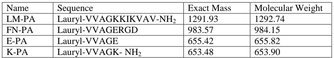
Name Sequence Exact Mass Molecular Weight
LM-PA Lauryl-VVAGKKIKVAV-NH2 1291.93 1292.74
FN-PA Lauryl-VVAGERGD 983.57 984.15
E-PA Lauryl-VVAGE 655.42 655.82
K-PA Lauryl-VVAGK- NH2 653.48 653.90
2.2.3 Preparation of Peptide Nanofiber Gels
Solutions were prepared by dissolving PAs in sterile double distilled water and their pH values were adjusted to 7.4. Each PA molecule was separately heated at
24
70 °C for 5 min and then mixed at a different volume ratio to have final neutral charge. Following 30 s sonication, mixture of PA combinations were reheated at 70 °C for another 5 min. Plates and coverslips were then coated with PA mixtures. With the annealing procedure, homogenous coating over the surface was achieved. After 30 min of incubation at 37 °C, coatings were left to dry overnight under sterile conditions.
2.2.4 Characterizations of Self-Assembled Peptide Nanostructures
Chemical and mechanical characterizations of peptide amphiphiles were performed using CD, SEM, TEM, and rheology.
2.2.4.1 Secondary Structure Analaysis by Circular Dichroism:
LM-PA, FN-PA and E-PA were mixed at 2 x 10⁻⁴ M concentration. Peptides were heated for 5 min, mixed, sonicated and heated again for 5 min before measurement. Following cooling of mixtures at room temperature, scanning was done between 190 nm to 300 nm using a digital integration time of 1 s, a band width of 1 nm and with standard sensitivity. Secondary structures of individual PA molecules were also analyzed by using 2 x 10⁻⁴ M concentration. Molar elipticity was calculated with the data obtained from measurements
2.2.4.1 Scanning Electron Microscopy:
SEM samples were prepared by mixing 1% (wt/V) LM-PA and FN-PA at 2:3 ratio and LM-PA and E-PA at 2:3 ratios to have neutral charge. All the samples were prepared with same annealing procedure. Before serial ethanol dehydration step, gels were put onto silicon wafers and incubated for 20 min cooling and stabilization. Then serial ethanol dehydration step was performed. After gradu al ethanol dehydration, gels were dried in a critical point dryer (Tousimis, Autosamdri-815B, Series C critical point dryer) and coated with 5 nm Au/Pd before imaging. PA gels were then observed with scanning electron microscopy (SEM, FEI Quanta 200 FEG) at 10 kV.
2.2.4.3 Transmission Electron Microscopy:
For analysis samples were prepared with same annealing procedure, 1 mM E -PA and 1 mM FN-PA were mixed with 1 mM LM-PA in 3:2 volumes and after heating and cooling, mixtures were put on a 200-mesh carbon TEM grid for 5 min