ROLE OF NUCLEAR MATRIX PROTEIN C1D IN REGULATION OF TRANSCRIPTION
A THESIS SUBMITTED TO
THE DEPARTMENT OF MOLECULAR BIOLOGY AND GENETICS AND THE INSTITUTE OF ENGINEERING AND SCIENCE OF
BİLKENT UNIVERSITY
IN PARTIAL FULFILLMENT OF THE REQUIREMENTS FOR THE DEGREE OF MASTER OF SCIENCE
BY
DİLHAN ÖNCEL AUGUST 2001
I certify that I read this thesis and that in my opinion it is fully adequate, in scope and in quality, as a thesis for the degree of Master of Science.
______________________ Prof. Dr. Ayşe Ayhan
I certify that I read this thesis and that in my opinion it is fully adequate, in scope and in quality, as a thesis for the degree of Master of Science.
______________________ Assist. Prof. Uğur Yavuzer
I certify that I read this thesis and that in my opinion it is fully adequate, in scope and in quality, as a thesis for the degree of Master of Science.
______________________ Assist. Prof. Uygar Tazebay
Approved for Institute of Engineering and Science.
___________________
ABSTRACT
ROLE OF THE NUCLEAR MATRIX PROTEIN C1D IN REGULATION OF TRANSCRIPTION
Dilhan Öncel
M.S in Molecular Biology and Genetics Supervisor: Assist. Prof. Uğur Yavuzer
August 2001, 88 pages
The nuclear matrix protein C1D is an activator of DNA-dependent protein kinase (DNA-PK), which is involved in non-homologous end joining and V(D)J recombination. Moreover, the nuclear matrix proteins C1D was shown to be phosphorylated in vitro by DNA-PK and its mRNA and protein levels have been demonstrated to be induced upon γ-irradiation. The available information suggests that C1D may play a role in DNA DSB-response pathways by targeting DNA-PKcs to nuclear matrix and matrix-associated DNA where DNA-PK may regulate several cellular processes. In an attempt to identify the biological function of C1D, the yeast two-hybrid system was employed and C1D was found to interact with a partial clone of TAFI 48, which is a subunit of the promoter selectivity factor SL1 required for the accurate initiation of transcription from the human ribosomal promoter. The possibility of an interaction between C1D and TAFI 48 was potentially important as DNA-PK was found to be capable of repressing Polymerase I transcription in vitro.
In this study, it was shown that C1D and TAFI 48 interact specifically in yeast, in vitro and in mammalian cells. It ıs belıeved that this interaction could have potential roles in regulating the repression of transcription from rDNA by DNA-PK in response to DNA double-strand breaks.
ÖZET
NÜKLEER MATRİKS PROTEİNİ C1D’NİN TRANSKRİPSİYONUN REGÜLE EDİLMESİNDEKİ ROLÜ
Dilhan Öncel
Moleküler Biyoloji ve Genetik Yüksek Lisans Tez Yöneticisi: Assist. Prof. Uğur Yavuzer
Ağustos 2001, 88 sayfa
Nükleer matriks proteini C1D, DNA uçlarının doğrudan bağlanmasında ve V(D)J rekombinasyonunda rol oynayan DNA bağımlı protein kinazın (DNA-PK) bir aktivatörüdür. Ayrıca nükleer matriks proteini C1D’nin , DNA-PK tarafından in vitro şartlarda fosforlanabildiği ve γ irradyasyonu sonrası mRNA ve protein seviyesinin arttığı gösterilmiştir. Elimizdeki bilgiler çift sarmal DNA kırıklarına tepki olarak C1D’nin DNA-PK’yı nükleer matrikse veya matrikse ilintili DNA’ya, birçok hücresel olayı kontrol etmek amacıyla yönlendirebileceğini önermektedir. C1D’nin biyolojik fonksiyonunu bulmaya yönelik bir denemede maya ikili hibrid sistemi uygulanmış ve C1D’nin insan ribosomal promotorlarından transkripsiyonun doğru başlaması için gerekli olan SL1’nın bir alt ünitesi olan TAFI 48’in bir kısmını kodlayan bir klon ile etkileştiği bulunmuştur. DNA-PK Polimeraz I transkripsiyonunu in vitro şartlarda bloke edebildiği için, C1D ve TAFI 48’in etkileşme ihtimali potensiyel bir önem arz etmektedir.
Bu çalışmada, C1D ve TAFI 48’in mayada, in vitro şartlarda ve memeli hücrelerinde spesifik olarak etkileşime girdiği gösterilmiştir. Bu etkileşimin çift sarmal DNA kırıklarının etkisiyle DNA-PK tarafından rDNA’dan transkripsiyonun bloke edilmesinin düzenlenmesinde potansiyel rolleri olduğu düşünülmektedir.
ACKNOWLEDGEMENT
This project was partially supported by a grant provided from TUBITAK (TBAG 1864 199 T101).
TABLE OF CONTENTS
SIGNATURE PAGE ii
ABSTRACT iii
ÖZET v
ACKNOWLEDGEMENT vii
TABLE OF CONTENTS viii
LIST OF FIGURES xi
LIST OF TABLES xii
ABBREVIATIONS xiii
INTRODUCTION 1
1. DNA DAMAGE 2
1.1. DNA Double strand Breaks 3 1.1.1. Homologous Recombination 4
1.1.2. Non-Homologous End Joining 5 1.2. Involvement of DNA-PK in NHEJ 6
2. EUKARYOTIC TRANSCRIPTION 9
2.1. Regulation of RNA Polymerase I and Connection with DNA-PK 11
3. DNA-PK AND THE NUCLEAR MATRIX 12
4. MATERIALS AND METHODS 16
4.1. Bacterial Strains 17
4.2. Growth and Maintenance of Bacteria 17
4.3. Yeast Strains 18
4.4. Growth and Maintenance of Yeast 18
4.5. Mammalian Cells 19
4.6. Oligonucleotides 19
4.7. Standard Solutions and Buffers 20 4.8. Recombinant DNA Techniques 22 4.8.1. Polymerase Chain Reaction 22
4.8.3. Purification of DNA Fragments by Agarose Gel
Electrophoresis 23 4.8.4. Restriction Enzyme Digestion of DNA 24
4.8.5. DNA Ligation 24
4.8.6. Plasmids 25
4.8.7. Recombinant Expression Constructs 29 4.9. Preparation of Competent Cells and Transformation of E.Coli 30
4.9.1. CaCl2method 30
4.9.2. Simple and Efficient Method 30 4.9.3. Transformation of E.Coli 31
4.10. Plasmid DNA Isolation 32 4.10.1. Small Scale Plasmid Isolation 32
4.10.2. Medium Scale Plasmid Isolation 33 4.10.3. Spectrophotometric Quantifictaion of DNA 33 4.11. Yeast Techniques 33
4.11.1. Small Scale Yeast Transformation 33 4.11.2. Liquid Culture Assay using ONPG as Substrate 34 4.12. Cell Culture Techniques 36 4.12.1. Thawing a frozen cell line 36
4.12.2. Sub-Culturing of Monolayer cells 36 4.12.3. Cryopreservation 37 4.12.4. Transient Transfections 37 4.13. Gel Electrophoresis 37 4.13.1. Agarose Gel Electrophoresis 37
4.13.2. SDS Polyacrylamide Gel Electrophoresis (SDS-PAGE) 38 4.14. Biochemical Techniques 39
4.14.1. Immunological Detection of Immobilized Proteins 39 4.14.1.1. Transfer of Proteins onto Membranes 39 4.14.1.2. Detection of Immobilized Proteins 40 4.14.1.3. Stripping and Reprobing Membranes 41 4.14.1.4. Peptide Neutralization of Primary antibody 41 4.14.2. Immunoprecipitation 41
4.14.3. Protein Purification of Glutathione-S-Transferase
tagged C1D protein 42 4.14.4. Protein Purification by Immobilised Metal Ion Affinity
Chromotagraphy 44
4.14.5. Protein Refolding 45 4.14.6. In vitro Transcription and Translation 45 4.14.7. Pull-Down Assays 46
4.14.7.1. Ni-NTA Pull-Down Assays 46 4.14.7.2. GST Pull-Down Assay using Immobilized
GST-C1D 47
4.14.8. Kinase Assays 48
4.15. Radiochemicals 49
4.16. Restriction and Modifying Enzymes 49 4.17. Chemicals, Kits and Special Consumables 49
4.18. Equipment 50
5. RESULTS 51
5.1. Introduction 52
5.2. Cloning of TAFI 48 in Various Expression Plasmids 53 5.3. Confirmation of C1D/TAFI 48 Interaction in Yeast Using
ONPG Assay 55
5.4. In vitro Interaction Assay 56 5.4.1. Purification of His-Tagged C1D under Denaturing
Conditions 57
5.4.2. Ni-NTA Pull-Down Assay 59 5.4.2. Purification of GST-C1D Protein under Non-Denaturing
Conditions 61
5.4.4. GST Pull-Down Assays using IVTT product of TAFI 48 62 5.4.5. GST Pull-Down Assay using TAFI 48 expressed in
Mammalian Cells 63
5.5. Kinase Assays 64
5.6. TAFI 48/C1D Interaction in Mammalian Cells 68 6. DISCUSSION 71 6.1. Future Perspectives 75
LIST OF FIGURES
Figure Page
Figure 1.1 Model for the role of Ku and DNA-PKcs in nonhomologous 8
end joining Figure 4.1 Restriction Map and Multiple Cloning Site (MCS) of pACT2 26
Figure 4.2 Map of pQE30 vector and its MCS 27
Figure 4.3 Map of pcDNA3.1/Myc-His vectors 28
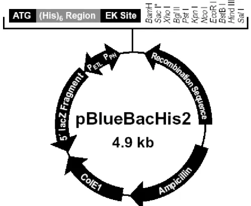
Figure 4.4 Map of pBlueBacHis2 28
Figure 5.1 Sample Digest Result 54
Figure 5.2 Yeast two hybrid Assay 59
Figure 5.3 Ni-NTA Immobilized C1D 58
Figure 5.4 Western blot detection of C1D 59
Figure 5.5 Ni-NTA Pull-Down Assay 60
Figure 5.6 Purification of GST and GST-C1D fusion protein 61 Figure 5.7 GST Pull-Down Assay using IVTT Product of TAFI 48 62 Figure 5.8 GST Pull-Down Assay Using TAFI 48 Expressed 64
in Mammalian Cells Figure 5.9 In Vitro Kinase Assay 66
Figure 5.10 Immuno-complex Kinase Assay 68 Figure 5.11 Immunoprecipitations from mammalian cells 70
LIST OF TABLES
Table Page Table 4.1 List of the E.coli strains used during the course of this study 17 Table 4.2 List of the yeast strains used during the course of this study 18 Table 4.3 Primer pairs used to amplify full-length open reading frame of 20 TAFI 48
Table 4.4 List of the constructs prepared during the course 29 of this study
ABBREVIATIONS
BIR break induced replication bp base pairs
BSA Bovine Serum Albumin
cDNA Complementary DNA DBD LexA DNA binding domain
DSB DNA double-strand break ddH2O deionized distilled water
DMEM Dulbecco’s modified Eagles’ medium DMSO dimethyl sulfoxide
DNA Deoxyribonucleic acid
DNA-PK DNA-dependent Protein Kinase dNTP deoxynucleotide triphosphate DTT dithiothreitol
EDTA diaminoethane tetra-acetic acid EtBr Ethidium Bromide
FCS Fetal Calf Serum
GST glutathione-S-transferase HA Hemagglutinin
HMG High Mobility Group
HR Homologous Recombination HRP Horse Radish Peroxide
ICR Internal Control Region Inr Initiator Sequence
IPTG Isopropylthio-b-D-galactosidase IR Ionizing Radiation
LB Luria-Bertoni Media MCS Multiple Cloning Site MMS Methyl-methoxy Sulfide NHEJ Non-homologous End joining NTA Nitrilo-tri-acetic acid
OD optical density
ONPG o-nitophenyl b-D-galactopyranoside PAGE Polyacrylamide Gel Eletrophoresis PBS Phosphate Buffered Saline
PCR Polymerase Chain Reaction PEG Polyethylene glycol
PMSF Phenylmethyl-sulfonyl fluoride Pol I Polymerase I
PSE Proximal Sequence Element RNA Ribonucleic Acid
RNAse Ribonuclease
RPA Replication Protein A Rpm rotation per minute SD synthetic dropout
SDS Sodium Dodecyl Sulfate
SDSA Synthesis-dependent Strand Annealing SEM Simple and Efficient Method
SL1 Selectivity Factor 1 snRNA Small Nuclear RNA SSA single strand annealing TAE Tris-Acetic Acid-EDTA TAF TBP-associated factor TB transformation buffer TBE Tris-Boric Acid-EDTA TBP TATA Box-binding Protein TBS Tris Buffered Saline
TBS-T Tris Buffered Saline with Tween
TEMED N,N,N,N-tetramethyl-1,2 diaminoethane Tris Tris (hydroxymethyl)-methylamine
UBF Upstream Binding Factor UCE Upstream Control Element UV Ultraviolet
XRCC x-ray cross complementing
1. DNA DAMAGE
Cells are constantly under threat from the cytotoxic and mutagenic effects of DNA damaging agents. These agents can either be exogenous or formed within cells. Environmental DNA-damaging agents include UV light and ionizing radiation, as well as a variety of chemicals contained in foodstuff and cigarette smoke. Endogenous damaging agents include methylating species and the reactive oxygen species that arise during respiration.
These DNA damaging agents may lead to two general types of abnormalities that, if left unrepaired, will eventually lead to mutation, which is a permanent change in DNA sequence. The first class of mutations involve single base changes, which does not cause a major structural distortion on the DNA. The second class on the other hand, induces distortions on the structure of DNA, which may interfere with transcription and replication processes, with deleterious effects on the cell survival.
To ensure the genetic stability and cell survival, cells are equipped with mechanisms to repair these structural abnormalities. The first reaction in response to DNA damage is to arrest the cell cycle to provide enough time for the repair process. This event also prevents transmission of the damaged DNA to the next generations. Following the cell cycle arrest, repair process is initiated, using one of the many repair pathways differing according to the type of damage. However, if the damage is beyond repair, then cells choose to commit suicide (apoptosis) to prevent the cell being divided with the heavily mutated DNA. In general terms there are three categories of DNA repair. First one is excision repair, which includes both base and nucleotide excision repair. The incorrect or
damaged base is removed either as a base (base excision) or as an (oligo)nucleotide (nucleotide excision), the single-stranded gap resulting from the excision is filled by a polymerase using the information of the other strand. Second one is mismatch repair, which relies on the parental strand to correct misincorporation of nucleotides during replication and heteroduplex regions produced by genetic recombination. The third category is double-strand break repair and unlike the other two pathways, presence of homology regions is not necessarily required for this type of DNA repair.
1.1. DNA Double-Strand Break (DSB)
The DNA double-strand break (DSB) is one of the most dangerous lesions that can occur within the cell. The presence of an unrepaired DSB will trigger the DNA-damage response systems of a cell to arrest its progression through the cell cycle and, sometimes, to cause apoptotic cell death (Huang et al. 1996). If repaired improperly, they can lead to chromosome translocations and cancer.
DNA DSBs can arise by exogenous agents such as ionizing radiation (IR) and certain chemotherapeutic drugs, or as a consequence of some physiological events such as endogenously generated reactive oxygen species, mechanical stress on the chromosomes, or when DNA replication forks encounter DNA single-strand breaks or other type of lesions. DNA DSBs are also generated to initiate recombination between homologous chromosomes during meiosis and occur as intermediates during developmentally regulated rearrangements, such as V(D)J recombination and immunoglobulin class-switch recombination (Smider and Chu 1997).
There are two general types of DSB repair: homologous recombination (HR) and non-homologous end joining (NHEJ).
1.1.1. Homologous Recombination
There are several types of homologous repair: gene conversion, break-induced replication and single-strand annealing. After a double-strand break (DSB) is created, the ends are resected and one of the 3' single-stranded ends can invade an intact template. Strand invasion requires the participation of the Rad51p strand exchange protein and a number of associated proteins, including Rad52p, Rad54p and Tid1p. During meiosis, these recombination proteins are joined by a second strand-exchange protein, Dmc1p. Strand invasion is believed to establish a modified replication fork, in which both leading and lagging-strand DNA synthesis occurs. As new DNA synthesis proceeds, branch migration displaces the two newly synthesized strands. The four main outcomes of homologous repair can be summarized as follows: (a) If the replication fork encounters the other end of the DSB, an intermediate containing two Holliday junctions can be formed, allowing gene conversions to be resolved both with and without crossing-over. (b) If the strands are completely displaced or if the leading strand pairs with the second end of the DSB, a simple synthesis-dependent strand annealing (SDSA) will occur, producing gene conversions without crossing-over (Pâques and Haber 1999; Fishman-Lobell et al. 1992). (c) If the second end of the DSB fails to engage, replication can proceed all the way to the end of the chromosome (or until it encounters a converging replication fork). This process is known as break-induced replication (BIR) (Bosco and Haber 1998; Morrow et al. 1997). (d) If resection proceeds far enough to expose complementary strands of homologous
sequences flanking a DSB, repair can occur by single-strand annealing (SSA), leading to a deletion of all intervening sequences (Sugawara et al. 2000).
1.1.2. Non-Homologous End Joining (NHEJ)
DSB ends can be repaired by several nonhomologous repair mechanisms, in which the DNA ends are joined with little or no base pairing at the junction (Moore and Haber 1996; Jeggo 1998). NHEJ of two DNA ends does not require an undamaged partner and does not rely on extensive homologies between the two recombining ends. In this process, sometimes after limited degradation at the termini, the two ends are ligated together. Consequently, NHEJ is prone to error, and small sequence deletions are usually introduced. End joining in yeast and mammals requires the same core set of proteins: the DNA end-binding proteins Ku70p and Ku80p, as well as DNA ligase IV and its associated Xrcc4 protein. Vertebrate cells also require DNA-PKcs, which together with the Ku70 and Ku80 forms the DNA-PK holoenzyme. DNA-PK is a nuclear serine/threonine kinase with a well-established role in NHEJ. Interestingly, no homologue has been identified in S.cerevisiae for DNA-PKcs. In mammals, the carboxy-terminal portion of Ku80 is essential for the activation of DNA-PK and this region is distinct from the one that interacts with Ku70 (Singleton et al. 1999). The requirement for this carboxy-terminal region in kinase activation is consistent with the absence of an analogous Ku80 carboxy-terminal tail in S.cerevisiae, which also lacks a homologous DNA-PKcs protein.
Although NHEJ has been considered the major pathway of DSB repair in mammalian cells, these two processes are in competition with each other. Very little is known in general about how cells choose which pathway to use. It has been suggested that
two DNA end-binding proteins, Rad52 and Ku compete for binding to DSBs and channel the repair of DSBs into HR and NHEJ, respectively (Van Dyck et al. 1999; Haber 1999).
1.2. Involvement of DNA-PK in NHEJ
The nuclear serine/threonine kinase, DNA-PK, is comprised of a catalytic subunit termed DNA-PKcs and two DNA binding componets, Ku70 and Ku80. The involvement of DNA-PK in DSB repair became evident from analyses involving a specific series of mutant rodent cell lines (Jackson and Jeggo 1995). Early studies in these mutant cells found them to be hypersensitive to irradiation (IR) and radiomimetic agents with little or no cross sensitivity to other types of DNA damaging agents and showed them to be defective in the repair of chromosomal DNA DSBs. Screening of these x-ray sensitive cell lines led to the identification of three distinct complementation groups, termed IR4, IR5, and IR7, with defects in both DSB repair and V(D)J recombination. Since efforts were undertaken to complement these rodent cell lines with human genes, the genes for the complementation groups were designated XRCC for x-ray cross complementing (Thompson and Jeggo 1995; Zdzienicka 1995). Later it was shown that, cells of IR5 and IR7 harboured inactivating mutations in the genes of Ku80 and DNA-PKcs, respectively, and that inactivation of Ku80 leads to a dramatic destabilization of both itself and Ku70 (Errami et al. 1996, 1998a, 1998b; Blunt et al. 1996; Danska et al. 1996; Araki et al. 1997; Singleton et al. 1997; Peterson et al. 1997; Fukumura et al. 1998; Priestley et al. 1998). It was therefore concluded that mutations in Ku80 and DNA-PKcs lead to IR sensitivity, that XRCC5 and XRCC7 encode Ku80 and DNA-PKcs respectively, and that DNA-PK is a crucial component of the mammalian DNA DSB repair apparatus (Priestley et al. 1998).
Although among these mutant cell lines, a mutation for Ku70 was not identified, later a group of cell line termed IR6 was established by knocking-out the Ku70 gene and was shown that these cells also carry the same phenotype as IR5 and IR7 and was defective in DNA-PK activity. Therefore, it was concluded that Ku70 is encoded by XRCC6 (Gu et al. 1997).
Although the precise molecular mechanism underlying the role of DNA-PK in DSB repair is not yet known, in the light of biochemical and genetics studies, some hypotheses have been proposed. The heterodimer of Ku80 and Ku70 binds with high affinity to DNA ends, suggesting an early role in damage recognition, processing or both. Ku binding probably protects the DNA ends from degradation until end joining is completed (Liang and Jasin 1996). In addition, Ku protein might play a role in juxtapositioning of two DNA ends since it can transfer between DNA fragments with complementary overhangs (Bliss and Lane 1997). On binding to the DNA ends, Ku may recruit DNA-PKcs. Synapsis of the two DNA ends might be mediated by two independent DNA binding sites on DNA-PK or by the association of two DNA-PK molecules (Chu 1997). The molecular architecture of DNA-PKcs suggests a structural role for the protein in the rejoining process. It has a potential DNA-binding groove and an enclosed cavity with three apertures through which single-stranded DNA could pass (Leuther et al. 1999). These findings are consistent with DNA-PKcs mediating the alignment of short stretches of single-stranded DNA prior to ligation.
DNA-PK has also an autophosphorylation activity (Chan and Lees-Miller 1996). It is hypothesized that this phosphorylation occurs in trans, so that the kinase assembled on one DNA end phosphorylates the other DNA-PK molecule assembled on the other DNA
end. Activity in trans would regulate the kinase activity so that processing of the DNA ends would occur only after synapsis of the two ends is accomplished (Chu 1997).
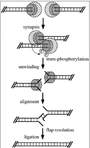
The autophosphorylation also causes the dissociation of the catalytic subunit from Ku (Chan and Lees-Miller 1996), and Ku acquires helicase activity (Cao et al. 1994; Tuteja et al. 1994). It is postulated that this helicase activity unwinds DNA ends in vivo so that exposed regions of microhomology can anneal by base pairing (Chu 1997). The unpaired DNA flaps could then be removed either by an exonuclease or a flap endonuclease. Gaps are then filled in by a DNA polymerase and the nicks sealed by ligase to complete the end-joining process (Figure 1, Chu 1997).
Once positioned at the DNA DSB, Ku and DNA-PKcs might then recruit other NHEJ factors essential for the repair process. It is possible that, as being a kinase, DNA-PK may also regulate the activity of the proteins in this repair complex by phosphorylation. In this regard, the plausible targets for DNA-PK include the single-stranded binding protein RPA (replication protein A), the DNA ligase IV cofactor Xrcc4, Ku, and DNA-PKcs itself (Karran 2000). However, DNA-PK has also been shown to phosphorylate many transcription factors, including the tumour suppressor p53 in vitro, which indicates that DNA-PK may also be involved in regulation of transcription. Indeed, DNA-PK has been demonstrated to repress Polymerase I transcription in vitro (Kuhn et al. 1995). All these findings suggest that DNA-PK may also regulate transcription at the sites of DNA damage and may be by repressing transcription and recombination, to allow enough time for the repair machinery to perform its function (Zhu et al. 1996).
2. EUKARYOTIC TRANSCRIPTION
In eukaryotes, transcription is carried out by three different RNA polymerases, RNA polymerase I, II, and III, each of which is dedicated to the transcription of different sets of genes. The genes in each class contain characteristic promoters, which often consist of two types of elements: the basal promoter elements and the modulator promoter elements. The basal promoter elements are sufficient to determine RNA polymerase specificity and direct low levels of transcription, whereas the modulator elements enhance or reduce the basal levels of transcription. None of the RNA Polymerases can recognize its target promoters directly. Instead, basal promoter elements are first recognized by specific transcription factors, which then recruit the correct RNA polymerase.
The RNA Polymerase I promoters, which direct transcription of the large rRNA genes, do not contain a TATA box (Reeder 1992). The human rRNA promoters are instead composed of a core element that overlaps the transcriptional start site and an upstream control element (UCE) located upstream of position –100 that stimulates transcription 10-to 100-fold.
RNA polymerase II promoters can be divided into two large classes: the mRNA promoters and the snRNA promoters. The mRNA promoters can in turn be divided into two classes depending on whether or not they contain a TATA box. The basal TATA-box containing mRNA promoters may consist of either the TATA box alone or the TATA box and an initiator sequence (Inr) that spans the transcriptional start site. The basal TATA-less mRNA promoters consist of just the Inr. Both TATA box and Inr appear to recruit the TBP-containing complex TFIID. TBP (TATA-box Binding Protein) on its own can, however, replace the TFIID complex to direct basal levels of transcription from TATA-containing promoters in vitro, suggesting that direct interactions exist between TBP and components of the basal transcription machinery.
The basal RNA polymerase II snRNA promoters consist of a proximal sequence element (PSE), which is also present in RNA polymerase III snRNA promoters. Transcription from these promoters also requires TBP, which is recruited by the PSE.
The RNA polymerase III promoters have been divided into three classes (Geiduschek and Kasavetis 1992). Classes 1 and 2 are gene-internal TATA-less promoters and consist of the internal control region (ICR) of 5S rRNA genes and the A and B boxes of tRNA and 7SL genes, Alu sequences, and viral genes such as the Ad2 VAI and VAII genes. Class 3 promoters are found in a number of vertebrate snRNA and cytoplasmic
RNA genes, including the U6, 7SK, hY1 and hY3, H1, and MRP/Th RNA genes (Hernandez 1992). They are located entirely upstream of the gene-coding sequences and are similar to the RNA polymerase II snRNA promoters except for the presence of a TATA box, which determines the RNA polymerase III specificity of the promoter. Transcription from both the TATA-less and TATA-containing RNA polymerase III promoters requires TBP, although in different complexes.
2.1. Regulation of RNA Polymerase I and Connection with DNA-PK
Human ribosomal RNA synthesis by RNA Polymerase I requires the upstream binding factor (UBF) and the promoter selectivity factor (SL1) for accurate initiation of transcription from the human ribosomal promoter (Learned et al. 1985; Learned et al. 1986; Bell et al. 1990). UBF binds in a sequence specific manner to both core and the UCE. In contrast, SL1 has no or little affinity for the rRNA promoter but binds cooperatively with UBF. SL1 consists of the TBP and three associated subunits, TAFI 110, TAFI 63 and TAFI 48 (Comai et al. 1992; Comai et al. 1994; Zomerdijk et al. 1994). TAFI 48 serves as a target for interaction with UBF and is required to form a transcriptionally active SL1 complex responsive to UBF in vitro (Beckmann et al. 1995). TAFI 48 also alters the ability of TBP to interact with TATA box elements, and the resulting complex fails to support transcription by RNA Polymerase II (Beckmann et al. 1995).
Transcription of ribosomal RNA genes by RNA polymerase I oscillates during the cell cycle, being maximal in S and G2 phases, repressed during mitosis, and gradually recovering during G1 progression. Silencing of cellular pre-rRNA synthesis during mitosis is caused by the inactivation of the Pol I-specific transcription initiation factor SL1 by
cdc2/cyclin B-mediated phosphorylation of TAFI 110 (Heix et al. 1998). Binding of SL1 to the core element of the ribosomal gene promoter is enhanced by the upstream binding factor (UBF), a member of the family of high mobility group (HMG) box proteins (Jantzen et al. 1990). Interestingly the chromatin associated high mobility group (HMG) proteins 1 and 2 have been shown to stimulate DNA-PK activity in vitro (Watanabe et al. 1994).
Mitotic phosphorylation has been demonstrated to impair the capability of SL1 to interact with UBF (Heix et al. 1998), indicating that phosphorylation of the Pol I-specific TBP-TAF complex is used as a molecular switch to prevent pre-initiation complex formation and rDNA transcription at mitosis.
DNA-PK was also found to be capable of repressing transcription by RNA Polymerase I in vitro (Kuhn et al. 1995; Labhart 1995). Transcriptional repression by DNA-PK is due to protein phosphorylation. DNA-PK inhibits transcription from both linear and circular templates, but the repression is more efficient on linear templates (Kuhn et al. 1995; Labhart 1995). Partial fractionation of the in vitro transcription system showed that a protein fraction containing transcription factor Rib1, the Xenopus equivalent of human SL1, mediated the repression of transcription by DNA-PK (Labhart 1995).
The effect of DNA-PK on Pol II and III has not been demonstrated so far. Moreover, the precise molecular mechanisms of Pol I repression by DNA-PK or whether DNA-PK is also capable of repressing Pol I transcription in vivo is not yet known.
3. DNA-PK AND THE NUCLEAR MATRIX
As summarised above, a wide range of functions have been proposed for DNA-PK many of which lack solid evidence. To date, although its involvement in DNA DSB repair
is well established, the molecular mechanisms that DNA-PK use while performing these functions are not yet clear. It is noteworthy to mention that DNA-PK is present at ~1x10⁵ to 5x10⁵ molecules per human cell (up to 1% of HeLa cell nuclear protein), which is far in excess of the number of DNA DSBs generated by physiological doses of DNA damaging agents and its levels do not appear to be regulated strongly by DNA-damaging agents (Lee et al. 1997). This could be because of the need for an immediate response to DNA damage and by having huge amounts of DNA-PK, the damaged ends could be detected immediately without spending time for the new protein synthesis. However, the DNA is not naked in the cellular environment and the chromatin context has to be considered when proposing models for how DNA-PK binds to DNA and gets activated to perform its kinase function. It is possible that DNA-PK may need an accessory protein that would allow its access to DNA wrapped in chromatin, which may prevent the exposition of broken DNA ends. Indeed, during DNA-PK purification from human cells, a substantial proportion (40 %) of DNA-PK was found to be associated with the insoluble fraction of the nucleus, which constitutes the nuclear matrix (U. Yavuzer, unpublished data).
Nuclear matrix by definition is the structure that can be isolated from cells after removal of soluble proteins, histones and most of the DNA. A group of proteins however, stay tightly attached to DNA and cannot be removed even by treating with harsh denaturants. Accumulating evidence indicate the association of nuclear matrix with several nuclear metabolic processes including DNA replication, transcription, RNA splicing, topoisomerase activity, nucleotide excision repair, and DNA DSB repair (Berezney 1984; Cockerill and Garrard 1986; Nelson et al. 1986; Verheijen et al. 1988; Jackson 1991;
Kaufman and Shaper 1991; Yasui et al. 1991, 1994; Korte and Yasui 1993; Johnston and Bryant 1994; Koehler and Hanawalt 1996).
Interestingly, attempts towards identifying the molecular mechanisms that DNA-PK employ to perform its function revealed that a nuclear matrix protein C1D interacts specifically with the putative leucine zipper region of DNA-PKcs. This interaction is interesting in the sense that C1D is capable of activating DNA-PK in the absence of free DNA ends (Yavuzer et al. 1998). Moreover, the nuclear matrix protein C1D is phosphorylated very efficiently by DNA-PK and its mRNA and protein levels have been demonstrated to be induced upon γ-irradiation (Yavuzer et al. 1998), suggesting that C1D, therefore the nuclear matrix may play a role in DNA DSB-response pathway in connection with DNA-PK. It is possible that C1D targets DNA-PK to the nuclear matrix and matrix-associated DNA in response to DNA DSBs. Indeed, the xrs-5 cells deficient in one of the subunits of DNA-PK, Ku80, exhibit irregularly shaped nuclear envelope and altered nuclear matrix compared to their wild-type controls (Korte and Yasui 1993, Yasui et al. 1991).
Recently, in this department, the yeast two-hybrid system was employed (Dincer et al. unpublished) to identify the interacting proteins with C1D in order to reveal the biological functions of the nuclear matrix and DNA-PK. It was found that C1D interacts with a group of proteins where TAFI 48 subunit of SL1 is one of them (unpublished data). Given the connection between DNA-PK, Pol I and C1D, this interaction was thought to be potentially interesting as verification of the interaction might shed light on to some of the molecular mechanisms of how DNA-PK performs its function.
The aim of this project therefore, is to demonstrate that C1D and TAFI 48 interact in yeast, in mammalian cells and in vitro specifically with the hope that further characterization of this interaction may provide clues about how transcription from rDNA is regulated by DNA-PK in response to DNA damage.
4. MATERIALS AND METHODS
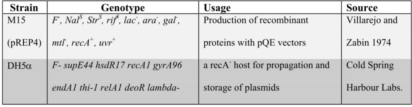
4.1. BACTERIAL STRAINSThe bacterial strains used in this study were DH5α and M15.
Table 4.1: List of the E.coli strains used during the course of this study
Strain Genotype Usage Source
M15 (pREP4)
F-, NalS, StrS, rif8, lac-, ara-, gal-, mtl-, recA+, uvr+
Production of recombinant proteins with pQE vectors
Villarejo and Zabin 1974 DH5α F- supE44 hsdR17 recA1 gyrA96
endA1 thi-1 relA1 deoR
lambda-a recA- host for propagation and
storage of plasmids
Cold Spring Harbour Labs.
4.2. GROWTH AND MAINTENANCE OF BACTERIA
Bacterial strains were stored in 50% glycerol at –70oC for long-term storage.
Overnight grown cultures to saturation were mixed with sterile glycerol with a ratio of 1:1, mixed to homogeneity, and stored at -70oC until required. Bacteria was recovered by scraping a small amount of cells from the frozen stock with an inoculation loop and streaking onto a LB-agar plate (supplemented with the appropriate antibiotics).
Liquid culture of plasmid-carrying E.coli was performed in LB (5 g NaCl, 10 g Bacto-tryptone, 5 g yeast extract, 1 ml of 1 M NaOH, ddH2O added to 1 L, sterilised by
autoclaving) with appropriate antibiotic selection. Liquid cultures were constantly agitated in a rotary shaking incubator (~200 rpm). When supplementing LB-agar (LB medium with 1.5% agar) with antibiotic, the LB-agar was melted and allowed to cool to 55oC before the
addition of antibiotic and pouring into 10 cm diameter plastic petri dish (Greiner Labortechnik). Plates were stored at 4oC and air-dried prior to use.
The specialised components of all media were obtained from Difco Laboratories Ltd.
4.3. YEAST STRAINS
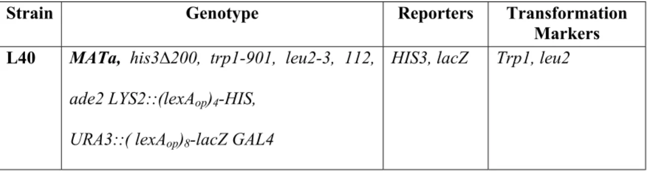
The yeast strain used in this study was L40.
Table 4.2: Features of the yeast strain L40
Strain Genotype Reporters Transformation
Markers L40 MATa, his3∆200, trp1-901, leu2-3, 112,
ade2 LYS2::(lexAop)4-HIS,
URA3::( lexAop)8-lacZ GAL4
HIS3, lacZ Trp1, leu2
L40 is a reporter host used when screening fusion libraries for proteins that interact with a target protein. This strain carries both LacZ and HIS3 reporters integrated to URA3 and LYS2 genes of the yeast genome (Hollenberg et al. 1995). The expression of the LacZ and HIS3 coding sequences are driven, respectively by minimal GAL1 and HIS3 promoters fused to multimerized LexA binding sites.
4.4. GROWTH AND MAINTENANCE OF YEAST
Yeast strains were stored in 25% glycerol at –70oC for long-term storage. Cultures grown in the appropriate Yc selective medium (1.2 g yeast nitrogen base without amino
acids, 5 g Ammonium sulfate, 10 g succinic acid, 6 g NaOH and 0.1 g of all the aminoacids apart from his, ura, trp and leu were dissolved in 1 L, and autoclaved) to saturation were mixed with sterile glycerol with a ratio of 3:1, mixed to homogeneity, and stored at -70oC until required. Yeast cells were recovered by scraping a small amount of cells from the frozen stock with an inoculation loop and streaking them onto a YPAD (or appropriate Yc) agar plate.
Liquid cultures of yeast were performed in YPAD (10 g yeast extract, 20 g peptone, 0.1 g adenine, ddH2O added to 900 ml, sterilised by autoclaving, cooled to ~55oC and 100
ml of 20% filter-sterilised glucose added) or in the appropriate Yc medium. Liquid cultures were constantly agitated in a rotary shaking incubator (~200 rpm) at 30oC.
4.5. MAMMALIAN CELLS
The only mammalian cell line used in this study was COS-7 (ATCC CRL-1651). This line was derived from the CV-1 cell line, African green monkey kidney origin, (ATCC CCL-70) by transformation with an origin defective mutant of SV40 that codes for wild-type T antigen.
4.6. OLIGONUCLEOTIDES
Primers used in this study were synthesised in the Beckman Oligo 1000M DNA Synthesiser (Beckman Instruments Inc. CA, USA) at Bilkent University, Department of Molecular Biology and Genetics (Ankara, Turkey).
Cloning Primers:
1. UY 21 TAFI 48 Full Length Forward PCR Primer:
5′ AGACGTCGACAAGGATCC ATG AGT GAT TTC AGT GAA GAA 3′
2. UY 22 TAFI 48 Full Length Reverse PCR Primer:
5′ AGACCTCGAGTCTAGA TCA GAG TCT TGG ATT TAC AAT 3′
3. UY 54 TAFI 48 Full Length Reverse PCR Primer:
5′ AGACTCTAGACT GAG TCT TGG ATT TAC AAT ACT 3′
Table 4.3: Primer pairs used to amplify full-length open reading frame of TAFI 48
Primer Pairs PCR Product Size Annealing Temperature
UY21/UY22 1384 bp 61oC
UY21/UY54 1379 bp 58oC
4.7. STANDARD SOLUTIONS AND BUFFERS TBE Buffer:
Working Solution: 10x Stock Solution (1lt): 45 mM Tris-borate 108 g Tris Base
1 mM EDTA. 55 g Boric Acid
40 ml 0.5 M EDTA, pH 8.0 TAE Buffer:
Working Solution 50x Stock Solution (1lt) 0.4 M Tris acetate 2 M Tris Base (242 g) 0.001 M EDTA 57.1 ml Glacial Acetic Acid 50 mM EDTA SalI SalI BamHI BamHI BamHI
Phosphate-buffered saline (PBS):
Working Solution, ~pH 7.3: 10x Stock Solution (1lt): 137 mM NaCl 80 g NaCl
2.7 mM KCl 2 g KCl
4.3 mM Na2HPO4.7H2O 11.5 g Na2HPO4.7H2O
1.4 mM KH2PO4 2 g KH2PO4
4x Tris-Cl/SDS, pH 6.8:
Tris base 18.2 g, SDS 0.4g, ddH2O to 100 ml, adjusted to pH 8.8 with HCl.
4x Tris-Cl/SDS, pH 8.8:
Tris base 6.05 g, SDS 0.4 g, ddH2O to 100 ml, adjusted to pH 6.8 with HCl.
10x SDS-PAGE Running Buffer:
Tris base 30.2 g, Glycine 144g, ddH2O to 1L.
Working Solution:
100 ml 10x stock 25 mM Tris 5 ml 20% SDS 192 mM Glycine 895 ml ddH2O 0.1% SDS
2x SDS PAGE Loading Buffer: 25 ml 4xTris-Cl/SDS pH 6.8 20 ml glycerol 3 g SDS 2 ml 2-ME 1 mg bromophenol blue Add ddH20 to 100 ml Transfer Buffer:
Working Solution (for proteins less than 150 kD): 25 mM Tris
192 mM Glycine 10% methanol
Working Solution (for bigger proteins): 50 mM Tris 384 mM Glycine 20% methanol 0.1% SDS 1x TE: 10 mM Tris, pH 8.0
4.8. RECOMBINANT DNA TECHNIQUES 4.8.1. Polymerase Chain Reaction
Polymerase chain reaction (PCR), a rapid procedure for in vitro enzymatic amplification of a specific segment of DNA (Mullis and Faloona 1987), was performed to amplify the coding sequence of TAFI 48. PCR reactions were performed in 0.2 ml ThermowellTM tubes (Corning Costar Corp.) using the GeneAmp PCR system 9600 (Perkin Elmer).
PCR reactions were carried out in a reaction volume of 50 µl containing 1-2 ng of template DNA, 5 µl 10x PCR buffer (Sigma), 5% DMSO (Sigma), 1.5 mM MgCl2
(Sigma), 0.2 mM of each dNTP (MBI Fermentas), 50 pmol of each primer and 1 unit Taq DNA polymerase (Sigma).
The reaction was preheated to 94oC for 5 minutes and then subjected to 30 cycles of denaturation (30 seconds at 95oC), annealing (1 minute at 61oC or 58oC depending on Tm degrees of the primers), and elongation (90 seconds at 72oC). At the end of the 30 cycles a final extension at 72oC for 10 minutes was also applied. PCR products were assessed by agarose gel electrophoresis and EtBr staining.
4.8.2. Phenol-chloroform extraction and ethanol precipitation of PCR products
This method was routinely used to purify and concentrate DNA preparations. The volume of the DNA solution was brought to 100 µl with ddH2O and an equal volume of
phenol was added on the solution to remove any protein contaminants. The mixture was vortexed vigorously and centrifuged at 13000 rpm for 2 minutes. The upper aqueous layer was removed, transferred into a new tube and an equal volume of chloroform was added to
remove any residual phenol. Once again the mixture was vortexed and centrifuged at 13000 rpm for 2 minutes. The upper aqueous layer was removed and transferred into a new tube. To the estimated volume of the solution, 1/10 volume of 3 M NaAc pH 5.2, and 3 volumes of ice-cold absolute ethanol was added. The solution was then frozen at –70oC for 10-30 minutes, followed by centrifugation at 13000 rpm for 20 minutes at 4oC. The supernatant was removed and the pelleted DNA was washed with 70% ethanol to remove salts and small organic molecules and dried at 37oC. Finally, DNA was resuspended in ddH2O at a concentration suitable for further experimentation and stored at –20oC.
4.8.3. Purification of DNA fragments by Agarose Gel Electrophoresis
The samples were loaded into a single well of a 0.8%-1% low melting temperature agarose gel containing 1µg/ml EtBr. After electrophoresis, the DNA bands were visualised under UV illumination and the desired bands were excised from the agarose gel with a surgical blade (Aesculap Surgical Industries). Agarose slices were suspended in about 300 µl of a buffer containing 10 mM Tris-HCl pH 8, 1 mM EDTA, and 100 mM NaCl. The gel slices were melted down by heating at 67oC for 20-30 minutes and while it was still at 67oC, an equal volume of phenol was added to the melted gel. The mixture was vortexed well and centrifuged for 15 minutes in a microfuge at 13000 rpm.
The melted gel material forms a thick precipitate at the interface. The aqueous phase was removed and was extracted once again with phenol, twice with an equal volume of chloroform and optionally once with an equal volume of water-saturated ether. Each extraction was done by vortexing for a few seconds and between extractions, aqueous phase was recovered by centrifugation in a microfuge for a few seconds at the maximum
speed. If used, residual ether after the last extraction was removed by heating the sample for 3 min at 67oC.
The DNA was precipitated with 3 volumes of absolute ethanol for 30 min to overnight at –70oC. The DNA was pelleted by centrifugation at the maximum speed for 30 min at 4oC, followed by washing once with 0.5 ml of 70% ethanol. The residual ethanol was removed by drying the pellet at 37oC for 2 minutes. The DNA pellet was resuspended in appropriate volume of ddH2O.
4.8.4. Restriction Enzyme Digestion of DNA
Depending on the amount of DNA (1-5 µg), restriction enzyme digests of DNA was carried out in a total volume of 20-50 µl with 10-50 units of restriction enzyme, volume of which was never greater than 1/10th of the total reaction volume. Appropriate buffers and incubation conditions were used for each digest as recommended by the manufacturer.
4.8.5. DNA Ligation
DNA fragments were ligated into plasmid vectors as described in Molecular Cloning (Maniatis et al. 1982). Prior to ligation, vector and insert concentrations were checked by agarose gel electrophoresis. For undirectional cloning, vector: insert ratio was set to 1:3 in molar terms and for directional cloning vector: insert ratio was kept close to 1 or slightly bigger than 1 in the ligation reactions. Ligations were performed in 15 µl reaction volumes containing 0.1-0.2 µg of plasmid DNA and the corresponding amount of insert in the presence of 4 Weiss units of T4 DNA ligase, 1mM ATP and standard ligation
buffer supplied by the manufacturer. The reaction was kept at room temperature for 4 hours or at 16oC overnight.
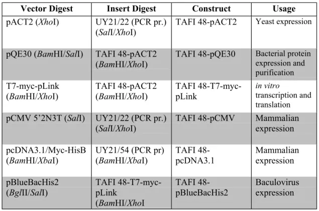
4.8.6. Plasmids
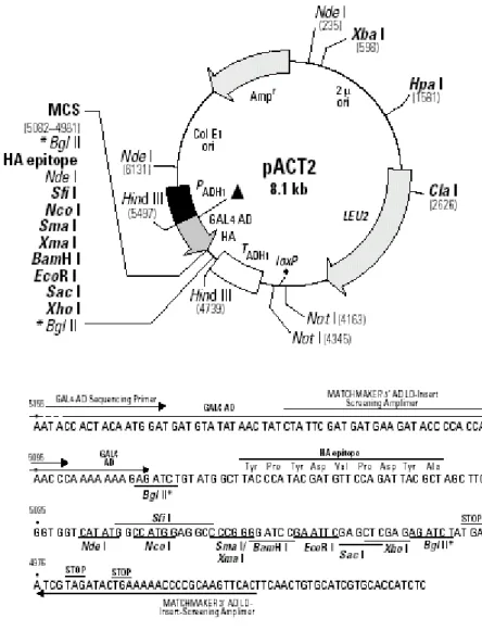
The plasmids used in this study to clone the full-length open reading frame of TAFI 48 are: pACT2, pQE30, T7-myc-pLink, pCMV 5’2N3T, pcDNA3.1/Myc-HisB and pBlueBacHis2. The available maps are presented in figures 4.1, 4.2, 4.3 and 4.4.
Figure 4.3: Map of pcDNA3.1/Myc-His vectors
4.8.7. Recombinant Expression Constructs
Constructs required for yeast, bacterial, mammalian and baculovirus expression, in vitro transcription and translation were prepared as follows. Inserts and plasmids were digested with appropriate restriction enzymes, were run on agarose gel for concentration determination and were ligated as described in the relevant section. Half of the ligation product was transformed into bacteria and recombinant plasmids were isolated (see sections 4.9. and 4.10.).
Table 4.3: List of the constructs prepared during the course of this study
Vector Digest Insert Digest Construct Usage
pACT2 (XhoI) UY21/22 (PCR pr.) (SalI/XhoI)
TAFI 48-pACT2 Yeast expression
pQE30 (BamHI/SalI) TAFI 48-pACT2 (BamHI/XhoI)
TAFI 48-pQE30 Bacterial protein expression and purification T7-myc-pLink
(BamHI/XhoI) TAFI 48-pACT2(BamHI/XhoI) TAFI 48-T7-myc-pLink
in vitro
transcription and translation pCMV 5’2N3T (SalI) UY21/22 (PCR pr.)
(SalI/XhoI) TAFI 48-pCMV Mammalianexpression pcDNA3.1/Myc-HisB (BamHI/XbaI) UY21/54 (PCR pr) (BamHI/XbaI) TAFI 48-pcDNA3.1 Mammalian expression pBlueBacHis2 (BglII/SalI) TAFI 48-T7-myc-pLink (BamHI/XhoI TAFI 48-pBlueBacHis2 Baculovirus expression
4.9. PREPARATION OF COMPETENT CELLS AND TRANSFORMATION OF E.COLI
4.9.1. CaCl2 Method
A single colony of an appropriate E.Coli strain was inoculated in 5 ml of LB (containing the appropriate antibiotics) and grown overnight at 37oC. The starter culture was diluted to an O.D600 of 0.2-0.3 in 50 ml of LB (containing the appropriate antibiotics)
and was grown further until the O.D600 reached to 0.6-0.7. The culture was cooled on ice
for 10 minutes and the cells were pelleted by centrifugation at 4000 rpm for 10 minutes at 4oC. The pellet was resuspended in 40 ml of ice cold filter-sterilised 100 mM CaCl2 and
incubated on ice for 1 hour. The cells were then pelleted as above and resuspended in 2 ml of ice cold sterile 100 mM CaCl2. The competent cells prepared by this way can be kept on
ice till transformation up to 24 hours (Cohen et al. 1972)
4.9.2. Simple and Efficient Method (SEM)
A single colony of appropriate E.coli strain was inoculated into 15 ml of LB (containing the appropriate antibiotics) and grown overnight at 37oC. The starter culture was diluted to an O.D600 of 0.2-0.3 in 250 ml of SOB medium (2% Bacto-tryptone, 0.5%
yeast extract, 10 mM NaCl, 2.5 mM KCl, 10 mM MgCl2, 10 mM MgSO4, sterilised by
autoclaving) and grown to an OD600 of 0.6 at 18oC with shaking at 200-250 rpm. The
culture was chilled on ice for 10 minutes, centrifuged at 2500 g for 10 minutes at 4oC. The
pellet was then resuspended in 80 ml of ice-cold TB (10 mM Pipes, 55 mM MnCl2, 15 mM
CaCl2, 250 mM KCl, pH 6.7, sterilised by filtration through 0.45 µm filter, stored at 4oC),
gently in 20 ml of TB. DMSO was added to a final concentration of 7%, mixed gently and incubated on ice for 10 minutes. Aliquots of this mixture were then immediately chilled in liquid nitrogen, and stored at –70oC up to 3 months without loss of transformation efficiency (Inoue et al. 1990).
4.9.3. Transformation of E.Coli
For transformation using competent cells prepared by CaCl2 method, DNA (less
than 100 ng) was added to 200 µl of competent cells in a microfuge tube, and incubated on ice for 30 minutes. The cells were then exposed to a 90 seconds heat shock at 42oC, chilled on ice for 1-2 minutes, and 800 µl of LB was added and followed by incubation at 37oC for
one hour. The culture was then pelleted, resuspended in 100 µl of LB, and was spread on LB-agar plates supplemented with appropriate antibiotics (50 µg/ml ampicillin and/or 25 µg/ml kanamaycin). The plates were left for drying, then inverted and incubated overnight at 37oC (Cohen et al. 1972).
For transformation with supercompetent cells, an aliquot of frozen competent cells was thawed on ice. 200 µl of the cells was mixed with plasmid DNA (less than 100 ng) in a 15 ml round bottom tube (Greiner Labortechnik), and incubated on ice for 30 minutes. The cells were then exposed to a 30 seconds heat shock at 42oC, chilled on ice for 1-2 minutes, 800 µl of SOC (SOB with 20 mM glucose) was added and the cells were grown at 37oC for
one hour with vigorous shaking at 250 rpm. The culture was then pelleted, resuspended in 100 µl of SOC, and was spread on LB-agar plates as described above (Inoue et al. 1990).
4.10. PLASMID DNA ISOLATION 4.10.1. Small Scale Plasmid Isolation
Small-scale preparation of plasmid DNA was performed by standard methods based on NaOH/SDS cell lysis and potassium acetate precipitation of cellular debris (Maniatis et al. 1982).
Cells were harvested by centrifugation (13,000 rpm, 3 minutes, at room temperature) from a 1.5 ml overnight culture of bacteria carrying the plasmid of interest. Following resuspension of the bacterial pellet in 100 µl of solution I (50 mM glucose, 25 mM Tris-Cl pH 8.0, 10 mM EDTA), 200 µl of freshly prepared solution II (200 mM NaOH, 1% SDS) was added and cells were lysed by gently inverting the tube a few times. Bacterial chromosomal DNA and proteins were precipitated by the addition of 150 µl of solution III (3 M potassium acetate, 11.5% glacial acetic acid). An equal volume of phenol: chloroform (1:1) mixture was added and the mixture was vortexed well. The phases were separated by centrifugation at 13,000 rpm for 5 minutes at room temperature and upper aqueous layer was transferred into a new tube. The DNA was precipitated from the aqueous phase by addition of 1 ml of cold absolute ethanol and the DNA was pelleted by centrifugation at 13000 rpm for 20 minutes at 4oC. After washing once with 70%
ethanol, the pellet was dried and resuspended in 30 µl of ddH2O with 10 µg/ml DNase-free
4.10.2. Medium Scale Isolation (Midi-Preparation)
Cells were grown in 50-100 ml of LB (containing the appropriate antibiotic) overnight to saturation and plasmid DNA was isolated by using Nucleobond AX 100 (Macherey-Nagel), according to the manufacturer’s instructions.
4.10.3. Spectrophotometric Quantification of DNA
The amount and the purity of DNA samples were determined by reading the absorbance of samples at 260 nm and 280 nm in a spectrophotometer (Beckman Instruments Inc., CA; USA). Nucleic acid samples displaying OD260 /OD280 ratio in the
range of 1.8 to 2.0 are regarded as highly pure. A value of OD260=1.0 corresponds to a
concentration of approximately 50 µg/µl for double stranded DNA, 40 µg/ml for single stranded DNA and 20 µg/µl for oligonucleotides (Maniatis et al. 1982).
4.11. YEAST TECHNIQUES
4.11.1. Small Scale Yeast Transformation
A single colony of L40 was inoculated in 10 ml of YPAD and grown overnight at 30oC with shaking at 200 rpm. The starter culture was diluted to an O.D600 of 0.2-0.3 in
50 ml of YPAD medium and was grown further until the OD600 reached to 0.6-0.7. All
subsequent steps were carried at room temperature. Yeast cells were pelleted at 2500 rpm for 10 minutes. After resuspending the pellet in 40 ml 1x TE, cells were repelleted and resuspended in 2 ml of LiAc/TE (100 mM LiAc in 0.5x TE), and incubated at room temperature for 10 minutes. Meanwhile 0.1 µg of DNA was dissolved in 8 µl of ddH2O
and 10 µl of salmon sperm DNA was added. 175 µl of competent yeast cells was dispensed to DNA tubes, mixed by flicking. 700 µl of 100mM LiAc/ 40% PEG-3350/1x TE was added and incubated at 30oC for 30 minutes on a rotating wheel. Subsequently, 88 µl of DMSO was added, followed by a heat shock at 42oC for 7 minutes. Cells were then
pelleted at full speed for 10 seconds, supernatant aspirated, and the pellet was resuspended in 1.0 ml of 1x TE. Cells were pelleted once more as above, resuspended in 100 µl of 1x TE, and were plated on the appropriate selective medium (Ito et al. 1983, Gietz et al. 1992).
4.11.2. Liquid Culture Assay Using ONPG as Substrate
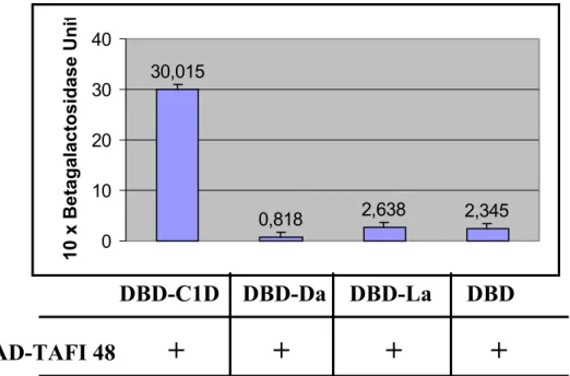
Liquid yeast cultures were assayed for β-galactosidase activity to verify and quantify the interaction between two proteins of interest. L40 cells were cotransformed with the plasmid expressing TAFI 48 in fusion with the activation domain of GAL4 (TAFI 48-pACT2) and each of the different baits expressing Lamin, Daughterless and C1D in fusion with a DNA binding domain (LexA DNA-binding domain (DBD) alone, DBD-Lamin, DBD-Daughterless, or DBD-C1D). 3 colonies were picked from each transformation plate and were grown in 4 ml of the appropriate selective liquid medium for 2 days. 2 ml of the saturated culture was transferred into 8 ml of YPAD. The cultures were incubated at 30oC for 3-5 hrs with shaking (230-250 rpm) until the cells grew to the mid-log phase (O.D600 = 0.5-0.8). The exact value of O.D600 was recorded when harvesting the
cells. 1.5 ml of culture was placed into each of three 1.5-ml microcentrifuge tubes. Cells were collected by centrifugation at 13000 rpm for 30 sec. After the supernatants were removed, the cells were resuspended in 1.5 ml of Z-buffer (60 mM Na2HPO4, 40 mM
supernatants were removed. Each pellet was resuspended in 300 µl of Z buffer so that the concentration factor was 5 fold. 0.1 ml of the cell suspension was transferred into a fresh microcentrifuge tube. Cells were lysed by freezing in liquid nitrogen for 30 seconds, followed by thawing in a 37oC water bath. The freeze/thaw cycle was repeated for two more times to ensure that the cells had broken open. A blank tube with 100 µl of Z buffer was set up. 0.7 ml of Z buffer (+ β-mercaptoethanol) was added to the reaction and blank tubes. The tubes were placed in a 30oC heat block. 160 µl of 4 mg/ml ONPG (Sigma) (prepared in Z buffer) was added to the reaction and blank tubes and the elapsed time until the yellow colour developed was recorded. When the colour developed, 0.4 ml of 1M Na2CO3 was added to the tubes to stop the reaction. The reaction tubes were centrifuged
for 10 minutes at 13000 rpm to pellet the cell debris and the supernatants were transferred into clean cuvettes. The spectrophotometer was calibrated against the blank tube at A420
and the O.D420 of the samples relative to the blank were measured. Finally, β-galactosidase
units was calculated according to the following formula: β-Galactosidase units =1000 x OD420 /(t xVx OD600)
Where: t = elapsed time (in min) of incubation V = 0.1 ml x concentration factor
1 unit of β-galactosidase is defined as the amount that hydrolyzes 1 µmol of ONPG to o-nitrophenol and D-galactose per min per cell (Miller 1972).
4.12. CELL CULTURE TECHNIQUES 4.12.1. Thawing a Frozen Cell Line
Frozen cell line stock in cryotube was transferred from liquid nitrogen into ice and then the frozen cells were thawed at 37oC in less than 2 minutes. After thawing, cells were transferred into a 15 ml falcon tube and 10 ml of warmed fresh culture medium (DMEM supplemented with 10% FCS and 1% Penicillin/Streptomycin, all purchased from Biochrom) was added dropwise. Cells were collected by centrifugation at 1500 rpm for 5 minutes at 4oC and the medium was removed by aspiration without disturbing the cell pellet. The pellet was then resuspended in 5 ml of fresh culture media and transferred into a 25 cm2 culture flask (Greiner Labortechnik). The culture flask was incubated at 37oC in a humidified atmosphere with 5% CO2 for cell growth (Doyle and Griffiths 1997).
4.12.2. Sub-Culturing of Monolayer Cells
When reached to 80% confluence, cells were sub-cultured into new flasks. The medium was aspirated and monolayer cells were rinsed quickly with a volume of 0.25% trypsin/0.03% EDTA solution (Biochrom) just enough to cover the surface. The solution was removed and an additional amount of trypsin-EDTA solution was added. After incubation at 37oC for 30 seconds, the trysin-EDTA solution was removed and the flask was allowed to sit at 37oC until the cells detached. 2-10 ml (depending on the size of the culture flask) of culture media was added and any cell remaining attached was suspended by gently pipetting. The medium was transferred to a fresh 15 ml falcon tube. Following centrifugation at 1500 rpm for 5 minutes at 4oC, the medium was removed by aspiration without disturbing the cell pellet. Then the pellet was resuspended in 5 ml of fresh culture
media and was dispensed into new culture flasks at the desired sub-culturing ratio. Additional fresh culture medium was added and the flasks were incubated at 37oC in a humidified atmosphere with 5% CO2 (Doyle and Griffiths 1997).
4.12.3. Cryopreservation
As in sub-culturing the cells were collected but instead of the culture medium, cells were resuspended in adequate amount of freezing medium (DMEM supplemented with 5% FCS and 10% DMSO) to give a cell density of 5 x 10⁶ cells /ml, dispensed into cryotubes and were frozen overnight at –80oC from where transferred into liquid nitrogen (Doyle and Griffiths 1997).
4.12.4. Transient Transfections
All transfections were performed with the FuGENE6 (Roche Molecular Biochemicals) transfection reagent according to the manufacturer’s instructions. One-day before transfection, 2 x 10⁵ cells in 4 ml total media were plated into a 60 mm culture dish
(Greiner Labortechnik). On the day of transfection, cells should be 50-80% confluent and most cell lines plated at a density of 2 x 10⁵ cells/ 4 ml in 60 mm culture dishes will achieve this confluency after overnight incubation at 37oC with 5% CO2.
4.13. GEL ELECTROPHORESIS 4.13.1. Agarose Gel Electrophoresis
DNA fragments were separated by gel electrophoresis using agarose at concentrations of 0.8-2.0% w/v in 1x TBE buffer. Samples were mixed with a fifth volume
of loading dye (8% deionised formamide, 1x TBE and 0.1% bromophenol blue) and the samples were loaded onto the gel. Electrophoresis was performed by running the gel at 100V. At the mid of the run, the gel was soaked briefly in 1x TBE containing 0.1% EtBr, and then was run further to completion. Gels were visualised by illuminating with UV light on a transilluminator at 302nm. The gel photos were captured with BioRad Multi-Analyst Software running on a PC and a hardcopy was produced by Lexmark Optra laser printer.
4.13.2. SDS Polyacrylamide Gel Electrophoresis (SDS-PAGE)
Electrophoretic separation of proteins under denaturing conditions was performed by following the methods described in Current Protocols (Ausubel 1987). Resolving gels were made up to the required concentration, 10-15%, from a 40% acrylamide stock solution (38% acrylamide, 2% bisacrylamide, Severn laboratories). The electrophoresis apparatus (Mini-Protean 3, BioRad) was assembled according to manufacturer’s instructions. Acrylamide stock solution (to the desired gel percentage) and 4x Tris-Cl/SDS pH 8.8 were mixed in ddH2O and 10% ammonium persulphate and TEMED (30 µl and 10
µl, respectively for a 5 ml solution) were added to assist polymerisation. The mixture was then poured into the gap between the glass plates so that sufficient space is left for the stacking gel and water saturated isobutanol was layered immediately onto the top of the gel. After the polymerisation, the isobutanol layer was removed, washed with water and the stacking gel (0.49 ml of acrylamide stock solution, 3.225 ml ddH2O, 1.25 ml 4x
Tris-Cl/SDS pH 6.8, 30µl of 10% ammonium persulphate, 5 µl of TEMED) was poured onto the top of the resolving gel. The comb was immediately inserted into the stacking gel
avoiding the formation of bubbles. After the stacking gel polymerised, the comb was carefully removed and the wells were rinsed with the running buffer to remove any unpolymerised acrylamide. The samples were mixed with appropriate volume of 2x SDS loading buffer, boiled at 100oC for 5 minutes to denature proteins, centrifuged at full speed for 5 minutes, and then loaded on to gel. Gels were run at 70V until the samples left the stacking gel. As the samples passed to the resolving gel the voltage was raised to 100V. After the electrophoresis, to detect the proteins, gel was carefully removed and stained in Coomassie Brilliant Blue (0.25% Coomassie Brilliant Blue G250, 45% methanol, 10% acetic acid) for 20 minutes, and destained in a solution of 30% methanol, 10% acetic acid, and 60% deionised distilled water for 1-4 hours. Alternatively gels were subjected to electrophoresis for the purpose of immunoblotting.
4.14. BIOCHEMICAL TECHNIQUES
4.14.1. Immunological Detection of Immobilized Proteins (Western Blotting) 4.14.1.1 Transfer of Proteins onto Membranes
Following SDS-PAGE, proteins were transferred from the gel to a nitrocellulose membrane (Schleicher & Schuell) using Mini Trans- Blot Cell (BioRad). Two sheets of 3 MM Whatman paper (Whatman International Ltd.) and one sheet of nitrocellulose membrane were cut to the dimensions of the gel to prepare the transfer stack. Nitrocellulose membrane was wetted in ddH2O by capillary action. The gel, the
membrane, Whatman papers and fiber pads were soaked in transfer buffer (25 mM Tris, 192 mM Glycine, 10% methanol or 50 mM Tris, 384 mM Glycine, 20% methanol and 0.1% SDS) for 15 minutes. Then the transfer stack was assembled in the following order
starting from the side that would face the cathode: fiber pad, one sheet of Whatman paper, the gel, the nitrocellulose membrane, one sheet of Whatman paper and finally the second fiber pad. While preparing the transfer stack, formation of any air bubbles between the layers was avoided. The transfer stack was then placed into the tank filled with transfer buffer and transfer was performed at 100V for 1 hour for proteins with molecular weights smaller than 150 kD and at 40V for 2 hours for bigger proteins.
4.14.1.2. Immunological Detection of Immobilized Proteins
After the transfer has been completed, the membrane was washed twice with TBS-T (TBS-TBS + 0.5% TBS-Tween-20), and incubated in 50 ml of blocking solution (10% non-fat dry milk in TBS-T) with continuous shaking for two hours at room temperature or overnight at 4oC. After blocking, the membrane was washed four times for 10 minutes each with TBS-T. The membrane was then incubated with the solution containing the primary antibody (diluted in 1% non-fat dry milk in TBS-T) for 1 hour. The membrane treated with the primary antibody was washed four times for 10 minutes each with TBS-T. The membrane was then treated with the HRP (horse reddish peroxidase) conjugated secondary antibody (diluted in 1% non-fat dry milk in TBS-T) for 1 hour and was washed as described before.
The immunodetection with HRP conjugated secondary antibody was carried out by using Amersham LIFE SCIENCE ECL Western blotting detection reagents according to the manufacturer’s instructions. The bands were visualised by exposing the membrane on photograph films.
4.14.1.3. Striping and Reprobing Membranes
This method was used to remove the primary and secondary antibodies completely from membranes. The membrane was submerged in stripping buffer (100 mM 2-mercaptoethanol, 2% SDS, 62.5 mM Tris-Cl pH 6.7) and the membrane was incubated at 65oC for 30 minutes with occasional agitation. The membrane was then washed four times for 10 minutes with large volumes of TBS-T at room temperature and all steps described above were applied for the immunodetection.
4.14.1.4. Peptide Neutralisation of Primary Antibody
For peptide neutralisation, primary antibody was combined with a five-fold (by weight) excess of blocking peptide in a small amount of PBS. The mixture was incubated for 2 hours at room temperature. Following neutralisation, antibody/peptide mixture was diluted in 1% non-fat dry milk in TBS-T and the protocol for immunological detection was performed as described above.
4.14.2. Immunoprecipitation
After thirty hours following transfection, the medium was aspirated and monolayer cells were rinsed twice with PBS at room temperature. All the following steps were carried out at 4oC using ice-cold buffers. 1.5 ml of PBS + 50 mM EDTA was added on monolayer cells and cells were incubated on a shaker for 10 minutes. After incubation, cells were scraped with a rubber scraper and were transferred into a microcentrifuge tube . Cells were collected by centrifugation at 2000 rpm for 5 minutes. The supernatant was removed; cells were resuspended in 0.1 ml of Lysis buffer (150 mM NaCl, 1 mM EDTA, 20 mM Tris pH
8.0, 0.5% NP-40, 10% glycerol, 1x CompleteTM protease inhibitors) and were incubated on a rotating wheel for 30 minutes. Following incubation cell lysates were spun down at 10,000g for 10 minutes to remove the insoluble cell debris. The supernatant, which is the total cell lysate, was removed into a new tube and was incubated with the appropriate antibody for 2 hours on a rotating wheel. Meanwhile 20 µl of resuspended volume of the appropriate agarose conjugate (10 µl of Protein A-agarose + 10 µl of Protein G-agarose) was washed three times with 0.5 ml of Lysis Buffer (10 minutes washing followed by 5 minutes centrifugation at 2500 rpm to remove supernatant). After 2 hours incubation, cell lysate/antibody mix was added onto equilibrated beads and was incubated for further 2 hours. The beads were collected at the end of the incubation by centrifugation at 2500 rpm for 5 minutes and the supernatant was removed. Then the beads were washed four times with 0.5 ml of Lysis buffer as described before. After the last wash, supernatant was discarded and the beads were resuspended in 20 µl of 2x SDS Loading Buffer. The samples were boiled for 5 minutes, centrifuged at full speed for 5 minutes and then loaded on to SDS PAGE gel. After electrophoresis, proteins were transferred to a nitrocellulose membrane and the immunological detection of the immobilized proteins was performed.
4.14.3. Protein Purification of Glutathione-S-Transferase Tagged C1D Protein
The pGEX vectors are designed so that foreign polypeptides can be expressed in E.coli in a form that allows them to be purified rapidly under nondenaturing conditions. Foreign polypeptides are expressed as fusions to the C terminus of glutathione-S-transferase (GST), a common 26 kD cytoplasmic protein of eukaryotes. The fusion protein typically remain soluble within the bacteria and can be purified from lysed cells because of
the affinity of the GST moiety for glutathione immobilised on agarose or sepharose beads. Recovery of the fusion protein is by elution with free reduced glutathione at reduced pH (Ausubel 1987).
A single colony of M15 strain of E.Coli transformed with pGEXTK2-C1D construct was inoculated in 5 ml of LB (supplemented by 50 µl/mg ampicillin and 25 µl/mg kanamaycin) and grown overnight at 37oC. The starter culture was diluted to an O.D600 of
0.2 in 50 ml of LB (containing the appropriate antibiotics) and was grown at 30oC till its O.D600 reached to 0.55. IPTG (MBI Fermentas) was added to a final concentration of 0.15
mM and cells were grown further for 3 hours at 30oC. Cells were then harvested at 4000 rpm for 20 minutes at 4oC, and then resuspended in 1 ml TBS-PMSF (1% Triton, 1mM PMSF, BSA 10 µg/ml, 1x CompleteTM protease inhibitors diluted in TBS). Cells were then
sonicated on ice giving 10 seconds bursts at 300 W by the aid of UP 50 Ultrasonic Processor (UniEquip), and were centrifuged at 8000 rpm for 30 minutes at 4oC.
Meanwhile, 30 µl of glutathione-sepharose-4B beads (Pharmacia Biotech) were equilibrated by washing with 0.5 ml of 50% TBS at 4oC (10 minutes incubation, 5 minutes centrifugation at 2500 rpm 4oC to remove supernatant) four times. The supernatant from the cell lysate was then loaded onto the equilibrated beads and rotated at 4oC for 30 minutes. Supernatant was then removed at the end of binding step and was preserved for checking. The beads were then washed with 0.5ml of TBS-PMSF four times as described before. Finally the beads were suspended in 50µl of TBS-PMSF and 8 µl of glycerol was added for storage at -20oC. 10 µl of beads with slurry was mixed with appropriate volume
of 2x SDS-PAGE loading dye. Samples were boiled at 100oC for 5 minutes, centrifuged at full speed for 5 minutes and analysed on SDS-PAGE.