THE ROLE OF LIPID-INDUCED INTEGRATED
STRESS RESPONSE IN METAFLAMMATION
AND ATHEROSCLEROSIS
A DISSERTATION SUBMITTED TO
THE GRADUATE SCHOOL OF ENGINEERING AND SCIENCE OF BILKENT UNIVERSITY
IN PARTIAL FULFILLMENT OF THE REQUIREMENTS FOR THE DEGREE OF
DOCTOR OF PHILOSOPHY IN
MOLECULAR BIOLOGY AND GENETICS
By Umut İnci Onat
THE ROLE OF LIPID-INDUCED INTEGRATED
STRESS RESPONSE IN METAFLAMMATION
AND ATHEROSCLEROSIS
By Umut İnci Onat June, 2019
We certify that we have read this dissertation and that in our opinion it is fully adequate, in scope and in quality, as a thesis for the degree of Doctor of Philosophy.
________________________ Ebru Erbay (Advisor) ________________________ Michelle Marie Adams ________________________ Murat Alper Cevher ________________________ Çağdaş Devrim Son ________________________ Erkan Yılmaz Approved for the Graduate School of Engineering and Science
__________________ Ezhan Karaşan
ABSTRACT
THE ROLE OF LIPID INDUCED INTEGRATED STRESS
RESPONSE IN METAFLAMMATION
AND ATHEROSCLEROSIS
Umut İnci OnatPh.D. in Molecular Biology and Genetics Advisor: Ebru ERBAY
June 2019
Chronic inflammation resulting from metabolic overloading of organelles (such as the endoplasmic reticulum (ER) and mitochondria that control cellular homeostasis) is a major cause of metabolic disorders including diabetes, obesity and atherosclerosis. ER is an organelle that plays a critical role in cellular metabolism through biosynthesis of lipids, protein maturation and secretion, and calcium storage. Furthermore, a stressed endoplasmic reticulum maintains cellular homeostasis by initiating a conserved stress response pathway that is known as Unfolded protein response (UPR). UPR is activated in response to diverse stimuli that disrupts ER functions and serves asva pro-survival mechanism to regain ER homeostasis. However, in prolonged or severe ER stress, chronic UPR can promote inflammation and apoptosis. Activated UPR, inflammation and necrosis are observed in and causally associated with atherosclerosis. UPR has three branches, one of which is initiated by the protein kinase RNA (PKR) like ER kinase (PERK) and signals to eukaryotic initiation factor 2a (eIF2a). This signaling arm of the UPR is also part of a larger, translational control pathway known as the integrated stress response (ISR). Activation of ISR has been observed in atherosclerosis and could promote atherosclerosis To study the contribution of ISR to atherogenesis, I took advantage of three small molecule inhibitors that can modulate this pathway. I also used a chemical-genetic approach, known as the Adenosine triphosphate (ATP) analog sensitive kinase (ASKA) technology, to interrupt PERK kinase activity. With these multiple tools, I was able to specifically interfere with ISR signaling at multiple molecular nodes in order to study the role of lipid-induced ISR in inflammation, inflammasome activation and atherosclerosis. I discovered that during lipid-induced ER stress, PERK to Activating transcription factor 4 (ATF4)
signaling resulted in transcriptional induction of a mitochondrial protease, Lon protease 1 (LONP1), which degrades PTEN induced putative kinase 1 (PINK1) and blocks Parkin-mediated mitochondria clearance (or mitophagy). This in turn causes an increase in mitochondrial reactive oxygen species (ROS) production, inflammasome activation and pro-inflammatory cytokine secretion such as interleukin-1b (IL-1b) in both mouse and human macrophages. I also discovered that these inhibitors are also effective in reducing hyperlipidemia-induced inflammasome activation in
Apolipoprotein E-deficient (Apoe/- ) mice and consequently, in preventing
atherosclerosis progression. These results point out that intercepting with ISR signaling in hypercholestrolemia can be considered as a novel therapeutic approach that could be developed against atherosclerosis.
Keywords: ER stress, integrated stress response, mitochondrial ROS, inflammasome,
atherosclerosis, ATP analog sensitive kinase allele, IL-1b
ÖZET
ENTEGRE STRES TEPKİSİNİN METAFLAMASYON VE
ATHEROSKLEROZDAKİ ROLÜ
Umut İnci Onat
Moleküler Biyoloji ve Genetik, Doktora Tez Danışmanı: Ebru ERBAY
Haziran 2019
Hücresel ve metabolik homeostazı sağlayan endoplazmik retikulum (ER) ve mitokondri gibi organellerde metabolik aşırı yüklenme sonucu oluşan kronik metabolik inflamasyon, tip II diyabet, obezite ve ateroskleroz gibi metabolik hastalıklara neden olur. Endoplazmik retikulum, lipid biyosentezi, protein olgunlaşması ve kalsiyum depolaması gibi hücresel metabolizmadaki kritik rollerine ilaveten katlanmamış protein yanıtı (KPY) olarak adlandırılan bir stres yanıtı yolu ile hücresel homeostazı korumaktadır. Hücresel homeostazı bozan çeşitli çevresel uyarıcılara cevap olarak, KPY hücresel fonksiyonları yeniden kazanmak için hayatta kalma yanlısı bir mekanizma olarak aktive edilir. Ancak, uzun süreli Endoplazmik Retikulum (ER) stresi durumlarında KPY hücreleri inflamasyon ve apoptik ölüme sürükler. Aktif KPY, artmış inflamasyon ve apoptoz, ER'i metabolik hastalığa bağlayan ve aterosklerozda aktif olan belirteçlerdir. KPY’in inhibisyonunun ateroskleroz gelişimini önlediği deneysel olarak gösterilmiştir. KPY’in bir kolu olan protein kinaz R benzeri ER kinaz (PERK) – ökaryotik inisiyasyon faktörü 2a (eIF2a) ve entegre stres tepkisi (EST) adlı bir başka stress yanıti ile örtüsmektedir. EST eif2a’ın fosforilasyonu ile genel proteine çevrim mekanizmasını bloke eder. EST’in belirteçlerinin aterosklerotik plaklarda aktive oldukları bilinmektedir ve aterosklerozun ilerlemesine neden olabileceği düşünülmektedir. Bu metabolik yolaklara ateroskleroz sürecinde ve makrofajlarda müdahale etmek için yakın zamanda keşfedilen üç küçük moleküler inhibitörden yararlandım. Bu yöntemde ADKA (ATP analoguna duyarlı kinaz aleli) teknolojisini PERK kinaza uygulayarak, kimyasal-genetik yöntem ilede bu kinazın aktivitesini ateroskleroz sürecinde ve makrojalarda kapatabildim. Bu değişik yöntemler ile EST stres yolağına birçok moleküler seviyede müdahale etme şansım oldu. Bu teknololojileri EST’nin, özellikle KPY ile örtüşen PERK-eIF2a yolağının makrofajlarda yağ yüklemesiyle artan
mitokondriyel reaktif oksijen türevlerinin (mtROT) üretimi, inflamazom aktivasyonu ve interlökin-1 beta (IL-1b) gibi inflamatuvar sitokinlerin salınımı ve bunların aterosklerozdaki rolünü incelemek için kullandım. Yağ kaynaklı ER stres koşullarında, PERK’den Aktifleştirici Transkripsiyon Faktörü 4 (ATF4)’e sinyalizasyon, Uyarılmış Putatif Kinaz-1(PINK1) sinyal yolunun, PINK1’ı parçalayan ve uyarılmış putatif kinaz 1 (PINK1)’ı parçalayan ve PINK1 / Parkin mitofaji yolağını bloke eden mitokondriyal proteaz Lon Proteaz 1 (LONP1)’ in transkripsiyon düzeyinde yapımını indüklediğini ve bunun sonucunda mtROT üretiminde, inflamazom aktivasyonunda ve interlökin 1- beta (IL-1b) salınımında bir artışa neden olduğunu keşfettim. Ayrıca, inhibitörlerin bu etkilerinin in vitro çalışmalar ile sınırlı
olmadığını, aynı zamanda hiperkolesterolemik apolipoprotein – E-/- (apoE-/-) farelere
tatbik edilmesiyle hiperlipideminin neden olduğu inflamazom aktivasyonunu sınırlandırdığını ve sonuç olarak aterosklerozun ilerlemesini baskıladığını keşfettim. Bu sonuçlar, hiperkolesterolemi ile artan EST sinyal yolağının baskılanmasının aterosklerozu azaltmak için terapötik bir yaklaşım olarak kullanılabileceğine işaret etmektedir.
Anahtar sözcükler: ER stres, entegre stres tepkisi, mitokondriyal reaktif oksijen
Acknowledgements
Firstly, I am very thankful to my supervisor Dr. Ebru Erbay for her scientific and personal guidance and support. I feel honored and very lucky to have had a chance to study in her lab. Dr. Erbay’s scientific lead and guidance and support during my scientific discoveries and publishing in one of the leading journals in cardiovascular research helped me surpass these challenging times.
I also want to thank Bilkent University for giving me the opportunity to do carry out my doctoral research under their academic roof. During my PhD, I improved my scientific knowledge, my personal skills, gained problem solving as well as detailed planning capabilities.
I appreciate the helps and scientific contributions of my thesis committee members Dr. Michelle Adams and Dr. Alper Murat Cevher. Furthermore, I would like to thank Dr. Erkan Yılmaz and Dr. Çağdaş Devrim Son for guidance during my dissertation and for their contribution as members of my thesis jury.
I am thankful for the contributions of our collaborators, Dr. Moshe Arditi, Dr. Roberta Gottlieb, Dr. Jon Sin, Dr. Kenichi Shimada and Dr. Shuang Chen, and for their assistance and support in difficult times during the revision of the studies for publication.
I also want to thank to my friends and colleges for their help and support during my Ph.D studies. I received a lot of support during my Ph.D. study in scientific manners as well as in friendship.
First of all, I want to thank Aslı Dilber Yıldırım for her friendship, help and contribution to the published work. She performed every task with great care and enthusiasm, and nothing I can say here can describe my great appreciation of her efforts.
I would like to also express my appreciation to Dr. İsmail Çimen for training me in animal experiments.
and giving me scientific perspective in immunostaining procedures.
I express my appreciations to Dr. Özlem Tufanlı Kireçcibaşı for her scientific lead, support and friendship during my Ph.D studies.
I also want to thank Dr. Hamid Sayed Muhammed, Zehra Yıldırım and Aslı Ekin Doğan for their support and friendship.
I would like to express my gratitude to Buket Gültekin for her help with problems regarding laboratory equipment, effort to supply research materials and for her friendship.
I want to thank to our veterinarian, Dr. Gamze Aykut for her technical support with animal experiments.
I cannot find enough words to express my gratitude and appreciation for the Zehra Veli’s help Whenever I was in trouble, unhappy or unmotivated, Zehra was always there for me as a great listener, friend and supporter. I did not know how to handle the unpredictability during the revision process prior to publication of my thesis work, but she was not there and supporting me. She is one of the most important persons in my life, who helped me keep going in the last two years.
I also want to thank for the funding that enabled me to complete my thesis; Scientific and Technological Research Council of Turkey (TUBITAK)-COST grant 113Z023, European Molecular biology Organization (EMBO) for Installation Grant and European Research Council (ERC) Starting Grant 336643.
And, most precious is my family. I want to express my deep gratitude and my love for my husband Dr. Onur Emre Onat for his outstanding support as a scientist, great friend and life partner. I would not be able to succeed or cope with the difficulties of a PhD study without him.
I also want to express my gratitude for my parents for bringing me this far, and doing every-thing in their power to support my education.
But most of all I want to thank god and my husband for their outstanding gift to me, my daughter, Zeynep. She is my strength, my love, and my dearie that gives me
emotional support and power. I am very lucky to have her. Her existence is the most powerful energy that led me go this far to reach this stage in my academic studies…
Contents
ABSTRACT ... II ÖZET ... IV ACKNOWLEDGEMENTS ... VII CONTENTS ... X LIST OF FIGURES ... XV LIST OF TABLES ... XIX ABBREVIATIONS ... XXCHAPTER 1. INTRODUCTION ... 23
1.1. The Endoplasmic Reticulum ... 23
1.1.1. Endoplasmic Reticulum Function... 24
1.1.2. The Endoplasmic Reticulum Stress and Unfolded Protein Response ... 26
1.2. Unfolded Protein Response Signaling ... 27
1.2.1. IRE1 ... 27
1.2.2. PERK ... 28
1.2.3. ATF6 ... 29
1.3. Integrated Stress Response ... 30
1.4. Endoplasmic Reticulum Stress and Immune Response ... 33
1.4.1. UPR Connection to Inflammatory Pathways ... 35
1.4.2. UPR Connection with Inflammasome Activation ... 37
1.5. Endoplasmic Reticulum and Mitochondria ... 40
1.5.1. Mitophagy and inflammation ... 42
1.6. Atherosclerosis ... 46
1.6.1. Initiation and Progression of Atherosclerosis ... 47
1.6.2. Endoplasmic Reticulum Stress in the Progression of Atherosclerosis ... 51
1.6.3. Therapeutic Approaches in Atherosclerosis ... 52
CHAPTER 2. MATERIALS AND METHODS ... 57
2.1. Materials ... 57
2.1.1. Reagents ... 57
2.1.2. Cell Culture Reagents ... 59
2.1.3. Antibodies ... 60
2.1.4. Study Specific Reagents and Kits ... 61
2.1.5. Solutions ... 62
2.1.6. Transgenic Mice Used ... 64
2.1.7. Diets used in mouse atherosclerosis studies ... 64
2.2. Methods ... 65
2.2.1. General Study Design ... 65
2.2.2. Cell Culture and Treatments ... 65
2.2.3. Mitochondria Isolation & Mitophagy Experiments ... 67
2.2.4. siRNA Electroporation ... 67
2.2.5. mtROS Staining and Quantification ... 67
2.2.6. Western Blot Analysis ... 68
2.2.7. RNA isolation and Quantitative Real Time Polymerase Chain Reaction... 68
2.2.8. Mouse Cytokine Measurements with Enzyme Linked Immunosorbent Assay... 69
2.2.9. Mouse Studies and Treatments ... 69
2.2.10. Plasma Lipids and Lipoprotein Analysis ... 70
2.2.11. Flow Cytometry ... 70
2.2.12. Immunohistochemistry ... 71
2.2.13. En face Aorta Lesion Analysis ... 72
2.2.14. Statistical Analysis ... 72
CHAPTER 3. IN VITRO RESULTS ... 73
3.1. Regulation of Inflammasome Activation ... 73
3.1.1. The Impact of Silencing PERK Under Lipid-induced ER Stress Conditions in Bone-marrow Derived Macrophages ... 73
3.1.2. Effect of Silencing PERK Kinase Activity Under Lipid-induced ER Stress Conditions in Bone-marrow Derived Macrophages ... 74
3.1.3. Alternative Approach Confirms of PERK Kinase Activity is
Necessary for Lipid-Induced Activation of Inflammasome Activation
in Macrophages and In Vivo ... 75
3.2. Contribution of the Integrated Stress Response to Inflammasome Activation ... 77
3.2.1. The Impact of eIF2a Phosphorylation on Inflammasome Activation ... 77
3.2.2. Delineating eIF2a to Inflammasome Signaling ... 79
3.3. The Impact of Silencing ISR on Lipid-Induced Activation in Human Macrophages ... 81
3.4. Regulation of Mitochondrial Oxidative Stress by the Integrated Stress Response ... 82
3.4.1. Regulation of Mitochondrial Reactive Oxygen Species (mtROS) Production by PERK ... 82
3.4.2. Regulation of Mitochondrial LON Protease by ISR Signaling ... 83
3.4.2.1. Regulation of Mitochondrial LON Protease by ISR in Mouse BMDMs ... 83
3.4.2.2. Regulation of Mitochondrial LON Protease by PERK in Human Macrophages ... 85
3.4.3. Regulation of Mitochondrial Reactive Oxygen Species (ROS) Production by LONP1... 86
3.4.4. Regulation of Inflammasome Activation by LONP1 ... 87
3.4.5. Regulation of PINK1 by PERK-eIF2a-LONP1 Signaling Axis ... 89
3.5. Regulation of Mitophagy by ISR ... 90
3.5.1. PINK1-Parkin Dependent Mitophagy Response ... 90
3.5.2. Confirming the Role of Mitophagy in Inflammasome Regulation by ISR ... 91
CHAPTER 4. PERK’S ROLE IN HIGH FAT-INDUCED INFLAMMATION AND ATHEROGENESIS IN VIVO ... 93
4.1. In vivo Administration of GSK2606414 to Inhibit PERK Kinase Activity in A Mouse Model of Atherosclerosis ... 93
4.1.1. General Study Design and Correlation of The Efficacy of GSK2606414 In Vivo ... 93
4.1.2. Analysis of Bodyweight, Blood Glucose and Plasma Insulin Levels in Animals ... 95
4.1.3. Analysis of Atherosclerotic Lesion Formation in PERK Kinase – Inhibited Apoe-/- Mice ... 96
4.1.4. The Impact of PERK Inhibition on Atherosclerotic Plaque Cellular Composition ... 98 4.1.4.1. Analysis of Aortic Sinuses for P-eIF2a and LONP1 Axis ... 98 4.1.4.2. Analysis of PERK Kinase Inhibitor’s Impact on Macrophage
and T Cell Content in the Aortic Sinus Atherosclerotic Plaque
Areas ... 100 4.1.4.3. Analysis of Inflammatory Cytokine Levels ... 101 4.1.4.4. PERK Kinase Inhibitor’s Impact on Aortic Plaque Collagen
Content ... 106 4.1.5. PERK Kinase Inhibitor’s Impact on Inflammatory Cell Numbers in the Circulation ... 108 4.1.6. The Impact of PERK Kinase Inhibitor on T Lymphocytes in the
Apoe-/- Mice in Western Diet ... 108 4.1.7. The Impact of PERK Kinase Inhibitor on Lipoprotein Profiles of
Apoe-/- Mice Fed with Western Diet ... 110 4.1.8. Analysis of Aortic Lesion Progression or Aortic in PERK Kinase
Inhibitor-Treated Mice ... 112
4.2. Modulating ATP Analog- Sensitive Kinase Allele of PERK In Vivo .... 113
4.2.1. General Study Design and Correlation of The Efficacy of 1-NAPP1 In Vivo ... 113 4.2.2. Analysis of Bodyweight and Blood Glucose ... 115 4.2.3. The Impact of PERK ASKA Kinase Inhibition by 1-NAPP1 on
Atherosclerotic Plaque Development ... 116 4.2.4. 1-NAPP1 Treatment Reduces Stress, Inflammation and
Atherosclerosis Progression in the Aortic Roots of
ASKA-Apoe-/- Mice ... 119 4.2.4.1. Analysis of Aortic Sinuses for P-eIF2a and LONP1 ... 119 4.2.4.2. The Impact of PERK Inhibition on Plaque Immune Cell
Content ... 121 4.2.4.3. PERK Kinase Inhibition Suppressed Hyperlipidemia-Induced
Inflammatory Cytokine Levels ... 122 4.2.5. The Impact of PERK Kinase Inhibition on Lipid Profiles of Mice ... 125
CHAPTER 5. EFFECT OF INTEGRATED STRESS RESPONSE IN
ATHEROSCLEROSIS ... 126 5.1. The Impact of ISRIB on Hyperlipidemia-Induced Inflammation and Atherosclerosis In Vivo ... 127
5.1.2. The Impact of ISRIB on Metabolic Parameters ... 129
5.1.3. The Impact of ISRIB on Atherosclerotic Plaque Development ... 129
5.1.4. The Impact of ISRIB on Plaque Immune Cell Composition ... 131
5.1.4.1. The Impact of ISRIB on Hyperlipidemia-Induced LONP1 Expression in Plaques ... 131
5.1.4.2. The Impact of ISRIB on Plaque Immune Cell Content ... 132
5.1.4.3. The Impact of ISRIB on Inflammatory Cytokine Levels Induced by Hyperlipidemia ... 133
5.1.5. The Impact of ISRIB on Circulating Immune Cell Counts ... 136
5.1.6. The Impact of ISRIB on Systemic Lipoprotein and Lipid Profiles in Apoe-/- Mice Fed with Western Diet ... 137
5.1.7. The Impact of ISRIB on Atherosclerosis Progression ... 138
5.2. The Impact of Higher Dose of ISRIB Administration on Atherosclerosis ... 139
5.2.1. The Impact of Higher Dose of ISRIB Administration on Metabolic Parameters ... 139
5.2.2. The Impact of Higher Dose of ISRIB Administration on Atherosclerosis ... 140
CHAPTER 6. DISCUSSION ... 141
CHAPTER 7. FUTURE PERSPECTIVES ... 147
BIBLIOGRAPHY ... 149
COPYRIGHT PERMISSIONS ... 172
List of Figures
Figure 1.1. Endoplasmic Reticulum Stress and Unfolded Protein Response...27
Figure 1.2. Schematic representation of ISR...33
Figure 1.3. ER Stress, Inflammation and Metabolic Disease...35
Figure 1.4. UPR Engages Inflammatory Pathways...36
Figure 1.5. Relationship Between PERK Signaling Pathway and Inflammation...37
Figure 1.6. Mitophagy Pathways...44
Figure 1.7. Mitophagy Reduces Inflammation and Inflammasome Activation...46
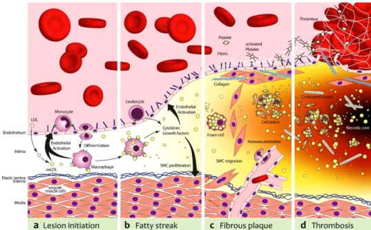
Figure 1.8. Four Stages of Atherosclerotic Lesion Formation...48
Figure 1.9. Role of UPR in the Progression of Atherosclerosis...52
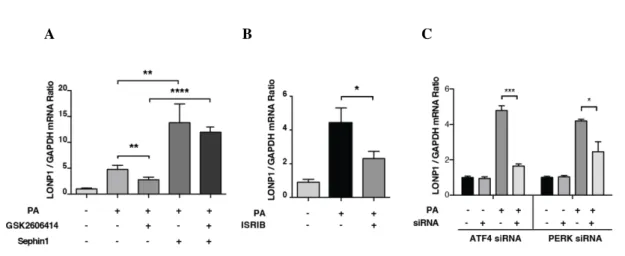
Figure 3.1. PERK expression silencing with siRNA can suppress lipid-induced NLRP3 inflammasome activation in BMDM cells...74
Figure 3.2 Inhibition of PERK kinase activity leads to suppression of lipid- induced NLRP3 inflammasome activation in BMDMs...75
Figure 3.3 Design of PERK_ASKA mice and successful breeding of homozygous mice...76
Figure 3.4 PERK kinase activity is critical for robust activation of the NLRP3 inflammasome by lipids was confirmed in peritoneal macrophages by an alternative approach involving chemical genetics...77
Figure 3.5 Sephin1 prolongs eIF2a phosphorylation and bypassess PERK inhibitor’s suppression of NLRP3 inflammasome...78
Figure 3.6. ISRIB inhibits NLRP3 inflammasome activation in lipid stressed BMDMs...79
Figure 3.7. ATF4 mediates lipid-induced NLRP3 inflammasome activation in BMDMs...80
Figure 3.8. ISR signaling plays a critical role in lipid-induced inflammasome activation in human macrophages...81
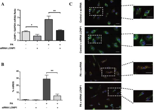
Figure 3.9. Regulation of lipid-induced mtROS production by ISR...83 Figure 3.10 Mitochondrial LONP1 is regulated by ISR in lipid-stressed
macrophages...84 Figure 3.11 Lipid-induced mitochondrial LONP1 is dependent on ISR signaling in human macrophages...85 Figure 3.12 Regulation of mtROS production by LONP1 ... 86 Figure 3.13. LONP1 regulates lipid-induced inflammasome activation kinase...88 Figure 3.14. PERK regulates inflammasome activation through mitochondrial LONP1 and PINK1...89 Figure 3.15. PERK-eif2a-LONP1 signaling axis regulates mitophagy...90 Figure 3.16. Inflammasome regulation via PERK–LONP1 axis is dependent on PINK1-PARKIN-mediated mitophagy...92 Figure 4.1. Inhibition of PERK kinase Activity In Vivo During Athersclerosis
progression...94 Figure 4.2. Metabolic characterization of 30mg/kg/day GSK2606414-treated
mice...95 Figure 4.3. PERK inhibition leads to reduction in atherosclerotic plaque formation in vivo...97 Figure 4.4. Loss of PERK kinase activity reduces of p-eIF2a and LONP1
levels in Apoe-/- mice in vivo...99 Figure 4.5. Loss of PERK kinase activity mitigates hyperlipidemia induced
immune cell accumulation in Apoe-/- mice...101
Figure 4.6. Inhibition of PERK kinase activity reduces hyperlipidemia-induced IL-1b production in aortic plaque macrophages...102 Figure 4.7. Inhibition of PERK kinase activity reduces hyperlipidemia-
induced cleaved caspase1 levels in aortic plaque macrophages...103 Figure 4.8. Inhibition of PERK kinase activity suppresses hyperlipidemia-
Figure 4.9. Analysis of VCAM-1 and Cell Death in Plaques from PERK
kinase-inhibited Apoe-/- Mice...106
Figure 4.10. Loss of Perk kinase activity does not affect smooth muscle cell
migration or collagen secretion...107 Figure 4.11. Loss of Perk kinase activity does not affect numbers of immune
cells in bloodstream...108 Figure 4.12. Inhibition of PERK kinase activity impacts Th lymphocyte
differentiation induced by hyperlipidemia in Apoe-/-...110
Figure 4.13. Plasma Lipid and Lipoprotein Profiles from 30mg/kg/day
GSK2606414-treated and control Apoe-/- mice...111
Figure 4.14. The impact of PERK kinase inhibitior on atherosclerosis
progression...112 Figure 4.15. Experimental Design for Monospecific Inhibition of ASKA mutant in vivo during atherosclerosis development...114 Figure 4.16. Bodyweight and glucose levels in 60mg/kg/day 1-NAPP1 injected Apoe-/- (control) vs ASKA-Apoe-/- mice in vivo...115
Figure 4.17. 1-NAPP1 does not effect atherosclerosis in control Apoe-/- mice...117
Figure 4.18. 1-NAPP1 administration to ASKA-Apoe-/- mice reduced
atherosclerosis development...118 Figure 4.19 PERK kinase activity is critical for hyperlipidemia-induced
eIF2a-LONP1 signaling in vivo...120 Figure 4.20. Inhibition of PERK kinase activity reduces hyperlipidemia-
induced macrophage infiltration in plaques...121 Figure 4.21. Inhibition of PERK kinase activity reduces hyperlipidemia-
induced inflammasome activation in mice...123 Figure 4.22. Inhibition of PERK kinase activity reduces hyperlipidemia-
Figure 4.23. Selective inhibition of PERK kinase activity with 60 mg/kg/day
1-NAPP1 does not affect blood lipid measurements in ASKA-Apoe-/- mice on
western diet...125
Figure 5.1. Experimental design and the efficacy of ISRIB administration in vivo...128
Figure 5.2. Analysis of metabolic parameters in ISRIB-treated Apoe-/- mice...129
Figure 5.3. Blocking ISR by ISRIB alleviates atherosclerosis in Apoe-/- mice...130
Figure 5.4. ISRIB supresses LONP1 expression in Apoe-/- aortic root sections...131
Figure 5.5. ISRIB supresses hyperlipidemia-induced inflammation in Apoe-/- mice...132
Figure 5.6. ISRIB supresses hyperlipidemia-induced IL-1b and cleaved caspase-1 levels in vivo...134
Figure 5.7. ISRIB administration reduces serum and plaque cytokine levels in Apoe-/- mice...135
Figure 5.8. Blockade of ISR with ISRIB does not alter circulating immune cells counts...136
Figure 5.9. ISRIB administration does not affect systemic lipid measurements...137
Figure 5.10. ISRIB Prevents Atherosclerosis Progression...138
Figure 5.11. Experimental design and metabolic characteristics of ISRIB administration in vivo...139
Figure 5.12. Blocking ISR by higher dose of ISRIB alleviates atherosclerosis...140
Figure 6.1. Lipid-induced ISR tegulates mitochondrial clearance, mitochondrial oxidative stress and subsequent inflammasome activation...146
List of Tables
Table 2.1. Chemicals and reagents that are used in laboratory generally...57
Table 2.2. Reagents, chemicals and kits used in cell culture...59
Table 2.3. Antibodies used in all procedures...60
Table 2.4. Study specific Reagents, chemicals and kits...61
Table 2.5. Solutions used in this study...62
Abbreviations
Abbreviation Explanation 1-NAPP1 1-(1,1-Dimethylethyl)-3-(1-Naphthalenyl)-1H-Pyrazolo [3,4-D] Pyrimidin-4-Amine Ab Antibody Ag AntigenAIM Absent in Melanoma-2 Receptors
APC Allophycocyanin
ASC Apoptosis Associated Speck-Like Protein Containing a
CARD
ATF Activating Transcription Factor
ATP Adenosine Triphosphate
BMDM Bone Marrow-Derived Macrophages
CARD Caspase Recruitment Domains
CDDO 2-Cyano-3,12-Dioxo-Oleana-1,9(11)-Dien-28-oic Acid
Methyl Ester
CHOP CCAAT-Enhancer-Binding Protein Homologous Protein
DAMPs Danger-Associated Molecular Patterns
DAPI 4',6-Diamidino-2-Phenylindole
DNA Deoxyribonucleic Acid
ECL Enhanced Chemiluminescence
EDTA Ethylenediaminetetraacetic Acid
eIF2α Eukaryotic Initiation Factor 2
ELISA The Enzyme-Linked Immunosorbent Assay
ER Endoplasmic Reticulum
ERAD ER Associated Degradation
FACS Fluorescence-Activated Cell Sorting
FBS Fetal Bovine Serum
FDA Food and Drug Administration
GADD34 Growth Arrest and DNA Damage 34
Abbreviation Explanation
GSK 2606414 7-Methyl-5-(1-{[3-(Trifluoromethyl) Phenyl] Acetyl}-2,3-
Dihydro-1H-Indol-5-Yl)-7H-Pyrrolo[2,3-D] Pyrimidin-4- Amine
HEPES 4-(2-Hydroxyethyl)-1-Piperazineethanesulfonic Acid
IFN Interferon
IRE1 Inositol-Requiring Enzyme-1
ISR Integrated Signal Response
ISRIB N,N'-Trans-1,4-Cyclohexanediylbis [2-(4-Chlorophenoxy)
Acetamide]
IκB Inhibitor of NF-Κb
i.p. Intraperitonel
KLF Kruppel-Like Factor
LDL Low Density Lipoprotein
LP Lipoprotein
LPS Lipopolysaccharide
MAM Mitochondria Associated Membranes
MAPK Mitogen-Activated Protein Kinase
MDP Muramyl Dipeptidemitochondrial Antiviral Signaling
Protein
MHC Major Histocompatibility Complex
MOMA Monocyte/Macrophage Marker Antibody
MSU Uric Acid Crystals
NK Naturel Killer
NLR Nucleotide Binding And Oligomerization Domain (NOD)-
Like Receptors
PAMP Pathogen-Associated Molecular Patterns
PBS Phosphate Buffer Saline
PDGF Platelet-Derived Growth Factor
PERK Protein Kinase RNA-Like ER Kinase
PMSF Phenyl-methane Sulfonyl Fluoride
PRRs Pattern Recognition Receptors
RNA Ribonucleic Acid
Abbreviation Explanation
siRNA Small Interfering RNA
SMC Smooth Muscle Cell
STAT Signal Transducer And Activator of Transcription
TBS Tris-Buffered Saline
Th Helper T Cell
TLR Toll-Like Receptor
TNF Tumor Necrosis Factor
TRAF TNF Receptor Associated Factor
Treg Regulatory T Cells
TUDCA Tauroursodeoxycholic Acid
TUNEL Terminal Deoxynucleotidyl Transferase dUTP Nick End
Labeling
TXNIP Thioredoxin Interacting Protein
UPR Unfolded Protein Response
UPRE Unfolded Protein Response Element
CHAPTER 1.
Introduction
1.1 The Endoplasmic Reticulum
Endoplasmic Reticulum (ER) is an organelle that is surrounded by a single, continuous membrane. ER membranes spread from the nucleus forming a network of membranes that reaches the plasma membrane. ER is also quite a dynamic organelle with branching tubules (1,2). ER can be subdivided into compartments according to specific functions. Those ER parts that associate with ribosomes during protein synthesis are known as ‘rough’, whereas the other non-ribosome associated parts are known as the ‘smooth’ ER. The ER plays a critical role in modification and folding of secreted and transmembrane protein, which relocate to the Golgi and to the plasma membrane. ER also makes membrane synapses/contacts with organelles such as the mitochondria. The contact sites between the ER and mitochondria are known as the Mitochondria Associated Membranes (MAMs). MAMs are known to have critical roles in metabolism as well as in initiating inflammatory responses (1,3). The functions of all the ER proteins are not known as the identity of ER proteins in the inter-organelle contact sites. The recent proteomic science advances and their application to discovering ER proteins and inter-organelle contact site proteins will generate a better understanding about this organelle and its functions and collaboration with other organelles in the cell (1).
1.1.1 Endoplasmic Reticulum Function
ER is a very important organelle in regulation of intracellular calcium levels as it is the major storage site for cellular calcium, from which controlled calcium release can occur in certain circumstances. Another major role of ER is in protein folding, modification and eradication of mis-folded proteins. Lastly, ER is important metabolic organelle with a major role in the synthesis of major lipid species.
Proper Protein Synthesis, Folding and Maturation
ER plays a critical role in folding of secreted and transmembrane proteins. Rough ER membranes harbor ribosomes that translate these proteins, but these ribosomes are transiently associated with ER membranes and they can be released into the cytosol post-translation. The growing polypeptide chains are transferred from these ribosomes into the ER lumen through membrane-spanning channels. The nascent proteins are folded properly in the ER lumen and and transferred back to ER membrane to be shipped to its target destinations such as plasma membrane, ER and other organelle membranes or to the Golgi, if the protein is to be secreted outside of the cell. ER is not only the place for proper protein folding but also for modifications such as oligomerization of proteins, disulfide bond formation and N-glycosylation, which are required for reaching the protein’s target destination or for function (2,4). If any of these processes go wrong, ER has its own mechanism to destroy these undesired forms of proteins called as ERAD (5).
Endoplasmic Reticulum - Associated Degradation (ERAD)
ERAD is a quality check pathway that prevents the accumulation of unwanted improper folded or immature proteins in ER lumen. Misfolded proteins in ER lumen are transferred back to cytosol, where ERAD acts. Any modifications on these proteins are removed before translocating to the cytosol and they are ubiquitinylated and tagged for proteasomal degradation.
Calcium (Ca2+) Metabolism
ER is a central player in cellular calcium homeostasis (6). As ER is the main calcium storage compartment of the cell, there is a huge difference between calcium
concentration between the ER, cytosol and extracellular compartments (~ 100µM, 100nM, 2nM, respectively) (7).
Calcium is a secondary messenger that regulate a multitude of cellular processes. Calcium is important for re-shaping the ER after neurotransmitter release or after a muscle contraction. Calcium also important for protein function, protein-protein, protein – nucleic acid, protein-organelle interactions (2,8). Another important impact of calcium is on cellular metabolism and homeostasis. For example, increased in calcium transfer into the mitochondria can regulate alter mitochondrial metabolism, membrane permeability, and ATP production. In turn mitochondria calcium concentration can determine cell death or survival (9-11).
As a calcium storing organelle, ER harbors many calcium channels (Sarcoplasmic
Reticulum Ca2+ ATPase (SERCA) pump), inositol 1,4,5-triphosphate receptors (IP3R),
and ryanodine receptors on its membranes that it uses for import and export of calcium based on the cellular need (2,7). Mitochondria also has calcium channels on its outer and inner membranes such as the Voltage Dependent Anion Channel (VDAC) family
of calcium transporters and the mitochondrial Ca2+ uniporter (MCU), respectively (12).
Membrane Lipid Biogenesis
ER is also an important organelle involved in lipid biogenesis. Two key-players of cell membranes, phospholipids and cholesterol, are both synthesized on ER. Together with Golgi and mitochondria, ER forms an endomembrane compartment that is responsible for lipid synthesis. Lipids synthesized on the ER are transported from ER membrane to these organelles, where further modifications take place (3).
To initiate the synthesis of lipid-bilayer cell membrane, two fatty acids are added to glycerol phosphate backbone (providing the diacylglycerol phosphate (DGP) precursor for phospholipids) on the cytosolic side of the ER membrane. Phosphatidylcholine (PC), the most abundant glycerophospholipid is synthesized in the ER and Golgi (3)..ER-mitochondria contact sites are also important locations where phospholipid biosynthesis enzymes such as phosphatidylserine decarboxylases are abundant. Mainly phosphatidylserine (PS) is transported to ER-mitochondria contact sites and PS is converted to phosphatidylethanolamine (PE) and
Phosphatidylcholine (PC) (13,14).
As ER is responsible for the lipid synthesis, it is also responsible for the regulation of genes that are required for lipid synthesis. ER exerts its effects by the Sterol Regulatory Element Binding Protein (SREBP) family of transcription factors that are normally found on the ER but in an inactive state. On the course of lipid synthesis, SREBP is first translocated to Golgi and then to nucleus to initiate transcription of the genes for lipid synthesis and uptake. These transcription factors a re-located to and modified in the Golgi because of their lipid-sensing capabilities. Then they move to the nucleus to play their role in initiating lipid synthesis and maturation (15-17).
1.1.2. The Endoplasmic Reticulum Stress and Unfolded Protein Response
Due to its role in protein folding and maturation of transmembrane and secretory proteins, ER has a tremendous amount of work-load. Unfolded or immature proteins are first transported to ER lumen through its membrane and folding and modifications by the help of chaperons, foldases and cofactors take place in the ER lumen (4). However, a portion of the proteins are not properly folded in the ER. ER has intrinsic mechanisms that sense the unfolded proteins and initiate the ERAD response that can eliminate these unwanted or potentially harmful proteins (18,19).
There are conditions that can cause ER dysfunction. Cells often endure metabolic changes that occur due to changing physiological or environmental conditions. These include decreased ATP levels, viral infections, oxidative stress, genetic abnormalities or increased amount of protein synthesis. Also in certain metabolic conditions that cause excess influx of cholesterol or fatty acids (such as in obesity and dyslipidemia) can also disrupt ER functions. The disturbance in ER functions is perceived as ER stress and initiates an elaborate stress signaling pathway that aims to cope with this (19,20). This signaling is known as the unfolded protein response (UPR) (21,22). UPR signaling is initiated by three ER resident transmembrane stress sensor proteins leading to transcriptional and translational changes in the cell. These are inositol-requiring-enzyme-1 (IRE1), protein kinase RNA (PKR) like ER kinase (PERK) and the activating transcription factor 6 (ATF6) (Figure 1.1). Misfolded proteins are sensed through protein-protein interactions with the stress sensors. These sensors normally
bind Binding-immunoglobulin protein / Glucose Regulated Protein 78 (Bip/Grp78) chaperone within the ER lumen. Upon ER stress, Bip dissociates from the sensors to associate with the misfolded proteins, initiating the UPR cascade. If ER stress is prolonged and the re-association of chaperone with stress sensors is delayed, then ER can adopt two measures: First ER can activate a survival response that involves transcriptional and translational mechanisms to induce the production of ER chaperon proteins to reduce misfolded proteins and re-gain ER homeostasis. However, if ER cannot re-establish cellular homeostasis by this measure, then the same stressor proteins engage apoptotic cascades to eliminate the stressed cell (21,23,24).
Figure 1.1 Endoplasmic Reticulum Stress and Unfolded Protein Response 1.2. Unfolded Protein Response Signaling
Unfolded protein response is directed by three ER transmembrane proteins, IRE1, PERK and ATF6.
1.2.1. IRE1
IRE1 is the first UPR kinase that was identified in yeast ER membranes and is conserved across the species. In mammals, however, there are two IRE1 homologs, namely IRE1a and IRE1b. The expression of IRE1b is limited only to respiratory and gastrointestinal tract, but IRE1a is ubiquitously expressed.
IRE1 is composed of three domains: luminal, transmembrane and cytosolic domains. The cytosolic domain harbors two enzymatic activities, namely kinase and endoribonuclease (RNase) activities. The domain that senses misfolded or unfolded proteins is the luminal domain of IRE1. Upon sensing, IRE1 oligomerizes and auto-phosphorylates. The RNase domain activation requires autophosphorylation. The RNase domain can initiate either death or survival pathways according to the nature and the duration of ER stress. The classically known IRE1 RNase domain substrate is X-box protein 1 (XBP1) messenger RNA(mRNA)(25). When IRE1 is activated, it splices XBP1 mRNA leading to deletion of a 26-base portion of an intron and frameshift. Spliced XBP1 (sXBP1) mRNA produces a larger protein, which is an active transcription factor. sXBP1 protein can activate transcription of genes such as chaperons or components of ERAD pathway to relieve ER stress (26,27). IRE1 was recently shown to also degrade mRNAs that associates with the ribosomes for translation. This leads to decrease in the protein load in the ER and is known as the IRE0dependent decay (RIDD) pathway (35,36).
1.2.2. PERK
PERK is another major transmembrane ER stress sensor protein. It is normally found on ER membrane as a monomer and bound to Bip/Grp78. PERK has three domains: The luminal domain mediates stress sensing and is the binding place for Bip/Grp78. There is a transmembrane domain that anchors PERK into ER membranes and PERK’s cytosolic domain harbors kinase activity (28). Under ER stress conditions, when Bip/Grp78 dissociates in order to bind misfolded proteins, the PERK monomers can oligomerize. And auto-phosphorylate itself at threonine-980. After this phosphorylation, the active PERK kinase can phosphorylate its substrate, eukaryotic initiation factor 2 a (eIF2a) on serine 52. Phosphorylation of eIF2a, leads to global translation attenuation via inhibiting cap dependent translation. An exception to this rule is a select group of genes that can be translated through upstream alternative open reading frame (ORF). The activating transcription factor-4 (ATF4) is one of the proteins made during PERK-induced translational attenuation and is an important transcription factor that activates the transcription of homeostatic genes involved in metabolism, oxidative stress, amino-acid import and synthesis, autophagy and survival. The outcome of PERK-ATF4 signaling can be different depending on the
degree of ER stress the duration of the ER stress (28-30). In early stages of stress, PERK-ATF4 signaling acts as a cytoprotective mechanism through inducing the Heme oxidase-1 (HO-1) antioxidant response. This occurs by the induction of activating transcription factor 5 (ATF5), autophagy related 7 (ATG7) and Unc-51 like serine threonine kinase-1 (ULK1). Another protein that plays important role in antioxidant response through PERK signaling is nuclear factor erythroid 2- related factor-2 (Nrf2) in conjunction with kelch like ECH – associated protein 1 (Keap1). Nrf2 is also shown to cross-talk with nuclear factor k B (NFkB); in the absence of Nrf2, NFkB is inhibited. PERK directly interacts with Nrf2 to exert antioxidant effects (31). However, if ER stress cannot be resolved, then PERK–ATF4 signaling pathway induces C/EBP-homologous protein (CHOP), which inhibits B-cell leukemia 2 (BCL-2) family proteins and induce BH3-only proteins, leading to apoptosis. eIF2a phosphorylation is temporary and it is dephosphorylated sometime after ER stress initiation by protein phosphatase-1 regulatory subunit015A (PP1R15A) or growth arrest and DNA damage-34 (GADD34), which is transcriptionally induced by CHOP. Therefore, the role of eIF2a dephosphorylation is instating ER homeostasis by re-activating the attenuated protein translation (32).
PERK has been shown to be activate the NFkB inflammatory pathway. PERK exert its effects on this pathway through a translational regulation mechanism, in which phosphorylation of eIF2a blocks translation of the inhibitory protein kB (IkB), in turn releasing NFkB transcription factor which goes to nucleus to activate pro-inflammatory genes (33).
In summary, PERK is a multifunctional kinase that can impact a multitude of cellular responses including inflammation, antioxidant effects, protein synthesis, autophagy, apoptosis and thereby, can contribute to cellular homeostasis and determine cell fate.
1.2.3. ATF6
ATF6 is the third transmembrane ER stress sensor that acts as a transcription factor when activated. ATF6 is a multi-domain protein consisting of a luminal domain, which binds to Bip/Grp78 to sense misfolded proteins. It also has a cytosolic domain, which has a bZIP motif (a DNA binding motif), and transmembrane domain, which contains
Golgi translocation signal to target Golgi for proteolytic cleavage (34,35). Activation of ATF6 takes place in the Golgi. After ER stress, ATF6 protein is transported to Golgi by ER –budded vesicles that contain the coat protein complex II (COPII) protein aiding in the translocation to Golgi. When ATF6 is transferred to Golgi, it undergoes two proteolytic cleavages by the membrane-bound transcription factor 1 and site-2 proteases (S1P and Ssite-2P). S1P cleaves the luminal domain and Ssite-2P cleaves the transmembrane domain, leaving the active cytosolic domain (ATF6-N) that contains bZIP motif. ATF6-N is transported from golgi to nucleus to act as a transcription factor for UPR target genes (36-38). These target genes are important for proper protein synthesis, increasing ER protein folding capacities (chaperons) and decreasing protein-folding load of ER (via ERAD) to activate pro-survival mechanisms (39-42).
1.3. Integrated Stress Response
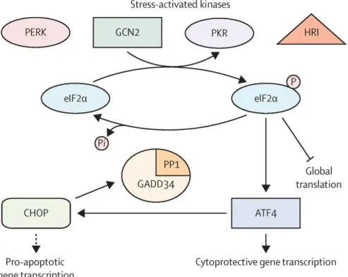
Integrated stress response (ISR) is a multiplex stress signal pathway that is activated in response to both physiological/metabolic demand and in pathological conditions(43-47). The main activators of ISR pathway include heme deprivation, amino acid deprivation, ER stress and viral infections (44,46-50). According to the type of the activator the proximal stress sensing protein (or eiF2a kinase) changes (51). For example, ER stress is sensed via PERK while heme deprivation is sensed via Heme-regulated eIF2a kinase (HRI). On the other hand, viral infections are sensed via the Double-stranded RNA-dependent proteins kinase (PKR) and amino acid deprivation is sensed via the General control non-derepressible-2 (GCN2) kinase (as shown in Figure 1.2) (52). The common and central player in the ISR is eIF2a. All 4 kinases mentioned can phosphorylate eIF2a on serine 51 to initiate the ISR pathway (43). As explained in the previous sections, phosphorylation of eIF2a causes global protein translation block and permits the translation of only select proteins such as ATF4, which favors survival and cellular homeostasis to be re-established (53). However, in severe or prolonged stress conditions ATF4 can switch to a pro-apoptotic gene activation program. As a result, phosphorylation of eIF2a and activation of ATF4 can have opposing effects on the cell depending on the duration of stress response (54-56).
RNAs (tRNA) initiates the ISR pathway in the absence of amino acids (58). It is also shown that glucose deprivation causes GCN2 activation (49). The reason for this may be the higher consumptions of amino-acids due to the absence of glucose to supply the energy needs of cells) (49).
PKR is activated by binding to double stranded RNA (dsRNA), which typically originates from the infecting virus. PKR auto-phosphorylates upon binding dsRNA on threonine 446, and this phosphorylation results in the activation of PKR and eIF2a phosphorylation by PKR(59-63). Consequently, PKR activation leads to the inhibition of viral protein synthesis along with a global translation attenuation (59,64,65). Also, PKR was shown to be activated by other stress conditions such as ER stress, oxidative stress or bacterial infections, but these mechanisms of activation are not clear (66-69). HRI is abundant in erythroid cells (70). HRI activation also happens by dimerization and auto-phosphorylation but upon heme deprivation (71,72). Normally heme bound to HRI and keeps the monomers in an inactive form, inhibiting its kinase activity (70,73). Upon heme deprivation, HRI actively dimerizes and initiates the ISR by phosphorylating eIF2a (70).
PERK is resident on the ER and senses the accumulation of unfolded or misfolded proteins (74,75). PERK also senses oxidative stress, energy, calcium levels, but how this happens is not clear (44). PERK can also be activated during glucose deprivation, whereas ATP deprivation leads to dysregulated ER calcium ATPase pump (76,77). eIF2a is the central player in ISR and also the major component of eIF2 complex, which consists of eIF2b and eIF2g in addition to eIF2a. eIF2 recognizes the translation start site (AUG codon) and binds to mRNA to initiate translation(78,79). eIF2 is part of the pre-initiation complex (PIC) for RNA translation(80,81). eIF2 is complexed with GTP when bound to mRNA in the PIC, but dissociates during translation initiation upon the hydrolysis of eIF2 bound GTP to GDP. The GDP-GTP exchange is executed by eIF2B. However, the activation of ISR and consequent phosphorylation of eIF2a inhibits the eIF2B regulated GDP-GTP exchange and this mechanism prevents 5’ cap dependent translation(78-82). There are a select group of proteins that are preferentially translated via alternative upstream-ORFs (uORF) and include ATF4,
ATF5, CHOP and Growth arrest DNA-inducible protein (GADD34 or PPPR15A) (82-84). ATF4, ATF5, and CHOP are regulatory proteins for cellular homeostasis and GADD34 de-phosphorylates eIF2a. Constitutive repressor of eIF2a phosphorylation (CreP)(PPPR15B) on the other hand is constitutively expressed and bound to GADD34, leading to continuous removal of the phosphate groups on eIF2a(85,86). However, GADD34 is not normally translated but induced as a consequence of ISR and downstream of ATF4. De-phosphorylation of eIF2a is crucial for survival, which requires re-activates the translation (87,88).
ATF4 is a central transcription factor in the ISR and is controlled by transcriptional, translational and post-translational manners during ISR (44). ATF4 is a basic leucine zipper domain (bZIP) type of transcription factor and can dimerize with a variety of partners for differential gene regulation by binding CCAAT-enhancer binding proteins / Activating Transcription factor (C-EBP/ATF) response elements (44,89-92). ATF4 can also form homo or hetero dimers with other DNA binding proteins such as CHOP. This formed hetero-dimer has been shown to activate the transcription of genes including ATF3, ATF5, PPP1R15A (89,93,94). The interaction partner determines how ATF4 will alter the cellular fate (such as survival vs death). There is little ATF4 present in non-stress conditions. It has two ORFs upstream of coding sequence. Normally ribosomes initiate translation of ATF4 through uORF 2, which overlaps with the coding sequence and this prevents the translation of ATF4 mRNA(95) . However, under stress conditions, translation is initiated from uORF1, from which bound ribosomes can detect the start codon in coding sequence (CDS) region of ATF4 (89,95,96). ATF4 is also regulated transcriptionally by factors such as NRF2 that induces ATF4 transcription during oxidative or ER stress(97). Another regulation of ATF4 is post- translational stability. It has a very short half-life - less than 1 hour (98,99). Post-translational modification of ATF4 mainly includes phosphorylation and ubiquitination. ATF4 is phosphorylated on four sites: Serine 219 and serine 224 phosphorylation both cause its proteasomal degradation(98). Serine 245 phosphorylation by ribosomal protein S6 (RPS6K) or serine 254 by protein kinase A (PKA) both induce the activity and stability of ATF4 (100,101).
Figure 1.2 Schematic Representation of ISR
In conclusion, ISR is a very important and complex signaling response that controls global translation and cell death pathways in response to diverse stress stimuli. It is mainly cell protective and seeks to regain cellular homeostasis, however, this pathway can switch the balance from survival to cell death depending on the nature and duration of stress.
1.4. Endoplasmic Reticulum Stress and Immune Response
Unfolded protein response is a critical homeostatic pathway in mammals, however, not all of the stress sensors proteins are conserved across the species. The most conserved and ancient protein in the UPR is IRE1, found in all eukaryotic species from yeast to mammals. However, other UPR sensors, PERK and ATF6, are not found in yeast (102,103). In plants, PERK is expressed, however only IRE1 and ATF6 participate in plant UPR activation. As in mammals, UPR in plants try to controls the protein load, however plants experience different stresses such as heat shock (103). Plant IRE1 is active in abiotic conditions and has a role in antibacterial host defense by increasing selective translation of foldases that aid in the maturation of plant host defense proteins (104,105)
Caenorhabditis elegans is an organism where UPR complexity increases. Here, all three branches of UPR are active (106). The mode of action of proteins are much more similar to mammals. All three arms work for preventing protein load, however, only IRE1 arm causes selective activation of host defense proteins in this organism (106,107).
In mammals, UPR is initially aims to re-instate homeostasis, but it can also engage inflammatory pathways in addition to pro-apoptotic pathways depending on the nature of the stress. Mammalian UPR is also activated with increased amounts of lipids and glucose and plays a role in metabolic stress conditions (108,109). UPR is also activated by pro-inflammatory cytokines (109,110).
Reactive oxygen species (ROS) is an agent that induces inflammation and inflammasome activation in macrophages. ER stress can increase ROS production from both the ER and the mitochondria during metabolic stress conditions (111,112).
ER stress leads to Ca2+ transport from the ER to mitochondria via the MAMs
(113,114). This transfer couples ER and mitochondria by potentiating ROS production.
ER stress and UPR is activated via metabolic stress such as in dyslipidemia and obesity. Also, increased inflammation in these metabolic conditions augments UPR activation. UPR activation in return disrupts metabolism and aggravates the inflammatory response, causing a feed-forward, chronic inflammatory process that underlies metabolic disease progression in obesity, atherosclerosis and diabetes (Figure 1.3) (110,115-118).
Figure 1.3 ER Stress, Inflammation and Metabolic Disease
1.4.1. UPR Connection to Inflammatory Pathways
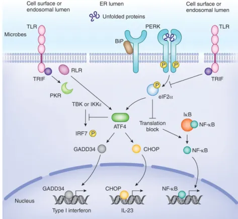
The UPR can engage Toll like receptor (TLR) signaling as well as the NFkB pathway shown in Figure 1.4 (119-121). Nuclear Factor Kappa B(NFkB) pathway is one of the major inflammatory pathways and all three branches of UPR activates NFkB pathway in response to ER stress to induce inflammation (122,123). NFkB is a transcription factor that is normally found in cytosol bound to its inhibitor, IkB, in an inactive state. The activation of NFkB requires first the phosphorylation of IkB by the IkB kinase (IKK) and dissociation of IkB from NFkB, allowing the free NFkB to translocate into the nucleus and initiate pro-inflammatory gene transcription (124). All three branches of UPR can activate inflammation through NFkB pathway, however by different mechanisms. Auto-phosphorylated IRE1a activates NFkB indirectly by recruiting
TNF receptor-associated factor 2 (TRAF2) to itself. TRAF2 then recruits IkB kinase
(IKK) and mediates the phosphorylation of IkB, in turn releasing NFkB to translocate into the nucleus for pro-inflammatory cytokine genes’ transcriptional activation (119). IRE1 can also activate NFkB pathway, via TRAF2-oligomerization domain
containing protein (NODs) to activate NFkB and induce cytokine production (125). PERK, activates NFkB through translational inhibition of IkB. NFkB and IkB are constitutively translated proteins; NFkB enables cells to initiate sudden inflammatory responses while IkB prevents an undesired inflammatory response. The translation inhibition of IkB, during PERK activation, results in the accumulation of larger numbers of NFkB free to translocate into the nucleus for pro-inflammatory cytokine gene transcriptional activation (124,126).
ATF6 activates NFkB pathway in a different way than both IRE1 and PERK. ATF6 induces mTOR pathway and inhibits Akt. Akt in turn phosphorylates IKK to inhibit IkB and release NFkB for further pro-inflammatory cytokine release (127-129)
Figure 1.4 UPR Engages Inflammatory Pathways (130)
PERK and IRE1 initiates inflammatory pathways through additional other mechanisms. IRE1 and PERK can both upregulate Thioredoxin interacting protein (TXNIP), which induces inflammasome activation and inflammatory cytokine release. PERK induces TXNIP through a selectively translated transcription factor (during ER stress) known as the activating transcription factor 5 (ATF5). On the other hand, IRE1 induce TXNIP through regulated IRE1 dependent decay (RIDD) pathway(131). IRE endoribonuclease activity degrades pre-miR17 and inhibits the biogenesis of miR17,
which normally suppresses TXNIP expression (132). IRE1 can also activate immune response through the Mitogen Activated Protein kinase (MAP kinase)Jun N Terminal Kinase1(JNK1)- Activator Protein 1 (AP-1) pathway, which is activated by TRAF2 association with autophosphorylated IRE1 (133,134). Also, the RNase substrate of IRE1, sXBP1, can regulate cytokine transcription (such as TNFa, IL-6 and IFNb) (135,136). PERK also activates inflammation through ATF4 and CHOP, which contribute to IFNb, IL-6, IL-8 and IL-23 transcription (119,137). ATF4 can also bind to IL-6 promoter to activate gene expression (138). Also PERK interaction with nuclear fator erythroid 2-related factor 2 (Nrf2) causes TNFa production (139) (Figure 1.5).
Figure 1.5 Relationship Between PERK Signaling Pathway and Inflammation
1.4.2. UPR Connection with Inflammasome Activation
Inflammasome
The pattern recognition receptors (PRR) are expressed in immune cells (such as macrophages, dendritic cells and neutrophils) as well as in epithelial cells and airway resident smooth muscle cells. These receptors bind to either damage associated
molecular patterns (DAMP) or pathogen associated molecular patterns (PAMP) and activate an innate immune response for clearance of damaged cells or pathogens. Extracellular ATP can serve as a DAMP that activates PRRs as does intracellular accumulation of reactive oxygen species (ROS), cholesterol crystals, uric acid crystals and intracellular release of the mitochondrial DNA. PAMPs include stimuli such as bacterial lipopolysaccharides (LPS), bacterial endotoxins, or pathogen associated RNA and DNA molecules. PRRs that recognize these PAMPs and DAMPs are many and differ in their function and structure. These PRRs are: Nucleotide binding and oligomerization domain (NOD) -like receptors (NLRs), absent in melanoma-2 receptors (AIMs) and pyrin. Upon recognizing the DAMPs and PAMPS, PRR leads to the assembly and activation of cytoplasmic inflammatory response complexes that are called ‘inflammasomes’ (140,141). NLR family of receptors can be subdivided into two according to the effector proteins. Nucleotide binding and oligomerization domain (NOD) -like receptors family of PRRs contains a pyrin domain (NLRP), whereas nucleotide binding and oligomerization domain (NOD) -like receptors family of PRRs contains a caspase activation and recruitment domain (CARD) (NLRC). NLR family of PRRs can activate the inflammasome complex by binding both PAMPs and DAMPs (140,141).
After assembly of inflammasome complex in the cytosol, pro-IL-1b and pro-IL-18 are subjected to proteolytic cleavage and maturation by the mature caspase-1(142). Mature IL-1b and IL-18 activate other inflammatory pathways and lead to pro-inflammatory cytokine release, recruitment of pro-inflammatory cells, and T cell polarization (143). Best known NLRP and NLRC members of inflammasomes are NLRP1, NLRP3 and NLRC4, which are activated via different stimuli. For example NLRP1 inflammasome is mainly activated by direct biding of the bacterial muramyl dipeptide (MDP) or bacterial anthrax toxin (144). NLRC4 inflammasome on the other hand needs an adaptor, the apoptosis inhibitory protein (NAIP), to sense and bind ligands such as bacterial flagellin (145,146). As opposed to NLRP1 and NLRC4, NLRP3 inflammasome has multiple activators that are DAMPS (such as cholesterol crystals, ATP, ROS, uric acid crystals, PAMPs (such as peptidoglycans, bacteria and virus originated nucleic acids, bacterial, fungal and viral toxins) as well as environmental effectors (such as aluminum crystals, asbestos and silica) (147).
Inflammasome activation is also detected in a variety of metabolic diseases such as the fatty liver disease, obesity, and diabetes, in which metabolites such as free cholesterol and fatty acids have been shown to activate NLRP3 (148).
The last member of inflammasome family, AIM2 inflammasome, also binds to caspase-1 through the apoptosis-associated speck-like (ASC) Pyrin Domain Containing-Caspase activation and recruitment domain (PYD-CARD) domain. Upon caspase-1 maturation and activation as in the other inflammasome complexes, cleavage and maturation of pro-IL-1band IL-18 occurs. AIM2 however, is activated via different stimuli such as cytoplasmic nucleic acids that can bind its hematopoietic interferon-inducible nuclear antigens with 200 amino acid repeats (HIN200) domain (149).
Among all the inflammasomes NLRP3 is the one that is most studied in literature. NLRP3 inflammasome activation in cell culture for experimental purposes requires two signals. First signal is needed for the activation of NFkB pathway and for transcriptional upregulation of pro-IL-1b and pro-IL-18. This first signal is called priming step. The most frequently used PAMPs for this purpose is lipopolysaccharide (LPS), which is a bacterial cell wall component that binds to TLR4 receptors to activate NFkB signaling pathway. The activation of NFkB pathway is also the trigger for NLRP3 inflammasome assembly on mitochondria-ER associated membranes (MAMs)(141,150,151). Second signal comes from the DAMPs such as extracellular ATP, cholesterol crystals, monosodium urate crystals (MSU) or increased levels of ROS. The second signal leads to translocation of the assembled NLRP3 inflammasome complex to the cytosol, which results in the activation of caspase-1 and cleavage and maturation of pro-IL-1b and pro-IL-18 (151,152).
ER stress, UPR and Inflammasome
Thioredoxin – interacting protein (TXNIP) has been recently shown to be induced by ER stress and UPR activation. TXNIP is a protein regulated by glucose metabolism and its expression is shown to be increased in diabetes. In literature, TXNIP was shown to induce NLRP3 inflammasome activation (131,132,153). Based on these publications, TXNIP induction occurs after prolonged ER stress and is one of the
molecular links that connects UPR signaling to inflammasome activation. IRE1a induces TXNIP through by cleaving and degrading pre-miR17, which is an inhibitor of TXNIP expression. By depleting miR17, IRE1 induces TXNIP expression during prolonged ER stress. As TXNIP causes inflammasome activation under ER stress, it is a link that connects IRE1 to inflammasome activation (132). In addition to this mechanism, IRE1a can directly induce NLRP3 inflammasome components transcription and activation under lipid induced ER stress in macrophages without altering TXNIP levels (154).
PERK also induces TXNIP under ER stress via promoting selective translation of ATF5, which binds to TXNIP promoter region to activate TXNIP’s transcription, providing a link between PERK and inflammasome activation (131). Another study revealed that NLRP1 inflammasome can be induced through ATF4, which is also downstream to PERK (155). On the other hand, another study described PERK is dispensable for inflammasome activation(156,157). While these contradictory findings exist, it is also not well understood how metabolic stress and in particular, saturated fatty acid-induced stress, is mediated to the inflammasome through UPR activation.
1.5. Endoplasmic Reticulum and Mitochondria
Mitochondria
Mitochondria are essential organelles in cells with important roles in maintaining cellular homeostasis not only by being a cellular energy power house but also as an organelle that is involved in regulating cellular metabolism and immune responses (158). These vital functions are conducted on mitochondria’s membrane and inter-membrane spaces. Mitochondria are comprised of outer inter-membrane, interinter-membrane space, inner membrane and the mitochondrial matrix. Citric acid cycle takes place in mitochondrial matrix whereas oxidative phosphorylation is carried out on the inner mitochondrial membrane (159). Mitochondria also has its own DNA, which encodes, together with the nuclear DNA, a total of 13 mitochondrial proteins (160).
As mitochondria is the place for respiration and subsequent oxidative phosphorylation,
and superoxide radicals (O-) are produced. These radicals are purged with the help of scavengers in healthy mitochondria, however, if mitochondrial dysfunction and resulting membrane potential loss occurs, these free radicals and ROS are released from mitochondria, leading to cellular oxidative stress. This oxidative stress can cause or potentiate stress in the neighboring ER s(111,112). The release of ROS through dysfunctional mitochondria as well as release of the mitochondrial DNA (mtDNA) from the disrupted mitochondrial membranes can also lead to inflammasome activation and further stress for the cell (161-163).
The Relationship of ER and Mitochondria
ER and mitochondria are similar as they are both dynamic organelles, which can modify their structures and their functions in response to changing environmental conditions and signals. ER and mitochondria are connected functionally through calcium and lipid metabolism and physically through ER-mitochondria membrane contact sites (MAMs) (13,164). Some of the MAM proteins identified include ion channels and calcium transporters (such as voltage dependent anion channels
(VDAC), inositol 1,4,5-triphosphate receptors (IP3Rs) and Ca2+ ATPases),
mitochondrial fusion proteins, and ubiquitin ligases. Among these, the mitochondrial fusion proteins help stabilizes the physical contacts between ER and mitochondria(165). Finally, both PERK and IRE1 can be found at the MAMs (166,167).
One of the most important duties of MAMs is Ca2+ transport between ER and
mitochondria. Some portion of the Ca2+ that resides in ER in normal conditions is released to the cytoplasmic space for mitochondrial uptake. Calcium is released by
IP3R calcium receptors, whereas mitochondrial uptake is done by VDAC or Ca2+
uniporters (168).
Another protein at the MAMS and for the stabilization of MAMs is a molecular chaperon known as mitochondrial Grp75, which stabilizes the IP3R at MAMs. These IP3R are which are very prone to proteasomal degradation, but Grp75 forms a bridge between IP3R and VDACs to stabilize the calcium transfer between ER and mitochondria (169). IP3Rs are stabilized on the mitochondria side with Grp75,
wherease, Sigma-1 Receptor (Sig1R) and Grp78/Bip stabilizes it from the ER side, allowing for a prolonged ER mitochondria calcium signaling (170).
Other poteins and chaperons that are found at the MAMs are calreticulin and calnexin as well as Phosphofurin Acidic Cluster Sorting Protein 2 (PACS2). Calreticulin and calnexin are ER molecular chaperons that regulate the IP3R controlled calcium
release. They also interact with Sarcoplasmic Reticulum Ca2+ ATPase (SERCA) pump
that regulates calcium import into the ER. Calnexin and PACS2 also serve to stabilizes the MAMs (171-173).
Prolonged ER stress leads to increase Ca2+ release from ER to mitochondria, disrupts
the mitochondrial membrane potential and leading to depolarization of inner membrane and the release of cytochrome c release into the cytosol. This in return, activates B-cell lymphoma 2 associated X (Bax)/ B-cell lymphoma 2 homologous antagonist killer (Bak)-regulated apoptosis. Also apoptosis protease -activating factor 1(Apaf1)/ caspase-9 related pathway of apoptosis is activated through this mechanism (174).
1.5.1. Mitophagy and Inflammation
Autophagy is an organelle clearance process in cell, basically eliminating non-functional or problematic organelles via formation of double-membrane encapsulated autophagosomes that fuse with lysosomes for lysosomal degradation of the cargo (175). Mitophagy is a form of autophagy that specifically clears the dysfunctional mitochondria (158). In mammals, two different pathways are used for mitophagical clearance of damaged mitochondria as shown in Figure 1.6. These are ubiquitin dependent and ubiquitin independent (receptor mediated) signaling cascades (158,176).
Ubiquitin Mediated Mitophagy
Phosphatase and tensin homolog (Pten)-induced putative kinase-1 (PINK1), a serine-threonine kinase, and Parkin, an E3 ubiquitin ligase, are two important effector proteins that regulate the Ubiquitin-dependent mitophagy. Disruption of mitochondrial membrane potential, increase in mitochondrial calcium concentration and enhanced release of mitochondrial ROS blocks constitutive degradation of PINK1 by


![[Tevfik Fikret ve Galatasaray futbol takımı oyuncularına ait fotoğraf]](data:image/gif;base64,R0lGODlhAQABAIAAAP///wAAACH5BAEAAAAALAAAAAABAAEAAAICRAEAOw==)