(CANCER RESEARCH 58. 1451-1455. April I. 1998]
Prognostic Value of P53 Gene Mutations in a Large Series of Node-negative
Breast
Cancer Patients
Nicole Palette, Marie-Pierre Paperin, Isabelle Treilleux, Anne-Christelle Gratadour, Nadine Peloux, Hervé
Mignotte,
Nigel Tooke, Esfir Löfman,Mats Inganäs,Alain Bremond, Mehmet Ozturk, and Alain Puisieux1
Unitéd'Oncologie Moléculaire. UnitéINSERM U453 ¡N.F., M-P. P.. M. O.. A. P.]. Departement ¡l'Anatomie et de Cytologie Pathologiques ¡I.T.. N. P.¡.ami Département ¡le Chirurgie Carcinologiatie ¡A-C.G., H. M., A. BJ. Centre LéonBérard,69373 Lyon Cedex 08, France: Pharmacia Biotech AB. S-75182 Uppsala. Sweden /N. T.. E. L, M. l.¡;and Department of Molecular Biology and Genelic\. Bilkent University. 06533 Ankara. Tnrkev IM. O.J
ABSTRACT
The most important subgroup ol breast cancer patients for which reliable prognostic factors are needed are women without axillary lymph node involvement. Although overall, these patients have a good prognosis, it is known that 20-30% will experience a recurrence of the disease. To determine the prognostic significance of P53 tumor suppressor gene mu tation, specimens from 113 primary breast cancers were evaluated for the presence of /'5.Õalterations, as detected by cDNA sequencing of the entire coding sequence of the gene. The median follow-up for patients was 105 months. P53 gene mutation was an independent prognostic marker of early relapse and death. Our results suggest that P53 gene mutations could be an important factor to identify node-negative patients who have a poor prognosis in the absence of adjuvant therapy. Prospective studies should be designed to determine which therapy should be performed in this subgroup of patients.
INTRODUCTION
Tumor involvement of the axillary lymph nodes has been the primary parameter in making decisions on adjuvant therapy for breast cancer patients because it is known that these patients have both a high risk for recurrence and a shorter overall survival time. However, 20-30% of lymph node-negative breast cancer patients will relapse with a local recurrence or distant metastasis ( 1). Reliable prognostic markers are needed to help clinicians identify this subset of patients and arrive at more rational treatment decisions. The importance of identifying high-risk patients at diagnosis is supported by the evidence of an improved survival after chemotherapy for specific subsets of patients with lymph node-negative breast cancer (2, 3). A variety of molecular genetic changes have been described in breast cancer. Oncogene and tumor suppressor gene alterations have been studied in an attempt to define the molecular correlation of prognosis and the clinical behavior of breast cancer phenotype (4). Among those, the P53 tumor suppressor gene has become the focus of intensive studies. The current and most powerful model of wild-type p53 function is one in which p53 monitors the genome for DNA damage (5). After treatment of cells with DNA damaging agents, p53 protein levels are increased by posttranslational stabilization and can transactivate var ious genes that may be related to cell cycle arrest or apoptosis (6). Arrest of cell cycle progression following DNA damage is thought to represent a basic protective mechanism preventing replication of damaged template DNA. If damage is irreparable, the cell may be driven to the apoptotic pathway, thus preventing replication of defec tive cells. Mutations in the P53 tumor suppressor gene are the most frequent known genetic alterations in all human cancers (7). Most of the biologically significant mutations impair the ability of p53 to participate to the maintenance of genomic stability. Consequently.
Received 9/2/97; accepted 2/2/98.
The coses of publication of this article were defrayed in part by the payment of page charges. This article must therefore be hereby marked advertisement in accordance with 18 U.S.C. Section 1734 solely to indicate this fact.
1To whom requests for reprints should be addressed, at Unitéd'Oncologie Molécu laire, UnitéINSERM U453. Centre LéonBérard.28 rue Laennec, 69373 Lyon Cedex 08. France.
tumors lacking normal p53 might be expected to be prone to delete rious mutations and to be more aggressive clinically. Many studies have examined the association between breast cancer prognosis and p53 protein expression in tumor cells (8-16). The use of IHC2 was based on the fact that missense mutations normally result in an increased half-life of the protein product and a consequent accumu lation of the mutant p53 protein in the nucleus. Differences in meth odologies and variability in the frequency and intensity of immuno-reactivity with p53-specific antibodies may explain discrepant results reported for the prognostic significance of p53 accumulation within the same tumor type (8, 11-15). In contrast, there have been only a few reports on P53 gene alterations in large series of breast cancers. Nevertheless, recent studies reported the prognostic value of P53 mutations in nonselected breast cancers comprising both node-nega tive and node-positive tumors (17. 18). In our study, the complete coding region of the P53 gene was sequenced from 113 node-negative breast cancers. In parallel, immunohistochemical studies were per formed using two monoclonal antibodies directed against p53. The P53 status was then related to prognosis for relapse-free survival and overall survival, with a median duration of follow-up of 105 months.
MATERIALS AND METHODS
Patient Population. Entered in this study were randomly selected tumor samples from 113 breast cancer patients treated and followed up at the Center LéonBérard.All patients had a primary breast tumor and hislopathologically verified carcinoma-free axillary lymph nodes. The median age of patients at the time of diagnosis was 59 years (range. 32-87 years). Patients had been surgically treated by mastectomy or lumpectomy and axillary dissection. Postoperative radiotherapy was given to 47 patients. Adjuvant chemotherapy consisting primarily of three courses of i.v. 5-fluorouracil. Adriamycin. and methotrexate was offered to all patients with a tumor >30 mm (n = 27 patients). Adjuvant hormonal therapy (tamoxifen) was given to postmeno-pausal patients having positive estrogen receptor tumors (n = 57).
The mean follow-up was 103 ±34 months for patients still alive (median follow-up. 105 months). All patients treated for breast cancer were seen on a regular basis every 6 months for 5 years: after 5 years, they were seen on a yearly basis. The follow-up evaluation consisted of a clinical evaluation, physical examination. X-ray procedures, and serum chemistry tests.
Tumor samples were collected at the Department of Pathology of the Center LéonBérard.Tumors were pure histological variants of invasive breast carci nomas comprising 98 ductal. 10 lobular. 2 mucinous. I tubular. 1 cribriform, and 1 squamous cell metaplastic carcinomas. Tumor si/.e was recorded as the maximum tumor diameter in a fresh mastectomy specimen. The median size of collected tumors was 25 mm (range. 7-70 mm). Histopathological type and grade of the carcinomas were reevaluated according to WHO criteria and Scarff-Bloom-Richardson grading, respectively (19). Results of the cytosolic estrogen and progesterone receptor assays (cutoff. 10 fmol/mg of protein), which had been performed on snap-frozen tumor samples by the dextran-coated charcoal method, were available from patient records in 112 cases. A sample was carefully selected from the most viable part of the tumor to be snap frozen in liquid nitrogen, transferred, and stored at —¿70°Cuntil analysis. A slide was generated to estimate the percentage of tumor cells. All samples studied contained 60% or more malignant cells.
: The abbreviations used are: 1HC. immunohistochemistry: RH. relative ha/ard. 1451
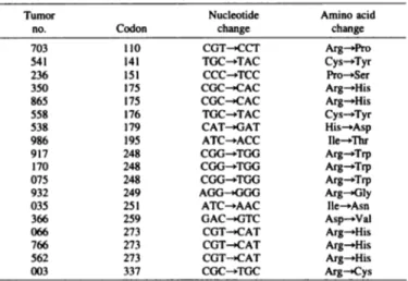
Table 1 Location and sequence change of P53 mutations Tumor no.7035412363308655585389X691717(1075»32035366(166766562003Codon11014115117517517617919524«248248249251254273273273337NucleotidechangeCGT^CCTTGC^TACCCC-»TCCCGC--CACCGC-^CACTGC-»TACCAT->GATATC^ACCCGG-^TGGCGG^TGGCGG^TGGAGG^GGGATC-»AACGAC-XÎTCCGT->CATCGT->CATCGT^CATCGC->TGCAmino acid changeArg— >ProCys^TyrPro— »SerArg->HisArg— »HisCys-»TyrHis— »AspIle—ThrArg->TrpArg-»TrpArg— »TrpArg— »GlyIle^-AsnAsp->ValArg-»HisArg— »HisArg— »HisArg->Cys
This retrospective study included 26% of tumors from node-negative pa tients treated at the Center LéonBérardbetween 1983 and 1989. The studied population was representative of node-negative breast cancer patients treated at the Center LéonBérardduring this period (median age of patients at the time of diagnosis. 57 years; median tumor size of invasive breast carcinomas, 22 mm; histopathological types: ductal carcinomas, 74%; lobular carcinomas, 8%; others, 18%).
IHC of Paraffin-embedded Tissue. Intracellular p53 protein levels were evaluated in 112 tumors by immunohistochemical analysis. Four-/xm sections from Bouin-fixed. paraffin-embedded blocks were deparaffmized in toluene, transferred to 100% and 95% ethanol, and air dried. They were then treated with 5% hydrogen peroxide for 20 min to exhaust endogenous peroxidase. Tissue sections were placed in sodium citrate solution (0.01 M. pH 6.0) and then incubated three times for 5 min each in a 800-W microwave oven. After preincubation in 1% BSA in PBS. sections were incubated for 90 min at 37°C with mouse monoclonal p53 antibodies DO-7 (Dako Corp.) or PAblSOl (Oncogene Science) diluted 1:50, washed, and reacted with streptavidin-biotin-peroxidase reagents (Dako) and diaminobenzidine chromogen. Sections were slightly counterstained with hematoxylin. The entire slides were microscopi cally evaluated separately by two investigators. The localization and the intensity of the staining and the percentage of positive-stained tumor cells were estimated by each investigator. When the values determined by the two investigators differed by greater than 5%, the score was decided by joint examination. A threshold of 5% cells staining was chosen to score tumors as IHC positive. In all series, a set of breast cancers with high levels of p53 was used as the positive control. Negative controls were obtained by omission of the primary antibody.
Sequence-based Analysis of P53. The method was essentially as described by Sjogren et al. (20). RNA was prepared from the frozen tumors under stringent conditions to avoid degradation and contamination. This was fol lowed by an enzymatic conversion of the RNA to cDNA. P53 was amplified from the tumor cDNA by the PCR using four overlapping primer pairs covering the complete coding region of the P53 gene. Biotin-labeled PCR products were generated with one of the primers (in each pair) being modified with a biotin molecule, which facilitated solid-phase sequencing. Solid-phase sequencing was carried out using AutoLoad solid phase sequencing combs and T7 DNA polymerase (Pharmacia Biotech). The sequencing products generated were analyzed using an automated laser fluorescence ALF DNA sequencer (Pharmacia Biotech). The limit for hétérozygotedetection in wild-type back ground was determined to be 25%.' All mutations were confirmed by ream-plifying the relevant cDNAs and sequencing the new PCR products.
Statistical Methods. Probability of survival was estimated with the Kaplan-Meier method. The equality between survival curves was tested with the log rank test. The Cox proportional hazards model was used to estimate the effect of P53 mutations on overall survival, with several prognostic factors taken into account simultaneously. The analyses were carried out using the
JMP software for Macintosh (version 3.2) from the SAS institute (Cortex AB, Solna, Sweden).
RESULTS
Detection and Characterization of P53 Mutations. A total of
113 breast cancer RNA samples were analyzed by cDNA sequencing. Mutations were detected in tumors from 18 (16%) patients (Table 1). All mutations were missense mutations. Thirteen of them were located within evolutionarily conserved domains of P53. G:C to A:T transi tions were predominant (12 of 18 mutations). This type of alterations is thought to be involved in spontaneous mutations (7). Codons 110,
175, 248, 273, and 337 (CGN), which encode arginine, accounted for 10 of the 18 mutations.
Immunohistochemical Analysis of p53 Protein Accumulation.
112 tumors from the total were examined by IHC for p53 protein overexpression using antibodies PAblSOl (p53IHC-PAbl801) and DO-7 (p53IHC-DO7). The localization and the intensity of the stain ing and the percentage of positive cells were assessed without knowl edge of clinical outcome and P53 mutation status. p53 labeling was restricted to tumor cell nuclei. Cytoplasmic staining, as reported by other authors (21), was not observed in our series. In agreement with previous reports (22, 23), we found a discrepancy between the im-munoreactivity of the different p53 monoclonal antibodies. Thirty tumors were positive using DO-7, although only 16 of them expressed a mutant P53 gene. Using PAblSOl, overexpression of the p53 protein was found in 13 tumors. Ten of these contained mutant P53. Thus, a significant fraction of IHC-positive tumors did not show any gene alterations by sequencing. It is noticeable that some of these tumors displayed up to 70% positive cells. Therefore, as previously reported, the proportion of p53-stained cells is not an exact represen tation of the number of cancer cells bearing a mutation within a tumor (24).
P53 Mutations and Pathobiological Variables. Table 2 shows the distribution of some tumors and patients characteristics by P53 status, x2 analysis revealed significant associations between the pres ence of P53 mutations and high histological grade (P < 0.001), lack of estrogen receptors (P = 0.0044), and lack of progesterone receptors (P = 0.0392). No correlation was observed between P53 gene muta tion and the age at diagnosis or tumor size.
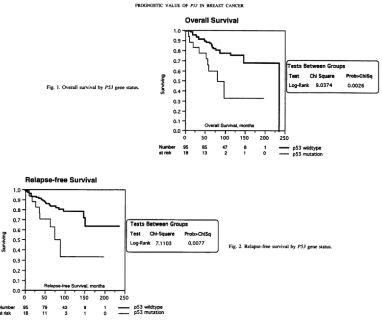
Survival Analysis. Kaplan-Meier curves demonstrated signifi
cantly shorter overall (P = 0.0026) and relapse-free (P = 0.0077) survival for patients who had a primary tumor with P53 alterations (Figs. 1 and 2). Only 60% of patients with P53 mutations were alive 5 years after surgery, compared with 88% for those lacking detectable mutation. Together with patient age, P53 status was the only factor that predicted survival. In univariate analyses, tumor size (RH, 0.98; P = 0.28). estrogen receptor (RH, 0.79; P = 0.42), progesterone receptor (RH, 0.65; P = 0.13). and histological grade (RH, 0.92; P = 0.78) had no significant power. The RH associated with a P53 mutation was 1.82 (95% confidence interval, 1.08-3.06; P = 0.02). The detection of a gene mutation by cDNA sequencing had a sub stantially greater prognostic value than the observation of a p53
Table 2 Distribution of Iunior and patient characteristics bv P53 ßenestatus
' N. Tooke and E. Löfman,unpublished data.
Covariate (no. of patients)Estrogen
receptor (112), a 10 fmol/mg Progeslerone receplor (112), alO fmol/mg Grade III versus grade I or II (1 10) Age, yr (113) Tumor size (100)PS3 mutationn %8 44.4 8 44.4 14 87.5 58.2 ±13.3° 29.47 ±6.74"Wild-type P53n %74 79.5 65 69.8 34 36.2 58.8 ±12.4" 27.39 ± 12.17"" Mean value ±SD. 1452
PROGNOSTIC VALUE OF P5.ÃŒIN BREAST CANCER
Overall Survival
Fig. 1. Overall survival by PSJ gene status.
Number at risk I.U -0.9- 0.8- 0.7- 0.6- 0.5- 0.4-0.3- 0.2-0.1 -^Overall S""Hurvival,monthsTests Between Groups Test Chi Square Prob>ChlSqLog-Rank 9.0374 0.00260.0 -| i | i | i | i | i 0 50 100 150 200 250ir 95 85 47 8 1 p53 wildtype 18 13 21 0 p53 mutation
Relapse-free Survival
Number al risk IJBâ„¢0.9-0.8-n 7 —¿U. i0.6 -0.5-0.4-0.3-0.2-0.1 -tj_VvY^-inihhRelapse-free
Survival, monthsI ' I ' 1 ' 1 'Tests Between GroupsTest Chi-Square Prob>ChiSqLog-Rank 7.1103 0.00770 SO 100 150 200 250:r 95wildtype1879 43 9 1 ^— p53 11 3 1 0 P53 mutationFig. 2. Relapse-free survival by P53 gene status.
protein accumulation by IHC. RHs associated with p53 accumulation detected by DO-7 and PAb 1801 were 1.53 (P = 0.05) and 1.63 (P = 0.10), respectively. As previously mentioned, in line with other investigators (25-27), we chose a threshold of 5% cells staining to score tumors as IHC-positive. This subjective choice was based on the decision not to take into consideration scattered single p53-positive cells that could reflect microenvironmental stresses in individual cells rather than a widespread change throughout the neoplastic cells. As shown in Table 3, depending on the antibody, the variation of the cutoff affected in a different way the prognostic significance of p53 protein accumulation. This confirms that p53 immunostaining de pends greatly on the conditions and reagents used in the test and illustrates the need for optimization of each individual antibody used in immunohistochemical studies.
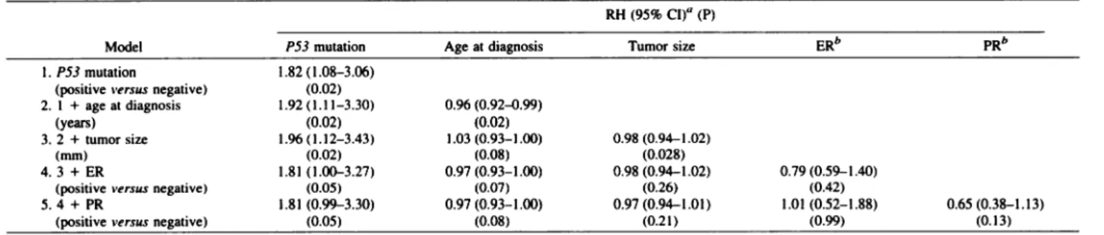
Next, we investigated whether P53 status maintained its prognostic value for survival in the presence of information provided by other variables. The variables used for the multivariate analysis were age at diagnosis, tumor size, and estrogen and progesterone receptors. Infor mation on 100 patients fulfilled the criteria set by the program. As shown in Table 4, P53 mutation was an independent prognostic factor for survival, with RHs of the same magnitude in both univariate and multivariate models. When the same parameters (age at diagnosis, tumor size, and estrogen and progesterone receptors) were considered, the prognostic value of p53 protein accumulation was statistically
significant only for p53IHC-PAbl801 (cutoff. 1%; RH, 1.83; P = 0.04). Tests using other thresholds of positivity and/or the DO-7 antibody had no prognostic significance in multiple regression analysis.
DISCUSSION
To date, seven independent studies, using different methodologies, evaluated the prognostic value of genetic alterations of the P53 tumor suppressor gene in invasive breast cancers (17, 18, 23, 26, 28-30). All but one (30) observed a strong association of P53 mutation with poor survival. The median clinical follow-up of these studies varied from
Table 3 Cav picpeffforal lui-ani ttnivariatf analysis of p53 protein accumulatimi Impact of the cutoff CJc = percentage of p53-positive cells) on the prognostic value of p53IHC-DO-7 and p53IHC-PAbl801.
Cutoff, 1%Cutoff. 5%Cutoff. 10%DO-7 RH (95% CD" <P)1.19(0.76-1.87)(0.45)1.53(0.99-2.39)(0.05)1.60(1.01-2.56)(0.05)PAb(P)2.01(95% CI)1801 RH (1.20-3.37)(0.008)1.63(0.91-2.96)(0.10)1.75 (0.91-3.39)(0.09)"
CI. confidence interval.
Table 4 Cox multivariale analysis of risk factors for overall sun'ival RHs obtained in the Cox models with increasing number of covariates.
Model1 . P53.mutation(positive versus negative)2. 1 + age at diagnosis(years)3. 2 + tumor size(mm)4. 3 + ER(positive irr.vu.v negative)5.4 + PR(positive VÎT.VH.Õnegative)P53 mutation1.82(1.08-3.06)(0.02)1.92(1.11-3.30)(0.02)1.96(1.12-3.43)(0.02)1.81 (1.00-3.27)(0.05)1.81 (0.99-3.30)(0.05)Age at diagnosis0.96 (0.92-0.99)(0.02)1.03(0.93-1.00)(0.08)0.97(0.93-1.00)(0.07)0.97(0.93-1.00)(0.08)RH (95% CI)" (P)Tumor size0.98(0.94-1.02)(0.028)0.98(0.94-1.02)(0.26)0.97(0.94-1.01)(0.21)ER6PR*0.79(0.59-1.40)(0.42)1.01(0.52-1.88) 0.65(0.38-1.13)(0.99) (0.13) ' CI, confidence interval; ER, estrogen receptor; PR, progesterone receptor.
'Cutoff. 10 fmo!/mg of protein.
24 to 60 months. To our knowledge, our study is the first one conducted on a large series of node-negative breast cancer patients (n = 113) with a prolonged clinical follow-up (median duration of follow-up, 105 months). A follow-up of patients over a long time course is of great importance considering the relative good prognosis of breast cancer in patients without axillary lymph nodes invasion. Using a cDNA-based sequencing method, we searched for mutations in the entire coding region of the P53 gene. The frequency of P53 missense mutations (16%) is in concordance with data reported by others (17). Although missense mutations are the most frequent alter ations in the P53 gene, nonsense mutations, deletions, and insertions have been recently reported at an unusual frequency in breast cancers (17, 18). The lack of detection of these types of mutation in our study could be either linked to the low number of detected mutations or due to a differential pattern of mutations among populations, as suggested previously (27, 31). p53 protein accumulation was detected in a number of tumors expressing wild-type P53. as assessed by cDNA sequencing. False-negative sequencing results may occur as a conse quence of contamination of tumor samples by adjacent normal tissue. However, the manner in which the tumor material was isolated in this study should have minimized such risk. Another explanation is the presence of mutations in noncoding regions of the gene. Alternatively, the accumulation of wild-type p53 may occur in response to a variety of stresses including DNA damage or hypoxia. Following the latter hypothesis, one could expect that the nuclear accumulation of wild-type p53 in tumor cells would be a predictive factor of favorable outcome. Further studies are necessary to test this hypothesis. How ever, it is noticeable that all four patients with a negative sequence tumor displaying >40% stained cells by IHC, using the DO-7 anti body, did not relapse and were still alive by the end of the study (mean follow-up of these patients, 104 months).
Our data clearly show that the detection of a P53 gene alteration constitutes a factor of poor prognosis. By the end of our study period, 50% of patients with mutant P53 had died, in contrast to 23% of patients with wild-type P53. More strikingly, among patients who did not receive adjuvant systemic chemotherapy, 50% (5 of 10) of the patients with mutant P53 died within 5 years after surgery, in contrast to only 9% (7 of 75) of the patients with wild-type P53. This observation indicates that among breast cancers usually classified as tumors of good prognosis, P53 mutations identify a subpopulation of aggressive tumors. Which therapy should be offered to these patients? Two recent nonrandomized studies, one based on cDNA sequencing and the other on IHC. provided indirect evidence of a beneficial effect from radiotherapy in preventing local relapse among node-negative patients with tumors displaying P53 abnormality (25, 32). In these studies, most of the patients were treated either by surgery only or by surgery and locoregional radiotherapy. Considering the possible im
pact on overall survival, the benefit from a systemic therapy (chem otherapy and/or hormonotherapy) should also be evaluated. It is known that antiestrogens and genotoxic drugs used in chemotherapy modulate p53 activity (33, 34). However, in spite of numerous studies, the relationship between the P53 status and the activity of anticancer agents remains unclear. There are theoretical arguments for both increased and decreased chemosensitivity in tumors with mutant P53 (35-37). In our study, based on the size of the tumor, 27 patients were treated with chemotherapy at the time of diagnosis. Eight of these patients had a tumor with mutant P53. Among them, six were still alive 5 years after surgery. The overall survival was not significantly different (P = 0.3547) from the patients with wild-type P53 who received a similar treatment. In contrast, P53 mutation was a signif icant prognostic factor for overall survival (P = 0.0065) among patients who did not receive systemic chemotherapy. Obviously, no conclusion can be drawn from these small subgroups of patients. Considering the high prognostic power of P53 mutations, prospective and randomized trials should be designed to evaluate the benefit of node-negative patients with a mutant P5.i-expressing tumor from specific adjuvant therapies.
ACKNOWLEDGMENTS
We thank Eric Tabone and Isabelle Richard for their technical assistance and Nathalie Borei for the preparation of the manuscript.
REFERENCES
1. Bonadonna. G. Conceptual and practical advances in the management of breast cancer. J. Clin. Oncol.. 7: 1380-1397, 1989.
2. Early Breast Cancer Trialist's Collaborative Group. Systemic treatment of early breast cancer by hormonal, cytotoxic or immune therapy: 133 randomized trials involving 31000 recurrences and 24000 deaths among 75000 women. Lancet, 339: 1-15. 1992.
3. Fisher, B., Dignam. J., Mamounas, E. P., Costantino, J. P.. Wickerham, D. L., Redmond. C. Wolmark N.. Dimitrov, N. V.. Bowman. D. M., Glass, A. G.. Atkins, J. N., Abramson. N.. Sutherland. C. M.. Aron. B. S., and Margolese. R. G. Sequential methotrexate and fluorouracil for the treatment of node-negative breast cancer pa tients with estrogen-receptor-negative tumors: eight-year results from National Sur gical adjuvant breast and bowel project (NSABP) B-13 and first report of findings from NSABP B-19 comparing methotrexate and fluorouracil with conventional cyclophosphamide, methotrexate, and fluorouracil. J. Clin. Oncol.. 14: 1982-1992,
1996.
4. Bieche, I., and Lidereau, R. Genetic alterations in breast cancer. Genes Chromosomes Cancer, 14: 227-251, 1995.
5. Lane, D. P. p53, guardian of the genome. Nature (Lond.). 358: 15-16, 1992. 6. Gottlieb, T. M.. and Oren, M. p53 in growth control and neoplasia. Biochim. Biophys.
Acta., 1287: 77-102, 1996.
7. Hollstein. M.. Sidransky, D., Vogelstein. B., and Harris. C. p53 mutations in human cancers. Science (Washington DO. 253: 49-53. 1991.
8. Varley, J. M., Brammar. W. J.. Lane, D. P., Swallow, J. E.. Dolan, C., and Walker. R. A. Loss of chromosome 17p 13 sequences and mutation of p53 in human breast carcinomas. Oncogene, 6: 413-421, 1991.
PROGNOSTIC VALUE OF P5.ÃŒIN BREAST CANCER
9. Isola. J.. Visakorpi. T., Holli. K.. and Kallioniemi. O. P. Association of overexpres- 24. sion of tumor suppressor protein p53 with rapid cell proliferation and poor prognosis in node-negative breast cancer patients. J. Nati. Cancer. Inst.. 84: 1109-1114, 1992. 10. Thor, A. D.. Moore, II, D. H., Edgerton, S. M., Kawasaki. E. S.. Reihsaus. E.. Lynch.
H. T., Marcus. J. N., Schwartz. L., Chen, L. C., Mayall. B. H., and Smith. H. S. 25. Accumulation of p53 tumor suppressor gene protein: an independent marker of prognosis in breast cancers. J. Nati. Cancer Inst.. 84: 845-855. 1992.
11. Allred, D. C, Clark, G. M., Elledge, R., Fuqua. S. A.. Brown, R. W., Chamness, G. C., Osborne, C. K., and McGuire, W. L. Association of p53 protein expression 26. with tumor cell proliferation rate and clinical outcome of node-negative breast cancer. J. Nati. Cancer. Inst., 85: 200-206, 1993.
12. Thor. A. D., and Yandell DW. Prognostic significance of p53 overexpression in node-negative breast carcinoma preliminary studies support cautious optimism. 27. J. Nati. Cancer. Inst., 85: 176-177, 1993.
13. Poller, D. N.. Mulchings. C. E.. Galea. M.. Bell. J. A.. Nicholson. R. A.. Elston. C. W., Blarney. R. W.. and Ellis. I. O. p53 protein expression in human breast carcinoma: relationship to expression of epidermal growth factor receptor. c-erbB-2 protein overexpression, and estrogen receptor. Br. J. Cancer. 66: 583-588. 1992.
14. Silvestrini. R.. Benini. E.. Grazia Daidone. M., Veneroni, S.. Boracchi, P.. Cappelletti. 28. V., Di Fronzo. G., and Veronesi. U. p53 as an independent prognostic marker in lymph node-negative breast cancer patients. J. Nati. Cancer Inst.. 85: 965-970. 1993. 15. Barnes. D. M., Dublin, E. A., Fisher, C. J., Levison, D. A., and Millis, R. R. 29.
Immunohistochemical detection of p53 protein in mammary carcinoma: an important new independent indicator of prognosis? Hum. Pathol.. 24: 469-476. 1993.
16. Elledge. R. M.. Clark. G. M.. Fuqua. S. A.. Yu. Y. Y.. and Allred. D. C. p53 protein 30. accumulation detected by five different antibodies: relationship to prognosis and heat shock protein 70 in breast cancer. Cancer Res., 54: 3752-3757. 1994.
17. Bergh. J.. Norberg. T.. Sjögren. S.. Lindgren. A., and Holmberg, L. Complete sequencing of the p53 gene provides prognostic information in breast cancer patients, 31. particularly in relation to adjuvant systemic therapy and radiotherapy. Nature Med., /.- 1029-1034. 1995.
18. Kovach. J. S.. Hartmann, A., Blaszyk. H., Cunningham. J.. Schaid. D., and Sommer, 32. S. S. Mutation detection by highly sensitive methods indicates that p53 gene muta tions in breast cancer can have important prognostic value. Proc. Nati. Acad. Sci. USA. 93: 1093-1096. 1996.
19. Elston, C. W.. and Ellis. I. O. Pathological prognostic factors in breast cancer. I. The 33. value of histological grade in breast cancer: experience from a large study with long-term follow-up. Histopathology, IV: 403-410, 1991.
20. Sjögren.S.. Inganäs.M., Norberg. T.. Lindgren. A.. Nordgren. H., Holmberg. L., and 34. Bergh, J. The p53 gene in breast cancer: prognostic value of complementary DNA sequencing versus immunohistochemistry. J. Nati. Cancer Inst., 88: 173-182, 1996. 21. Moll, U. M.. Riou. G.. and Levine, A. J. Two distinct mechanisms alter p53 in breast 35.
cancer: mutation and nuclear exclusion. Proc. Nati. Acad. Sci. USA, 89: 7262-7266, 1992.
22. Cattorelti, G., Rilke, F., Andreola. S.. D'Amato. L., and Delia. D. p53 expression in 36. breast cancer. Int. J. Cancer. 41: 178-183. 1988.
23. Andersen, T. I., Holm, R.. Nesland. J. M.. Heimdal, K. R.. Ottestad. L., and Börresen, A. L. Prognostic significance of TP53 alterations in breast carcinoma. Br. J. Cancer, 37. 68: 540-548. 1993.
Jacquemier, J., Moles, J. P., Penault-Llorca, F.. Adelaide, J., Torrente. M.. Viens. P.. Birnbaum. D., and Theillet. C. p53 immunohistochemical analysis in breast cancer with four monoclonal antibodies: comparison of staining and PCR-SSCP results. Br. J. Cancer. 69: 846-852. 1994.
Silvestrini. R., Veneroni. S.. Benini. E.. Daidone. M. G., Luisi. A., Leutner. M.. Maucione A.. Kenda. R.. Zucali. R.. and Veronesi. U. Expression of p53, glutathione S-transferase-ir. and Bcl2 proteins and benefit from adjuvant radiotherapy in breast cancer J. Nail. Cancer Inst.. 89: 639-645, 1997.
Thorlacius. S.. Thorgilsson. B.. Björnsson. J.. Tryggvadottir. I... Börresen. A. L., Ogmundsdottir. H. M.. and Eyfjörd.J. E. TP53 mutations and abnormal p53 protein staining in breast carcinomas related to prognosis. Eur. J. Cancer, MA: 1856-1861, 1995.
Blaszyk, H., Hartmann, A.. Tamura. Y.. Saitoh. J. M., Cunningham. J. M.. McGovern. R. M., Schroeder. J. J., Schaid. D. J.. Li. K.. Monden, Y., Morimoto. T., Komaki. K.. Sasa. M.. Hirata. K.. Okazaki. M.. Kovach, J. S.. and Sommer. S. S. Molecular epidemiology of breast cancers in northern and southern Japan: the frequency. clustering and patterns of p53 gene mutations differ among these two low-risk populations. Oncogene. 13: 2159-2166. 1996.
Elledge. R. M.. Fuqua. S. A.. Clark. Ci. M.. Pujol. P., Allred. D. C. McGuire, W. L. Prognostic significance of p53 gene alterations in node-negative breast cancer. Breast Cancer Res. Treat.. 26: 225-235, 1993.
Seshadri, R.. Leong. A. S., McCaul, K.. Figaira, F. A.. Setlur. V.. and Horsfall. D. J. Relationship between p53 gene abnormalities and other tumour characteristics in breast-cancer prognosis. Int. J. Cancer. 69: 135-141. 1996.
Caleffi. M., Teague, M. W., Jensen. R. A.. Vnencak-Jones. C. L.. Dupont. W. D., and Pari, F. F. p53 gene mutations and steroid receptor status in breast cancer. Clinico-pathologic correlations and prognostic assessment. Cancer (Phila.l. 73: 2147-2156. 1994.
Sommer. S. S . Cunningham. J.. McGovem. R. M.. Saitoh. S., Schroeder. J. J.. Wold. L. E.. and Kovach. J. S. Pattern of ft53 gene mutations in breast cancers of women of the midwestem United States. J. Nati. Cancer Inst., 84: 246-252. 1992.
Jansson, T., Inganäs.M.. Sjögren. S.. Norherg. T.. Lindgren. A., Holmberg, L„and Bergh. J. p53 status predicts survival in breast cancer patients treated with or without postoperative radiotherapy: a novel hypothesis based on clinical findings. J. Clin. Oncol., 13: 2745-2751. 1995.
Guillot. C., Falene, N.. Courtois. S.. Voellzel. T., Garcia, li.. Ozturk. M., and Puisieux, A. Alteration of p53 damage response by lamoxifen treatment. Clin. Cancer Res., 2: 1439-1444, 1996.
Kastan. M. B., Onyekwere, O.. Sidransky. D.. Vogelslein, B., and Craig, R. W. Participation of p53 protein in the cellular response to DNA damage. Cancer Res., 51: 6304-6311. 1991.
Lowe, S. W.. Bodis. S., McClatchey. A. M.. Remington. 1... Ruley. H. 1:., Fischer, D. A.. Housman. D. E.. and Jacks. T. p53 status and the efficacy of cancer therapy m vivo. Science (Washington DC). 266: 807-810. 1994.
Fan, S., Smith, M. L., Rivet, D. J.. II. Duba. D.. Zhan. Q., Kohn. K. W., Fornace, A. J., Jr.. and O'Connor. P. M. Disruption of p53 function sensitizes breast cancer MCF-7 cells to cisplatin and pemoxifylline. Cancer Res.. 55: 1649-1654. 1995. Hawkins. D. S.. Demers. G. W.. and Galloway. D. A. Inactivation of p53 enhances sensitivity to multiple chemotherapeutic agents. Cancer Res.. 56: 892-898, 1996.