99
Journal of Neurological Sciences [Turkish] 31:(1)# 39; 099-106, 2014
http://www.jns.dergisi.org/text.php3?id=745 Research Article
C1-C2 Posterior Segmental Fixation For Traumatic Atlantoaxial Instability
Olcay ESER1, Ergün KARAVELIOĞLU2, Mehmet Erhan TÜRKOĞLU3
1
Balıkesir University, Department of Neurosurgery, Balıkesir, Türkiye 2Afyon Kocatepe University, Department of Neurosurgery, Afyonkarahisar, Türkiye 3Düzce University,
Department of Neurosurgery, Düzce, Türkiye Summary
Cervical spinal trauma is one of the most frequent causes of disability. Upper cervical spinal trauma, caused by more than one mechanism, is a complex injury which may show various neurological statements. Atlantoaxial fractures are seen frequently after motor vehicle accidents and falls from height. Various treatment modalities defined in the upper cervical spine trauma and surgical treatment technique, so there is still unclear. Seven patients, six male and one female, were surgically treated for symptomatic atlantoaxial instability. The major cause of instability was; motor vehicle accident in six patients and falling from height in one patient. C1-C2 posterior fixation and posterolateral fusion was performed in 7 cases. The fusion and C1-C2 stability were achieved in all cases after 6 months. C1-C2 posterior fixation and fusion technique is a reliable and safe method to treat atlantoaxial instability when followed the given instruction by spinal surgeons. But, further prospective clinical studies will be needed to determine the effectiveness of this technique.
Key words: Atlantoaxial instability, C1 lateral mass- C2 pedicle screw, instrumentation Travmatik Atlantoaksiyel Instabilitede C1-C2 Posterior Segmental Fiksasyon Özet
Servikal travmalar sakat kalmanın en sık nedenlerinden biridir. Birden fazla mekanizmayla oluşan üst servikal travma değişik nörolojik bozukluklar gösterebilen kompleks bir yaralanmadır. Atlantoaksiyal kırıklar sıklıkla trafik kazlarından ve yüksekten düşmelerden sonra görülür. Üst servikal travmalarda değişik tedavi modaliteleri ve cerrahi tedavi teknikleri tanımlanmıştır fakat hala tedavi yöntemi karışıktır. Altı erkek bir bayan toplam 7 hasta semptomatik atlantoaksiyal instabilite nedeniyle cerrahi olarak tedavi edildi. Altı hastada atlantoaksiyal instabilite nedeni trafik kazası, bir hastada ise yüksekten düşmeydi. 7 hastada C1-C2 posterior fiksasyon ve posterolateral füzyon yapıldı. 6 ay sonra tüm hastalarda C1-C2 stabilizasyon ve füzyon sağlandı. C1-C2 posterior fiksasyon ve füzyon tekniği, spinal cerrahların verdiği açıklamalara uyulduğu sürece atlantoaksiyal instabiliteleri tedavi etmek için emniyetli ve güvenli bir metottur. Bu tedavinin etkinliğini belirlemek için daha fazla prospektif klinik çalışmalara ihtiyaç vardır.
Anahtar Kelimeler: Atlantoaksiyel instabilite, C1 lateral kitle-C2 pedikül vidası,
100
INTRODUCTION
Fracture of the cervical spine (C0-C1-C2) are frequent(17). More than 60% of spinal injuries involve the cervical spine(24). Fracture of C1 account for approximately 2-15% of acute cervical fractures, while fractures of C2 account for approximately 17-25%(17). Odontoid fractures are
consisting of 10-15 % of all cervical spine fractures while 60 % of them are type-2 fractures. Also combined fractures of C1 and C2 constitute 3% of all cervical spine fractures(5). Cervical spinal trauma is one of the most frequent causes of disability. Upper cervical spinal trauma, caused by more than one mechanism, is a complex injury which may show various neurological statements.
Atlantoaxial fractures are seen frequently after motor vehicle accidents and falls from height.
Various treatment modalities defined in the upper cervical spine trauma and surgical treatment technique, so there is still unclear. Modern sublaminar wiring technique which is first described by Brooks and Jenkins(1) in 1978 is easy to perform but non union is a well known complication(15). Furthermore sublaminar wiring technique can not provide immediate stabilization(2). Transarticular
atlantoaxial fixation technique, described by Magerl in 1987, is another choice(21). Disadvantages of this technique are; high risk of vertebral artery injury, screw malpozition, instrumentation failure(3,4) and the necessity of a complete reduction of the
C1-C2 before stabilization procedure(3,18,19,27). The technique described by Harms has been widely accepted because it minimizes the risk of injury to the vertebral artery, allows intraoperative reduction and achieve a good fusion rate(13).
The present study describes our clinic's experience with posterior atlantoaxial stabilization using polyaxial screw-rod
system in traumatic patients. Also we investigated the clinical and radiological result of posterior atlantoaxial stabilization.
MATERIAL AND METHODS
Between May 2010 and June 2012, totally seven patients, six male and one female, were surgically treated for symptomatic atlantoaxial instability. In seven patients C1 lateral mass and C2 pedicle screw fixation was performed. Also in 4 patients, the screws were extended to subaxial cervical spine such as C3 and C4 (lateral mass) where necessary. Also in one patient, non-union of a dens fracture after anterior screw fixation was seen and posterior C1-C2 fixation was performed. The average age of the patients was 60 (range 25 to 79) years. The mean follow-up period was one year. The major cause of instability was; motor vehicle accident in six patients and falling from height in one patient. The complains of patients were persistence posterior cervical pain with radiation to the suboccipital area. Preoperative neurological examination was normal in all patients except one with left sided hemiplegia (Table 1). All patients' neutral plain cervical radiographs, Computed Tomographic (CT) scans and Magnetic Resonance Images (MRI) were obtained preoperatively. Preoperative CT scans used to perform the measurement of lateral mass and C2 pedicle screw. Also postoperative cervical plain radiographs and CT scans of the instrumented spinal levels were evaluated for correct screw position (Figure 1 and 2).
Surgical Techniques
The patient was placed in the prone position after the general anesthesia. The neck was held in neutral position by a three-pin head clamp. Reduction of C1-C2 facet joints were achieved with the aid of C-arm fluoroscopy intraoperatively. After preparing the surgical area in the usual fashion, a posterior paramedian incision, including C1 posterior arch and the upper edge of C4 or C5 lamina was made.
101 lateral radiography. Exposure of lateral margins of the facet joints from C2 to C4 or C5 was provided with subperiostal dissection of the cervical paraspinal muscles. The suboccipital bone and the lamina of C1 were identified. Dorsal root ganglion of C2 and venous plexus were dissected. The C1 lateral mass entry point, in the middle of the junction of the C1 posterior arch and the midpoint of the posterior inferior part of the C1 lateral mass, was marked just below the posterior arch by pushing the C2 root caudally with a hook. The sagittal cranio-caudal direction is determined by pointing to the C1 anterior tubercle on lateral C-arm view. The screw follows a posterior to anterior direction with 50-100 of convergence. Venous plexus bleeding was controlled by hemostatic sponge and cottonoids. Following drilling of the entry point of C1
tapping through the entire vertebral body, polyaxial screw was inserted bicortically into the lateral mass of C1. C2 polyaxial pedicule screw was inserted through the cranial and medial quadrant of the isthmus surface of C2 by 200 to 300 with respect to the medial and cephaled directions. Posterolateral fusions were performed with using allograft.
Posterior cervical fusion was extended to C3 vertebra in two patients because of ligamentous injury such as interspinous and supraspinous ligaments. Also posterior cervical fusion was extended to C4 in one patient because of C3-C4 retrolisthesis and facet fracture.
All patients were mobilized on the first postoperative day with a Philadelphia collar.
Table 1: Data and characteristics of patients
Case Age Sex Trauma Pre-op
Symptomos Diagnosis Surgery 1 38 M Motor vehicle accident Left Hemiplegia
Atlantoaxial rotator dislocation, transverse ligament injury
C1-C2 posterior fusion
2 60 M Fall Down Neck Pain Tip 2 odontoid fracture C1-C2 posterior
fusion
3 65 F Motor vehicle
accident
Severe Neck Pain
Tip 2 odontoid fracture, atlantoaxial dislocation
C1-C2 posterior fusion
4 65 M Motor vehicle
accident
Neck Pain Tip 3 odontoid fracture,
transverse ligament injury
C1-C2 posterior fusion 5 79 M Motor vehicle accident Severe neck pain
Jefferson fracture, Tip 2 odontoid fracture
C1-C2-C3-C4 posterior fusion
6 75 M Motor vehicle
accident
Neck pain Tip 3 odontoid fracture, C2
horizontal fracture and transver ligament injury
C1-C2-C3 posterior fusion
7 75 M Motor vehicle
accident
Neck Pain Tip 2 odontoid fracture, C1
posterior arch fracture
C1-C2-C3 Posterior fusion and anterior Odontoid fixation
102
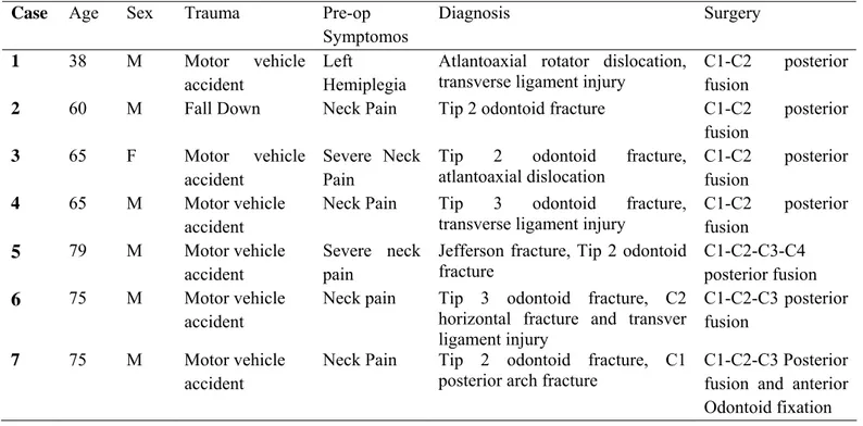
Figure 1: A-B Preoperative CT scan of case 4. C Post operative control cervical radiographs.
103
RESULTS
All the patients were admitted to the hospital after the trauma and atlantoaxial instability was diagnosed before surgery by radiological investigations. 7 patients were treated with C1-C2 posterior fixation with posterolateral fusion. In one case, anterior odontoid screw fixation was performed previously. Because of non-union after 3 months, posterior C1-C2 fixation with posterolateral fusion was performed in this patient.
The mean operation time was 150 minutes (90-210 minutes); mean blood loss was 450 ml (180-820 ml).
All of the patients had been used Philadelphia collar after the operation for 6
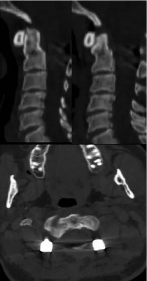
weeks. There were no mortality and morbidity. There were no neurological or vascular complications associated with all surgical procedures. Also any postoperative complication such as wound infection was observed. All patients relieved from posterior cervical pain with suboccipital radiation. The neurologic sequela of one patient did not improve post operatively and the patient was transferred to rehabilitation unit. The patients were followed up with clinical and radiographic examination at 3, 6 and 12 months (Figure 3). Satisfactory screw positions were noted on postoperative CT scans. We did not perform any revision surgery. The fusion and C1-C2 stability were achieved in all cases after 6 months.
104
DISCUSSION
It is well known that the atlantoaxial segment is the most mobile region of the vertebral column. Approximately 50 % of the rotation of the cervical spine occurs at the C1-C2 joint. The stability of atlantoaxial joint is mainly provided by the ligamentous structures and dens. Various conditions such as trauma, inflammatory disease and congenital malformations implicated in the development of atlantoaxial instability. Instability of atlantoaxial joint can cause to pathologic motion with neurologic sequela. Atlantoaxial arthrodesis that reduces deformity and provides stability is a life saving procedure. Because instability of atlantoaxial joints cause sudden death due to compression over the spinal cord. Because of the clinical importance and complex anatomy, different fixation and fusion techniques have been reported in the literature for the treatment of atlantoaxial instability. Formerly, a posterior wire stabilization technique which was described by Brooks and Jenkins was used to treat atlantoaxial instability(16). Although this technique is simple, safe and less dangerous to the nerve root and vertebral artery, it requires an intact C1 arch. Also it can not provide immediate stability, limits the flexion and extension of the cervical spine. Non-union rates is up to 30% even with halo vest immobilization and neurological injuries because of sublaminar passage of wires
(2-4,12-15-17,19-24,27). C1-C2 transarticular screw fixation
techniques which was described by Magerl, requires reduction of C1-C2 before stabilization. Also prominent anatomy such as vertebral foramen precludes placement of the screws in approximately 20% of patients so it is important to evaluate preoperative CT scans before the screw placement(22). The probability of neurovascular compromise during implantation of transarticular screws is very high(12). Also the risk of
vertebral artery injury is an incidence of 2.2% per transarticular screw and 4.1% per operated patient in Magerl technique(3,13). Recently, Goel et al. reported a new technique, involving C1 lateral mass and C2 pedicle screw which is popularized by Harms et al.(9,10,13). The advantages of Harms technique are; a) minimal risk of vertebral artery and spinal cord injury b) it can be applied if C1-C2 is not reduced or posterior elements of C1-C2 are absent or present c) it is not required postoperative using of halovest. Also this technique can be incorporeted with other cervical spine fusion constructs. The bony fusion rates range from 75% to 80% in posterior wire stabilization technique and 90% in Magerl technique while fusion rate is 100% in Harms technique. Also in our cases, we achieved the %100 C1-C2 bony fusion. In all types of C1-C2 posterior stabilization, it is possible to damage the vertebral artery especially in patients with high-riding vertebral artery. In our series, there was no vertebral artery injury. Also either Harms or Goel did not reported vertebral artery or spinal cord injury after screw placement while individual authors reported vertebral artery injury using Magerl technique in 3,7-8,2% of patients(11,14). The authors attribute this to
better direct visualization of screw insertion than in the Magerl technique(9,13). It is important to know the anatomy and variations of C1-C2 complex prior to surgery. Preoperative CT scans, especially three-dimensional CT reconstruction is mandatory to evaluate venous and vertebral artery anatomy, bone structure and also for safe screw insertion in the C1-C2 complex(23,28).
Recent years using the navigation system for the risk of vessel and spinal cord injuries in spinal surgery is much popular. But using navigation system in craniocervical junction surgery has some disadvantages because of high mobility of
105 navigation system, bending of instruments and non-rigid connection between the reference base and the actual surgical site are other disadvantages of intraoperative navigation system in craniocervical junction surgery(26). Due to this advantages, most neurosurgeons use conventional intraoperative fluoroscopy. While Liu et al(18). were reported that C1 lateral mass screws could be inserted without fluoroscopy with microscope assistance on a series of 24 consecutive patients, Simsek et al.(25) demonstrated that unicortical C1 lateral mass screws could be inserted safely and rapidly without fluoroscopy guidance in 17 consecutive patients.
Bleeding from the venous plexus around the greater occipital nerve may occur during the dissection of the C1-C2 inter-laminar space for C1 screw insertion. Bipolar coagulation in combination with tamponade is necessary to control bleeding. Also prompt insertion of screw into the C1 lateral mass is useful for stopping bleeding. Caudal retraction or sectioning of the C2 ganglion for C1 screw placement is controversial. The C2 ganglion occupies 76% of the foraminal height of the C2, between the arch of the atlas and the lamina of the axis so sectioning of the C2 ganglion and bipolar coagulation of the venous plexus provide wide exposure of the lateral mass entry point, thus, facilitating the placement of the C1 lateral mass screws(25). While Harms et al(13). avoided leaving the nerve intact and retracting it caudally; Goel et al(10). reported that patients tolerated the dissection of the nerve quite well. Harms et al.(13) reported that the risk of injury or
irritation of the C2 nerve root can be avoided by traction of the root inferiorly during screw insertion. However Goel et al(10). reported no significant clinical symptoms with sectioning the C2 ganglion sharply to gain wide exposure of the C1 lateral mass entry point on 160 patients. We performed subperiostal dissection
nerve intact in all cases.
CONCLUSION
C1-C2 posterior fixation and fusion technique is a reliable and safe method to treat atlantoaxial instability when followed the given instruction by spinal surgeons. This study has some limitations. First, in this study, the patients were not randomized and there were limited number of patients. Also all patients had trauma. There were no patients with inflammatory disease, cancer metastasis or congenital malformations. Also the relatively short follow-up time is the second study limitation. Therefore, further prospective clinical studies will be needed to determine the effectiveness of this technique.
Correspondence to:
Ergün Karavelioğlu
E-mail: [email protected]
Received by: 12 March 2013 Revised by: 30 October 2013 Accepted: 06 December 2013
The Online Journal of Neurological Sciences (Turkish) 1984-2014
This e-journal is run by Ege University Faculty of Medicine,
Dept. of Neurological Surgery, Bornova, Izmir-35100TR
as part of the Ege Neurological Surgery World Wide Web service.
Comments and feedback: E-mail: [email protected] URL: http://www.jns.dergisi.org
Journal of Neurological Sciences (Turkish) Abbr: J. Neurol. Sci.[Turk]
106
REFERENCES
1. Brooks AL, Jenkins EB. Atlanto-axial arthrodesis by the wedge compression method. J Bone Joint Surg Am 1978;60(3):279–84 2. Calısaneller T, Yılmaz C, Özdemir Ö, Caner H.
Posterior atlantal lateral mass fixation technique with polyaxial screw and rod fixation system. Turkish Neurosurgery 2008;18(2): 142-148
3. Coric D, Branch CL Jr, Wilson JA, Robinson JC. Arteriovenous fistula as a complication of C1–2 transarticular screw fixation: Case report and review of the literature. J Neurosurg 1996;85:340–3.
4. Coyne TJ, Fehlings MG, Wallace MC, Bernstein M, Tator CH. C1–C2 posterior cervical fusion: Long-term evaluation of results and efficacy. Neurosurgery 1995;37:688–92; discussion 692
5. Dickman CA, Hadley MN, Browner C, Sonntag VK. Neurosurgical management of acute atlas-axis combination fractures. A review of 25 cases. J Neurosurgery 1989;70:45-49.
6. Dickman CA, Crawford NR, Paramore CG. Biomechanical characteristics of C1-2 cable fixations. J Neurosurg 1996;85:316–22. 7. Farey ID, Nadkarni S, Smith N. Modified
Gallie technique versus transarticular screw fixation in C1–C2 fusion. Clin Orthop 1999:359;126–35.
8. Glaser JA, Whitehill R, Stamp WG, Jane JA. Complications associated with the halo-vest. A review of 245 cases. J Neurosurg 1986;65:762–9.
9. Goel A, Laheri V. Plate and screw fixation for atlantoaxial subluxation. Acta Neurochir (Wien) 1994;129:47–53.
10. Goel A, Desai K, Mazumdar D. Atlantoaxial fixation using plate and screw method: a report of 160 treated patients. Neurosurgery 2002;51:1351–6.
11. Haid Jr RW, Subach BR, McLaughlin MR, Rodts GE Jr, Wahliq Jb Jr. C1–C2 transarticular screw fixation for atlantoaxial instability: a 6-year experience. Neurosurgery 2001;4:65–8.
12. Hajek PD, Lipka J, Hartline P, Saha S, Albright JA. Biomechanical study of C1–C2 posterior arthrodesis techniques. Spine (Phila Pa 1976) 1993;18:173–7.
13. Harms J, Melcher PR. Posterior C1-C2 fusion with polyaxial screw and rod fixation. Spine 2001;26(22);2467-71.
14. Hott JS, Lynch JJ, Chamberlain RH, Sonntag VK, Crawford NR. Biomechanical comparison of C1–2 posterior fixation techniques. J Neurosurg Spine 2005; 2:175–81.
15. Jacobson ME, Khan NS, An HS. C1-C2 Posterior Fixation: Indications, Technique, and Results. Orthop Clin N Am 2012; 43: 11–18. 16. Jeon SW, Jeong JH, Choi GH, Moon SM,
Hwang HS, Choi SK. Clinical outcome of posterior fixation of the C1 lateral mass and C2 pedicle by polyaxial screw and rod. Clin Neurol Neurosurg. 2012 Jul;114(6):539-44.
17. Longo UG, Denaro L, Campi S, Maffulli N, Denaro V. Upper cervical spine injuries: indications and limits of the conservative management in Halo vest. A systematicreview of efficacy and safety. Injury 2010;41(11);1127-35.
18. Liu G, Buchowski JM, Shen H, Yeom JS, Riew KD. The feasibility of microscope-assited ‘free-hand' C1 lateral mass screw insertion without fluoroscopy. Spine 2008;33:1042-49.
19. Madawi AA, Casey AT, Solanki GA, Tuite G, Veres R, Crockard HA. Radiological and anatomical evaluation of the atlantoaxial transarticular screw fixation technique. J Neurosurg 1997; 86:961–968
20. Madawi AA, Solanki G, Casey AT, Crockard HA. Variation of the groove in the axis vertebra for the vertebral artery. Implications for instrumentation. J Bone Joint Surg Br 1997; 79:820–823.
21. Magerl F, Seemann PS. Stable posterior fusion of the atlas and axis by transarticular screw fixation. In: KehrP, Weidner A, editors. Cervical spine 1:Strasbourg 1985. New York: Springer-Verlag; 1987. p. 322–7.
22. Paramore CG, Dickman CA, Sonntag VK. The anatomical suitability of the C1-2 complex for transarticular screw fixation. J Neurosurg 1996;85:221–4.
23. Resnick DK, Lapsiwala S, Trost GR. Anatomic suitability of the C1–C2 complex for pedicle screw fixation. Spine 2002;27:1494–8. 24. Shin JJ, Kim SJ, Kim TH, Shin HS, Hwang YS,
Park SK. Optimal use of the halo- vest orthosis for upper cervical spine injuries. Yonsei Med J. 2010;51(5):648-52.
25. Simsek S, Yigitkanli K, Seckin H, Akyol C, Belen D, Bavbek M. Freehand C1 lateral mass screw fixation technique: our experience. Syrgical Neurology 2009;72:676-81.
26. Tessitore E, Bartoli A, Schaller K,Payer M. accuracy of the freehand fluoroscopy-guided placement of C1 lateral mass and C2 isthmic screws in atlanto-axial instability. Acta Neurochir 2011; 153:1417-25.
27. Wright NM, Lauryssen C. Vertebral artery injury in C1-2 transarticular screw fixation: results of a survey of the AANS/CNS section on disorders of the spine and peripheral nerves.American Association of Neurological Surgeons/Congress of Neurological Surgeons. J Neurosurg 1998; 88:634–640
28. Yoshida M, Neo M, Fujibayashi S, Nakamura T. Comparison of the anatomical risk for vertebral artery injury associated with the C2-pedicle screw and atlantoaxial transarticular screw. Spine 2006;31:E513–7.