287
Case Reports
Relapsed acute myeloid leukemia
presenting with myocardial hypertrophy
and constrictive pericardial physiology
Aynur Acıbuca*, Mahmut Yeral**, Nazım Emrah Koçer***, Zafer Koç****, Hakan Güllü*
Departments of *Cardiology, and **Hematology, ***Pathology, ****Radiology, Faculty of Medicine, Başkent University, Dr. Turgut Noyan Practice and Research Center; Adana-Turkey
Introduction
Extra medullary relapse (EMR) after stem cell transplantation can be seen up to 20% of acute leukemia patients (1). Moreover, EMR rarely occurs in the heart and there are few cases reporting cardiac relapses in leukemia (2-5).
Despite exceedingly rare incidence during clinical follow-up, cardiac leukemic involvement can frequently detected micro-scopically in up to 40% of patients on postmortem examination (6). The ante mortem diagnostic process can be challenging because cardiac symptoms secondary to infections or chemo-therapeutics are similar to the symptoms related with leukemic cardiac infiltration (7). We report a case of cardiac relapse in myeloid leukemia presenting with myocardial hypertrophy and constrictive cardiac physiology, that was diagnosed by pericar-dial biopsy.
Case Report
A 54-year-old male presented to our cardiology department with new onset dyspnea and lower extremity swelling for 3 weeks. There was no history of heart disease. The patient had undergone allogenic peripheral blood stem cell transplanta-tion because of acute myeloid leukemia (AML) type M4 nearly one and a half years ago. Thereafter, he had suffered from EMR and graft versus host disease (GVHD) and had received proper treatment. The bone marrow was in remission during clinical visits.
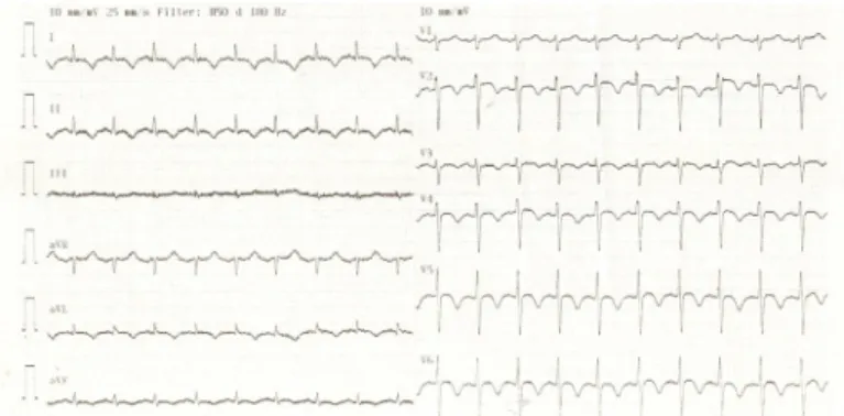
On admission, electrocardiography (ECG) revealed T-wave inversion in the precordial and inferior leads (Fig. 1). The chest radiogram showed increased cardiothoracic ratio. There was no remarkable abnormality except thrombocytopenia (40.00
×
103/µL) in blood tests. The troponin level was normal. Transthoracic echocardiography (TTE) displayed new extensive myocardial hypertrophy, increased pericardial thickness, massive peri-cardial effusion, and left ventricular systolic dysfunction com-pared with TTE done 10 months previously, which showed no evidence of structural heart disease. Pericardiocentesis was
performed because of tamponade signs. The effusion was an exudate and atypical cells were not detected during cytologic examination.
Despite the removal of pericardial effusion, myocardium was observed to be constrictive as if surrounded by a shield leading to systolic dysfunction in echocardiographic views (Video 1-3). The most striking point was diffuse myocardial hy-pertrophy with no clear reason. Also, the patient was still suf-fering from shortness of breath and pedal edema. Therefore, a cardiac magnetic resonance imaging (MRI) was planned to differentiate opportunistic infection, storage disease, or other possible etiologies. The MRI illustrated pericardial and bilateral pleural effusions, pericardial thickening, diffuse left and right ventricular hypertrophy, myocardial and pericardial late gado-linium enhancement, and diffuse left ventricular hypokinesis (Fig. 2 a-2g), which signs were interpreted by the radiologist to may appear as a result of GVHD, leukemic infiltrates, or infec-tious diseases. Therefore, a pericardial biopsy was performed to make an accurate diagnosis. While awaiting the results of pericardial biopsy, blood smear unexpectedly showed atypical mononuclear cells; therefore, a bone marrow biopsy was per-formed. Pathologic examination of pericardium displayed ex-tensive leukemic infiltration. Thereafter, microscopic examina-tion of bone marrow confirmed the recurrence of AML. Despite reinitiation of chemotherapy, the patient died 10 days after the diagnosis.
Discussion
The signs and symptoms of leukemic cardiac involvement are nonspecific and can change according to the infiltrated part of the heart. Therefore, diagnostic process can be challenging. Leukemic infiltration can be presented with heart failure, pericardial effusion, and rarely constrictive pericarditis (4, 5, 8). Cardiac infiltrates of AML can be diffuse as well as massy (3, 9).
In this case, we suspected myocarditis because of systolic dysfunction and remarkable cardiac hypertrophy.
Figure 1. Electrocardiogram illustrate T wave inversion in precordial and inferior leads
Case Reports Anatol J Cardiol 2019; 21: 287-91
288
Also, concomitant pericarditis could explain pericardial signs. However, polymerase chain reaction analysis of common isolated cardiotropic viruses was negative and the troponin level was normal. Therefore, we ruled out active viral myocarditis.
The second likely cause of cardiac signs and symptoms was chemotherapeutic agents. However, he had not been exposed to cardiotoxic chemotherapeutics. His MRI images, illustrating abnormal heterogeneous myocardial infiltration with late gadolinium enhancement can provide a hint to rule out cardiotoxicity that cause diffuse homogenous myocardial involvement (4).
Cardiac amyloidosis was also considered in the differential diagnosis, but it was ruled out because there was no sign of low-voltage in ECG, TTE did not show “speckled” pattern, and MRI findings were not consistent with those of amyloidosis (10). Besides, new myocardial hypertrophy could not be explained by hypertensive heart disease, aortic stenosis, or
hypertrophic cardiomyopathy, when considering clinical and imaging features.
Our case differs from the other cases in terms of the use of only pericardial biopsy to reveal the diagnosis of AML relapse. We preferred pericardial biopsy to endomyocardial biopsy (EMB) because myocardial involvement was heterogeneous and EMB might not reveal a definitive diagnosis in case of being taken from an area without leukemic infiltrates. Besides, myocardial invasive procedures have more potential complications compared to pericardial interventions.
Conclusion
To sum up, when we encounter a patient with hematologic malignancy presenting with heart failure, we should consider leukemic cardiac involvement in addition to cardiotoxic drugs
Figure 2. (a) Axial GE T2 weighted image shows pericardial [yellow arrow] and bilateral pleural [yellow arrow] effusion.
(b) Cine short axis view shows biventricular myocardial [yellow arrow], parietal [white arrow] and visceral pericardial thickening and subendocardial [green arrow] and subepicardial [red arrow] hyperintense rim related with myocardial invasion.
(c) Short axis TSE T2-weighted image shows midwall hyperintense areas [yellow arrow] related with myocardial invasion. (d) Late gadolinium-enhanced four-chamber view shows epicardial [yellow arrow] and pericardial [white arrow] rim enhancement. LA - left atrium, LV - left ventricle, RA - right atrium, RV - right ventricle
a b
Case Reports
Anatol J Cardiol 2019; 21: 287-91
289
or opportunistic infections. Also, leukemic infiltration should be considered in differential diagnosis of cardiac hypertrophy in this patient group. Repetitive cases with different clinical futures will increase our awareness and improve our understanding about leukemic heart disease. Prompt accurate diagnosis and urgent therapy may improve the clinical process of this disease.
Informed consent: An informed consent was obtained from the pa-tient’s wife.
Video 1. Parasternal long axis view illustrating severe myocardial hypertrophy, moderate pericardial effusion, and limitation of myocardial motion.
Video 2. Parasternal short-axis view showing extensive myocardial hypertrophy, moderate pericardial effusion, and systolic dysfunction.
Video 3. Apical four-chamber view displaying disproportionate myocardial hypertrophy, and limitation of diastolic expansion movement of the ventricles.
References
1. Clark WB, Strickland SA, Barrett AJ, Savani BN. Extramedullary relapses after allogeneic stem cell transplantation for acute my-eloid leukemia and myelodysplastic syndrome. Haematologica 2010; 95: 860-3. [CrossRef]
2. Jung MH, Lee YH, Lee KY, Jung HO, Youn HJ. Leukemic infiltration presenting as myocardial hypertrophy after complete remission of acute myeloid leukemia. Echocardiography 2017; 34: 136-8. 3. Facenda-Lorenzo M, Sánchez-Quintana A, Quijada-Fumero A,
Laynez-Carnicero A, Breña-Atienza J, Poncela-Mireles FJ, et al. Cardiac Relapse of Acute Myeloid Leukemia after Allogeneic He-matopoietic Stem Cell Transplantation. Case Rep Oncol Med 2016; 2016: 5091021. [CrossRef]
4. Chang K, Kim DY, Lee KH, Huh J, Kang JW, Shin DY, et al. An iso-lated cardiac relapse after allogeneic hematopoietic stem cell transplantation for acute lymphoblastic leukemia. Korean J Intern Med 2017; 32: 753-7. [CrossRef]
5. Hori T, Suzuki N, Mizue N, Hatakeyama N, Takamuro M, Tsutsumi H. Relapse of T-cell all after stem cell trans- plant presenting as hypertrophic cardiomyopathy: the value of non-invasive diagnos-tic imaging in detecting cardiac leukemia. Pediatr Blood Cancer 2006; 46: 108-11. [CrossRef]
6. Roberts WC, Bodey GP, Wertlake PT. The heart in acute leukemia: A study of 420 autopsy cases. Am J Cardiol 1968; 21: 388-412. 7. Barbaric D, Holley D, Lau KC, McCowage G. It is ALL in the heart: A
patient with acute lymphoblastic leukemia and cardiac infiltration at time of diagnosis. Leuk Lymphoma 2002; 43: 2417-9.
8. Butany J, Nair V, Naseemuddin A, Nair GM, Catton C, Yau T. Car-diac tumours: diagnosis and management. Lancet Oncol 2005; 6: 219-28. [CrossRef]
9. Makaryus AN, Tung F, Liu W, Mangion J, Kort S. Extensive neoplas-tic cardiac infiltration in a patient with acute myelogenous leuke-mia: role of echocardiography. Echocardiography 2003; 20: 539-44. 10. Mohty D, Damy T, Cosnay P, Echahidi N, Casset-Senon D, Virot P, et al. Cardiac amyloidosis: updates in diagnosis and management. Arch Cardiovasc Dis 2013; 106: 528-40. [CrossRef]
Address for Correspondence: Dr. Aynur Acıbuca, Başkent Üniversitesi Tıp Fakültesi,
Dr. Turgut Noyan Uygulama ve Araştırma Merkezi, Kardiyoloji Anabilim Dalı,
01250 Adana-Türkiye Phone: +90 322 327 27 27/12094 Fax number: +90 322 327 12 74 E-mail: [email protected]
©Copyright 2019 by Turkish Society of Cardiology - Available online at www.anatoljcardiol.com
DOI:10.14744/AnatolJCardiol.2019.64011
Echocardiographic imaging of systolic
anterior motion caused by extremely
elongated posterior mitral leaflet in
hypertrophic cardiomyopathy
Berhan Keskin, Ali Karagöz, Mahmut Buğrahan Çiçek, Ahmet Karaduman, Gökhan Kahveci
Department of Cardiology, Koşuyolu Kartal Heart Training and Research Hospital; İstanbul-Turkey
Introduction
Systolic anterior motion is frequently observed in hypertro-phic cardiomyopathy (HCM) and causes left ventricular outflow tract (LVOT) obstruction, and systolic anterior motion (SAM) is generally seen at the anterior mitral leaflet (AML) (1). However, an isolated SAM of the posterior mitral leaflet (PML) can be un-usually seen. Full coaptation of mitral leaflets and an absence of mitral regurgitation are distinct features of this pattern, and elongation of PML is also essential. We present a case with this unusual pattern to increase awareness about this unique mecha-nism in HCM.
Case Report
A 66-year-old asymptomatic male visited our clinic for a rou-tine cardiac examination. He did not have any history of cardiac or systemic disease and had 3/6 systolic ejection murmur on the second intercostal space, which was heard on auscultation. Electrocardiography (ECG) showed nonspecific ST-T changes and left ventricular hypertrophy findings. His midesophageal, three-chamber view on transesophageal echocardiography showed extremely elongated PML (3.63 cm) (Fig. 1). Parasternal short-axis view demonstrated extensive hypertrophy of the left ventricle (Video 1). His interventricular septum thickness was 2.1 cm, and asymmetric septal hypertrophy can be seen in the para-sternal long-axis view (Video 2). His LVOT gradient was 30 mm
![Figure 2. (a) Axial GE T2 weighted image shows pericardial [yellow arrow] and bilateral pleural [yellow arrow] effusion.](https://thumb-eu.123doks.com/thumbv2/9libnet/3975376.52644/2.914.66.854.134.707/figure-axial-weighted-pericardial-yellow-bilateral-pleural-effusion.webp)
