doi:10.37201/req/085.2019
an indication that banknotes may contribute to the spread of pathogens and antimicrobial resistance. Therefore, we may need to start using alternative products instead of banknotes. Key-words: Paper currency; Bacterial contamination; Antimicrobial resis-tance genes; Staphyloccoccal enterotoxins
¿Deberíamos dejar de usar los billetes? Análisis
microbiológico
RESUMEN
Objetivo. Los patógenos se pueden transmitir a los
bille-tes debido a los hábitos antihigiénicos personales. El objetivo del estudio fue buscar los posibles patógenos en los billetes que circulan en el mercado y también observar su resistencia antibacteriana así como sus diversos factores de virulencia uti-lizando métodos genotípicos y fenotípicos.
Material y métodos. Se recogieron al azar un total de
150 muestras de billetes entre agosto de 2017 y marzo de 2018. Se utilizaron los sistemas VITEK para la identificación y las pruebas de sensibilidad a los antimicrobianos, respectiva-mente. Los genes de resistencia a los antimicrobianos (mecA,
van, betalactamasas de espectro ampliado [BLEA] y
carbapene-masas) y los genes de virulencia estafilocócica (SE, pvl y tsst -1) se determinaron mediante PCR a tiempo real.
Resultados. Se detectó la presencia de cepas de Staphylo-coccus aureus, StaphyloStaphylo-coccus coagulasa negativos (SCN), En-terococcus spp, bacterias gramnegativas, bacterias
gramnega-tivas no fermentagramnega-tivas y Candida spp en un 48%, 54,7%, 56%, 21,3%, 18,7% y 4% de los billetes, respectivamente. Se observó la presencia de S. aureus resistente a meticilina, Enterococcus resistentes a vancomicina y gramnegativos productores de BLEA en un 46,8%, 1,3% y 28,7%, respectivamente. Los ge-nes Pvl, tsst-1 y SE se encontraron en un 2,8/4,9%; 1,4/1,2% y 100/87,8% de las cepas de S. aureus/SCN, respectivamente. El gen sea fue el gen enterotoxigénico más frecuente. Los genes
Should we leave the paper currency? A
microbiological examination
1Beykent University, School of Medicine, Department of Medical Microbiology, Istanbul, Turkey 2Bahcesehir University, School of Medicine, Department of Medical Microbiology, Istanbul, Turkey
3University of Health Sciences, Umraniye Education and Research Hospital, Medical Microbiology, Istanbul, Turkey 4Beykent University, School of Medicine, Department of Public Health, Istanbul, Turkey
5Istanbul University-Cerrahpasa, Cerrahpaşa School of Medicine, Department of Medical Microbiology, Istanbul, Turkey 6Istanbul University-Cerrahpasa, Cerrahpaşa School of Medicine, Department of Histology and Embryology, Istanbul, Turkey 7University of Health Sciences, Haydarpasa Numune Education and Research Hospital, Medical Microbiology, Istanbul, Turkey
Mehmet Demirci1
Yiğit Celepler2
Şölen Dincer3
İrem Yildirim2
Hatice Nur Çiğrikci2
Nursena Kalyenci2
Necmi Namal4
Hrisi Bahar Tokman5
Emine Mamal6
Sebahat Aksaray7
Orhan Cem Aktepe2
Müzeyyen Mamal Torun2
Article history
Received: 23 October 2019; Revision Requested: 4 December 2019; Revision Received: 6 December 2019; Accepted: 7 January 2020; Published: 17 February 2020
ABSTRACT
Objetives. Pathogens can be transmitted to banknotes
due to the personal unhygienic habits. The aim of study was to find the possible pathogens on the banknotes circulating in the market and also to present their antibacterial resistance and their various virulence factors using genotypic and phe-notypic methods.
Material and methods. A total of 150 samples of
bank-notes were randomly collected between August 2017 and March 2018. VITEK systems were used for identification and antimicrobial susceptibility testing respectively. Antimicrobial resistance genes (mecA, van, extended-spectrum β-lactama-se [ESBL] and carbapenemaβ-lactama-ses) and staphyloccoccal virulence genes (staphyloccoccal enterotoxins [SEs], pvl, and tsst-1) were determined using with real-time PCR.
Results. Staphylococcus aureus, coagulase-negative
staphylococci (CoNS), Enterococcus spp., Gram-negative en-teric bacteria, non-fermentative Gram-negative bacteria and
Candida spp. were detected 48%, 54.7%, 56%, 21.3%, 18.7%,
and 4%, respectively. Methicillin-resistant S. aureus, vancomy-cin-resistant enterococci and ESBL producing Gram-negative were found 46.8%, 1.3%, and 28.7%, respectively. Pvl, tsst-1, and SEs genes were found in a 2.8/4.9%, 1.4/1.2%, and 100/ 87.8% of the S. aureus/CoNS strains, respectively. The sea gene was found the most common enterotoxigenic gene. blaTEM,
blaSHV, blaCTX-M-2, blaCTX-M-1, blaKPC, and blaOXA-48 were found
55.8%, 46.5%, 41.2%, 18.6%, 18.6%, and 18.6%, respectively in Gram-negative strains.
Conclusion. These results is very important to highlight
hygienic status of paper currencies. This can be considered as Correspondence:
Mehmet Demirci
Beykent University School of Medicine
Department of Medical Microbiology, 34520, Istanbul, Turkey. Phone: +905337106295.
number of studies on the dissemination of antibiotic resistance by paper money in the literature. At the same time, it has been determined that there are no studies investigating the species of staphylococci that can be carried by paper money and inves-tigating the important virulence factors of staphylococci such as PVL, TSST-1 and SEs.
This study was planned in order to determine the micro-organisms that can be transported with Turkish currency bank-notes in Istanbul and to determine their role in the spread of antibiotic resistance and the potential effects of money on the spread of toxin genes by investigating the toxin genes of staphylococci.
MATERIAL AND METHODS
Bacterial isolates. A total of 150 samples of Turkish
bank-notes involving six denominations (5, 10, 20, 50, 100 and 200), 25 samples each, were randomly collected from hospital cafe-teria, canteen of medical faculty, supermarkets near the hos-pital and restaurants, banks, buyers in open-air markets, and filling-stations in Istanbul, the most populated in Turkey from August 2017 to August 2018. The banknotes were obtained by using aseptic sampling method and banknotes were placed in a sterile polyethylene bag. The bag was sealed and the individ-ual was given a replacement banknote, then all the collected samples were taken to the medical microbiology laboratory at the Medical School in Istanbul. Each banknote was placed in 10-mL of thioglicolat broth and shaken for 5–10 min on and subsequently incubated at 35-37°C for 48 hours. For isolation of bacteria, a sterile, cotton-tipped swab was introduced in the incubated thioglicolat broth and was then inoculated onto blood agar plates, Chromagar methicillin-resistant S. aureus (MRSA) and MacConkey agar plates and incubated at 35-37°C for 48 hours. For routine identification procedures automatized systems VITEK MS (BioMerieux, France) was used [4, 11]. For identification of fungi, a loopful of incubated nutrient broth was inoculated onto Sabouraud dextrose agar plates and incu-bated at 22-25°C for 48–72 hours. Identification of fungal iso-lates was based on growth characteristics and the lacto-phenol cotton blue reaction [4]. The isolates were stored separately in tryptic soy broth medium with 15% glycerol at −80°C for fur-ther phenotypic and genotypic analysis.
Phenotypic antibiotic susceptibility patterns of the ısolates. Phenotypic antimicrobial susceptibility testing was
performed by VITEK 2 Compact (BioMerieux, France), and inter-pretation was done according to EUCAST-2016 guidelines [11]. MRSA isolates were defined as MRSA using a cefoxitin 30-μg disk screening test and PCR (for mecA gene). S. aureus ATCC 25923 was used as quality control [11].
Suspected isolates of Enterococcus spp. were screened for vancomycin resistance. The concentration of vancomycin in vancomycin screening agar was 6 mg/L. A swab which was dipped in a suspension of the isolate and then was deposited as a spot on the agar surface and it was incubated for 24 hours at 35°C. Any growth after 24 hours was interpreted as
vancomy-blaTEM, blaSHV, blaCTX-M-2, blaCTX-M-1, blaKPC, y blaOXA-48 se
encontra-ron 55,8%, 46,5%, 41,2%, 18,6%, 18,6%, y 18,6%, respectiva-mente en cepas gramnegativas.
Conclusión. Estos resultados son muy importantes para
resaltar el estado higiénico de los billetes. De este modo, los billetes pueden contribuir a la propagación de patógenos y de la resistencia a los antimicrobianos. Por lo tanto, es posible que debamos comenzar a utilizar productos alternativos a los bi-lletes.
Palabras clave: Papel moneda; Contaminación bacteriana; Genes de resis-tencia, Antimicrobianos; Enterotoxinas estafilocócicas
INTRODUCTION
The hygienic status of banknotes has been a topic of spec-ulation since the late 1800s [1]. In vitro culture studies have
established that microbial contamination of paper currency is widespread, and that money represents an important hu-man-microbe interface. Microbial contamination of paper money can ocur by money counting machines, atmosphere, dust, soil, storage process, during usage or production pro-cess [2]. Contamination during use is most often caused by handwashing after the toilet or false hand washing, by saliva counting, coughing and sneezing in hands. As a result, paper money is contaminated with microorganisms from the human hand, mouth and even in the gastrointestinal tract microbiota. As a result of the exchange of these contaminated banknotes among people, microorganisms begin to spread, contributing to the spread of both antibiotic resistance and many virulence factors and they pose a risk to public health [2, 3]. Research-es show that the most common microorganisms carried with paper money were enteric bacteria such as Salmonella spp.,
Shigella spp., Klebsiella spp. and Escherichia coli, Pseudomonas aeruginosa, Acinetobacter spp. and other non-fermentative
Gram-negative bacilli, Staphylococcus aureus and other var-ious Gram-positive cocci and varvar-ious types of fungus such as
Candida spp., Aspergillus spp., Penicillium spp. [2]. Humans are
the most important source of Staphylococcus spp, especially S.
aureus and S. epidermidis but also S. hominis, S. haemolyticus, S. saprophyticus, S. capitis, S. warneri, S. simulans and S. cohnii.
The pathogenic capacity of these Staphylococcus spp. that can be easily transmitted to paper money is attributed to a combi-nation of invasive properties, production of extracellular fac-tors (like toxins) and antibiotic resistance. Staphylococcal tox-ins with superantigens characteristic include Panton-Valentin Leucocidin (PVL), toxic shock syndrome toxin 1 (TSST-1), ex-foliative toxins (ETA to ETD) and staphylococcal enterotoxins (SEs) [4]. Staphylococcal food poisoning (SFP) is caused by the ingestion of food containing SEs produced by enterotoxigen-ic strains of coagulase-positive staphylococci (CPS), mainly S.
aureus, although other CPS strains, such as S. hyicus, may also
be enterotoxigenic [4, 5]. Recently, the enterotoxigenic poten-tial of coagulase-negative staphylococci (CoNS) species in food poisoning has also been recognized [5-8]. There are various publications which investigated the microorganisms carried by currency banknotes [1-3, 9, 10]. However, there is limited
for detection of mecA (table 1). As positive controls, S. aureus ATCC BAA-41 was used. Light Cycler 480 Probe Master kit
(Ro-che Diagnostics GmBH, Mannheim, Germany) was used with these primers and probes on Light Cycler 480 II (Roche Diag-nostics GmBH, Mannheim, Germany) instrument according to the manufacturer’s instructions [12]. Real-time PCR profile was
used; denaturation step at 95°C for 10 min, followed by 45 cycles, of 10s at 95°C, 30s at 55°C, 1s at 72°C.
Molecular detection of van genes in Staphylococ-cus spp., EnterococStaphylococ-cus spp., and ESBL genes in Gram- negative strains. Primers of vanA, vanB, vanC1, vanC2-C3 genes for Staphylococcus spp. and Enterococcus spp. and beta
lactamase & carbapenemase (blaCTX-M1, blaCTX-M2, blaKPC, blaOXA-48,
blaSHV and blaTEM) genes for Gram-negative strains were
provi-ded from Integrated DNA Technologies (IDT, Coralville, IA) (ta-ble 1) [13-16]. Light Cycler 480 Sybr Green I Master kit (Roche
Diagnostics GmBH, Mannheim, Germany) was used with these primers on Light Cycler 480 II (Roche Diagnostics GmBH, Man-nheim, Germany) instrument according to the manufacturer’s cin resistance [4,11]. For quality control, was used Enterococcus
faecalis ATCC 29212 as a susceptible control and Enterococcus faecium ATCC 51299 as a resistant control.
Isolates of Gram-negative bacilli were inoculated on MH-agar plates. Discs containing respectively ceftazidime (30 μg), cefotaxime (30 μg), ceftriaxone (30 μg) and aztreonam (30 μg) disks were placed 20 mm (center to center) away from a disc containing a 20 μg amoxicillin/10 μg clavulanic acid disk before overnight incubation at 37°C. Extended-spectrum β-lactama-se (ESBL) production was considered positive when the clavu-lanate mediated enhancement of the activity of an indicator drug produced a keyhole effect and regarded as a phenotypic confirmation of the presence of ESBL [11].
Molecular detection. Template DNA was prepared by a
simple and rapid boiling procedure from suspension of S.
au-reus colonies [12]. DNA was collected and stored at -20°C until
real-time PCR runs.
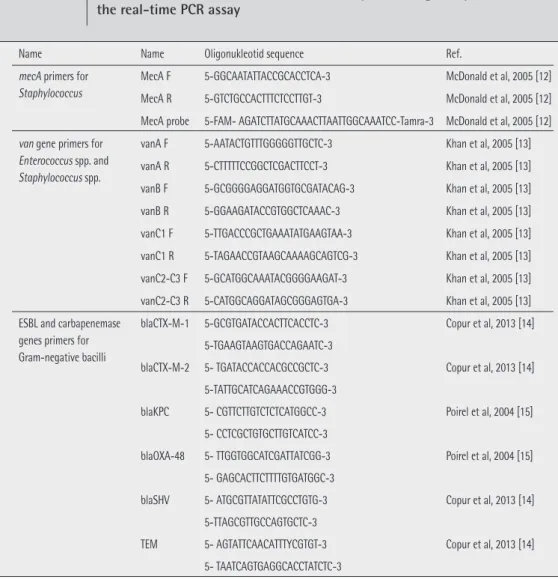
a) Molecular detection of staphyloccoccal mecA genes. Real-time polymerase chain reaction (PCR) was used
Name Name Oligonukleotid sequence Ref.
mecA primers for Staphylococcus
MecA F 5-GGCAATATTACCGCACCTCA-3 McDonald et al, 2005 [12] MecA R 5-GTCTGCCACTTTCTCCTTGT-3 McDonald et al, 2005 [12] MecA probe 5-FAM- AGATCTTATGCAAACTTAATTGGCAAATCC-Tamra-3 McDonald et al, 2005 [12] van gene primers for
Enterococcus spp. and Staphylococcus spp.
vanA F 5-AATACTGTTTGGGGGTTGCTC-3 Khan et al, 2005 [13] vanA R 5-CTTTTTCCGGCTCGACTTCCT-3 Khan et al, 2005 [13] vanB F 5-GCGGGGAGGATGGTGCGATACAG-3 Khan et al, 2005 [13] vanB R 5-GGAAGATACCGTGGCTCAAAC-3 Khan et al, 2005 [13] vanC1 F 5-TTGACCCGCTGAAATATGAAGTAA-3 Khan et al, 2005 [13] vanC1 R 5-TAGAACCGTAAGCAAAAGCAGTCG-3 Khan et al, 2005 [13] vanC2-C3 F 5-GCATGGCAAATACGGGGAAGAT-3 Khan et al, 2005 [13] vanC2-C3 R 5-CATGGCAGGATAGCGGGAGTGA-3 Khan et al, 2005 [13] ESBL and carbapenemase
genes primers for Gram-negative bacilli
blaCTX-M-1 5-GCGTGATACCACTTCACCTC-3 Copur et al, 2013 [14] 5-TGAAGTAAGTGACCAGAATC-3
blaCTX-M-2 5- TGATACCACCACGCCGCTC-3 Copur et al, 2013 [14] 5-TATTGCATCAGAAACCGTGGG-3
blaKPC 5- CGTTCTTGTCTCTCATGGCC-3 Poirel et al, 2004 [15] 5- CCTCGCTGTGCTTGTCATCC-3
blaOXA-48 5- TTGGTGGCATCGATTATCGG-3 Poirel et al, 2004 [15] 5- GAGCACTTCTTTTGTGATGGC-3
blaSHV 5- ATGCGTTATATTCGCCTGTG-3 Copur et al, 2013 [14] 5-TTAGCGTTGCCAGTGCTC-3
TEM 5- AGTATTCAACATTTYCGTGT-3 Copur et al, 2013 [14] 5- TAATCAGTGAGGCACCTATCTC-3
Table 1 Primers used for mecA, van, ESBL and carbapenemase genes presence in
30s at 55°C, 1s at 72°C and melting curves; 5s at 95°C, 60s at 65°C, and 97°C cont. reading). As positive controls, S. aureus ATCC 13565 (sea, sej), S. aureus ATCC 14458 (seb), S. aureus ATCC 19095 (sec, seh), S. aureus ATCC 23235 (sed, seg, sei), S.
aureus ATCC 27664 (see), S. aureus ATCC 25923 (pvl), S. aureus
ATCC 51650 (tsst-1) were used. As a nontoxigenic control S.
aureus ATCC 6538 was used.
RESULTS
Of the 150 samples of Turkish currency banknotes on which bacteriological analysis was conducted, 81% were found to be contaminated with several microbial species. The spectrum of microbial species were detected at rates of;
S. aureus 48% (46.8% MRSA and 1.2% MSSA), CoNS 54.7%, Enterococcus spp. 56%, enteric bacteria 21.3%,
non-fermen-tative Gram-negative bacteria 18.7% and Candida spp. 4%. A wide distribution of pathogens occurred from the different points included (table 2). The highest microbial contamina-tion was obtained in the Turkish currency banknotes from the hospital cafeteria, followed by the cafeteria of medical faculty students. Others were with order supermarkets and restau-instructions. Real-time PCR profile was used; denaturation
step at 95°C for 10 min, followed by 35 cycles of amplification; 10s at 95°C, 30s at 52°C, 1s at 72°C and melting curves; 5s at 95°C, 60s at 65°C, and 97°C cont. reading). E. faecium ATCC 51559, E. faecalis ATCC 51299, E. gallinarum ATCC 49573, and
E. casseliflavus ATCC 25788 strains were used as a positive
control for vanA, vanB, vanC1, and vanC2-C3 genes respecti-vely. K. pneumoniae ATCC 700603 and E.coli ATCC 25922 were also used as a control of beta lactamase and carbapenemase genes.
b) Molecular detection of SEs, pvl and tsst -1 genes.
Real-time polymerase chain reaction (real-time PCR) was used for detection of specific genes to confirm their identities (such
as SEs, pvl, and tsst-1 gene) via the primers previously described Peck et al [17]. Light Cycler 480 Sybr Green Master kit (Roche Diagnostics GmBH, Mannheim, Germany) was used with these primers on Light Cycler 480 II (Roche Diagnostics GmBH, Man-nheim, Germany) instrument according to the manufacturer’s instructions. 0.5 uM primers were added in reactions of final concentrations. Real-time PCR profile was used; denaturation step at 95°C for 10 min, followed by 40 cycles, of 10s at 95°C,
Microorganisms Paper currencies [n=25 each other] Total [n=150]
5£ 10£ 20£ 50£ 100£ 200£
Bacillus spp. 20 17 14 14 12 13 90 (60%)
Corynebacterium spp. 4 4 1 2 1 2 14 (9.3%)
Staphylococcus aureus 14 12 10 11 10 15 72 (48%)
Coagulase negative staphylococci (CoNS) 21 17 15 14 9 6 82 (54.7%)
Streptococcus spp. 1 1 0 0 0 0 2 (1.3%) Micrococcus spp. 2 1 1 0 0 0 4 (2.7%) Enterococcus spp. 8 18 12 15 11 20 84 (56%) Neisseria spp. 1 1 1 0 0 0 3 (2%) Escherichia coli 2 0 0 0 0 2 4 (2.7%) Enterobacter cloacae 10 2 1 1 1 0 15 (10%) Pantoea agglomerans 1 0 3 3 2 0 9 (6%) Klebsiella pneumoniae 2 1 0 0 1 0 4 (2.7%) Klebsiella oxytoca 1 1 0 0 0 0 2 (1.3%) Pseudomonas aeruginosa 4 2 2 1 1 0 10 (6.7%) Pseudomonas putida 2 2 0 0 1 1 6 (4%)
Acinetobacter baumannii complex 9 3 1 0 0 2 15 (10%)
Candida spp. 0 0 3 2 0 1 6 (4%)
Total 102 82 64 63 49 62 422
Table 2 Frequency distribution [%] of microorganisms isolated from
comycin resistance genes were not detected. Enteric bacteria
isolated from banknotes were 21.3%. Enterobacter cloacae was the first line of enteric bacteria with 46.9%. The others were Pantoea agglomerans 28.2%, E. coli 12.5%, K.
pneumoni-ae and K. oxytoca 12.5%, respectively. When the
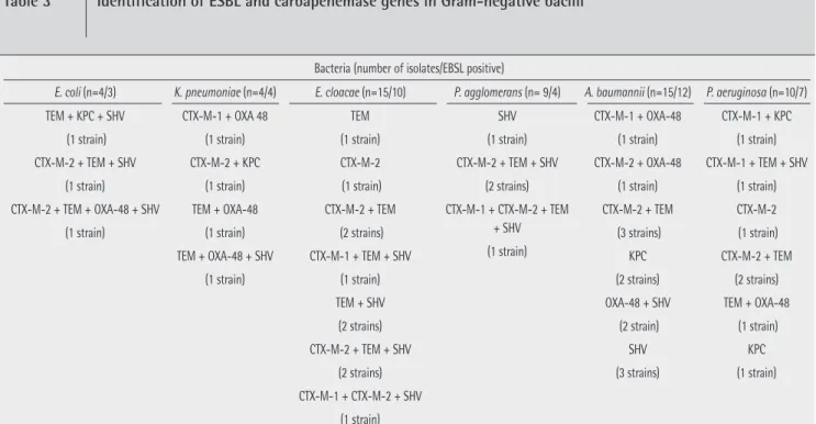
antimicrobi-al resistance in enteric bacteries was examined ampicillin was found to be with the highest resistance rate as 81%. Resistance rates to other antibiotics were determined as follows: ceftazi-dime 75%, cefuroxime and cefuroxime + axetil combination of 65.6% cefoxitine 62.5%, cefepime 78%, ceftriaxone 9.4%, ertapenem, meropenem, imipenem 12.5%, amikacin 25%, gen-tamicin 22%, ciprofloxacin 40.6%, tigecycline 3% trimethop-rim-sulfamethoxazole 25%, colistin 6.3%. MDR in enteric bac-teria was 40.6%. ESBL enzyme genes were found to be 66.7% in enteric bacteria (table 3). Non-fermentative Gram-negative rods isolated from banknotes were 18.7%. Among the non-fer-mentative bacteria, Acinetobacter baumannii complex ranked first with 53.6%. The others were P. aeruginosa 35.7%, P.
puti-da 10.7% and P. stutzeri 7.2% respectively. Antimicrobial
re-sistance rates of Pseudomonas spp. were as piperacillin 50%, piperacillin+ tazobactam 40%, ceftazidime 40%, ceftriaxone 30%, imipenem 10%, amikacin 20% and ciprofloxacin 30%. Antimicrobial resistance rates of Acinetobacter baumannii complex were as piperacillin 53.3%, piperacillin+ tazobactam 40%, ceftazidime 66.7%, ceftriaxone 33%, imipenem 26.7%, amikacin 33% ve ciprofloxacin 46.7%. MDR was 60% in P.
aeruginosa and 76% in A. baumannii complex. ESBL enzyme
genes were found to be 65.6% in enteric bacteria and 76% in non fermentative Gram-negative bacteria. The distribution by species was E. coli 75%, K. pneumoniae 100%, E. cloacae 66.7%, P. agglomerans 44.4%, P. aeruginosa 70% and A. bau-rants around the hospital, banks, buyers in open-air markets
and filling-stations. In the Turkish currency banknotes, the most intensive bacterial contamination was found in 5£, fol-lowed by 10£, 20£, 50£ and 100£, respectively. When look-ing at 200£banknotes, the contamination rate was found to be higher than 100£. The species of Staphylococcus spp. 154 produced in the highest proportion were S. aureus 48% and CoNS 54.7%. The distribution of CoNS were S. epidermidis 46.7%, S. haemolyticus 20%, S. hominis 12.2%, S. capitis 11%,
S. warneri 4.9%, S. lugdunensis 3.7%, S. caprae 2.4% and S. saprophyticus 2.4%. The mecA gene was observed in 90.3% of S. aureus and in 73% of CoNS isolates. When the antibiotic
re-sistance of Staphylococcus spp. were examined; the rere-sistance rates in MRSA strains were erythromycin 66.7%, clindamycin 22.2%, gentamicin 16.7%, trimethoprim+sulfamethoxazole (SXT) 16.7%; In S. epidermidis, erythromycin 34.3%, clindamy-cin 17.2%, gentamiclindamy-cin 5.9%, ciprofloxaclindamy-cin 5.9% and SXT 5.9%; in S. haemolyticus erythromycin 72.2%, clindamycin 44.4%, tigecycline 38.9%, ciprofloxacin 38.9% and linezolid 38.9%; in
S. hominis erythromycin 16.2% and SXT 16.2%; in S. capitis
gentamicin 20%. None of the staphylococci strains were found to have quinupristin/dalfopristin and vancomycin resistance. The rate of multi-drug resistance (resistance to more than three antibiotics-MDR) was found as 40.3%.
The second most frequently isolated 84 Enterococcus spp. (56.7%) was the distribution of bacteria in the species E.
fae-cium 35 (41.7%), E. faecalis 8 (9.5%), E. casseliflavus 21 (25%)
and other Enterococcus spp. 10 (11.9%), respectively. Van-comycin resistance was determined by both phenotypic and genotypic methods in two origins, one E. faecium and one E.
casseliflavus (2.4%). The resistance gene was vanA. Other
van-Bacteria (number of isolates/EBSL positive)
E. coli (n=4/3) K. pneumoniae (n=4/4) E. cloacae (n=15/10) P. agglomerans (n= 9/4) A. baumannii (n=15/12) P. aeruginosa (n=10/7)
TEM + KPC + SHV (1 strain) CTX-M-2 + TEM + SHV (1 strain) CTX-M-2 + TEM + OXA-48 + SHV (1 strain) CTX-M-1 + OXA 48 (1 strain) CTX-M-2 + KPC (1 strain) TEM + OXA-48 (1 strain) TEM + OXA-48 + SHV (1 strain) TEM (1 strain) CTX-M-2 (1 strain) CTX-M-2 + TEM (2 strains) CTX-M-1 + TEM + SHV (1 strain) TEM + SHV (2 strains) CTX-M-2 + TEM + SHV (2 strains) CTX-M-1 + CTX-M-2 + SHV (1 strain) SHV (1 strain) CTX-M-2 + TEM + SHV (2 strains) CTX-M-1 + CTX-M-2 + TEM + SHV (1 strain) CTX-M-1 + OXA-48 (1 strain) CTX-M-2 + OXA-48 (1 strain) CTX-M-2 + TEM (3 strains) KPC (2 strains) OXA-48 + SHV (2 strain) SHV (3 strains) CTX-M-1 + KPC (1 strain) CTX-M-1 + TEM + SHV (1 strain) CTX-M-2 (1 strain) CTX-M-2 + TEM (2 strains) TEM + OXA-48 (1 strain) KPC (1 strain)
in S. lugdunensis, 50% in S. caprae and 50% in S.
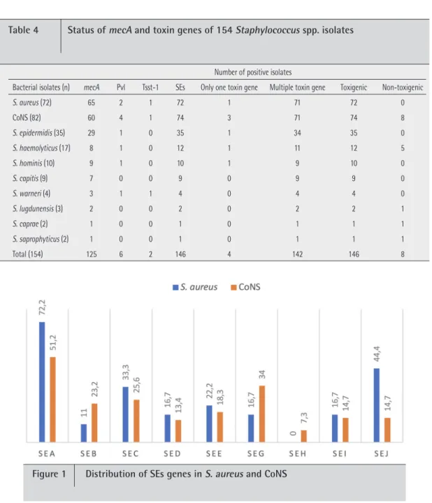
saprophyt-icus. The distribution of toxin genes were pvl 2.8%, tsst-1
1.4% and SEs 100% in S. aureus, pvl 4.9%, tsst-1 1.2% and SEs 87.8% in CoNS (table 4). The distribution of SEs genes in S.
aureus were as sea 72.2%, seb 11%, sec 33.3%, sed 16.7%, see
22.2%, seg 16.7%, sei 16.7% and sej 44.4%, the seh gene was not found. The distribution of SEs genes in CoNS were as sea 51.2%, seb 23.2%, sec 25.6%, sed 13.4%, see 18.3%, seg 34%,
seh 7.3%, sei 14.7% and sej 14.7% (figure 1). Comparing with
that of CoNS, the sea gene was found statistically significantly high in S. aureus strains (p<0.05) and comparing with that of
S. aureus strains, the seb, seg and seh genes were found
statis-tically significantly high in CoNS strains.
mannii 80% (table 3). CTX-M type ESBL enzyme genes were
found to be 43.8% in enteric bacteria and 40% in non fermen-tative Gram-negative bacteria. The distribution by species was
E. coli 50%, K. pneumoniae 50%, E. cloacae 46.7%, P. agglo-merans 22.2%, P. aeruginosa 50% and A. baumannii 33.3%.
In our study, blaKPC was found as 6.2 % in enteric bacteria and
as 12 % in non-fermentative bacteria. The distribution by spe-cies was E. coli 25%, K. pneumoniae, 25%, P. aeruginosa 20% and A. baumannii 13.3%. OXA-48 enzyme genes were found
to be 12.5 % in enteric bacteria and 20% in non-fermentative Gram-negative bacteria. The distribution by species was E. coli 25%, K. pneumoniae 75%, P. aeruginosa 10% and A.
bauman-nii 26.7%. The availability of toxin genes were 100% in S. au-reus, 100% in S. epidermidis, 70.6% in S. haemolyticus, 66.7%
Number of positive isolates
Bacterial isolates (n) mecA Pvl Tsst-1 SEs Only one toxin gene Multiple toxin gene Toxigenic Non-toxigenic
S. aureus (72) 65 2 1 72 1 71 72 0 CoNS (82) 60 4 1 74 3 71 74 8 S. epidermidis (35) 29 1 0 35 1 34 35 0 S. haemolyticus (17) 8 1 0 12 1 11 12 5 S. hominis (10) 9 1 0 10 1 9 10 0 S. capitis (9) 7 0 0 9 0 9 9 0 S. warneri (4) 3 1 1 4 0 4 4 0 S. lugdunensis (3) 2 0 0 2 0 2 2 1 S. caprae (2) 1 0 0 1 0 1 1 1 S. saprophyticus (2) 1 0 0 1 0 1 1 1 Total (154) 125 6 2 146 4 142 146 8
Table 4 Status of mecA and toxin genes of 154 Staphylococcus spp. isolates
that the rate of methicillin resistant S. aureus was 90.3% and methicillin resistant in CoNS is 73.2%. The highest antibiotic resistance in staphylococcus was erythromycin (72.2%), and clindamycin (44.4%) resistance in S. haemolyticus; gentamicin resistance (20%) in S. capitis. Tigecycline (38.9%), ciprofloxacin (38.9%) and linezolid (33.3%) resistance were found only in S.
haemolyticus strains. None of the staphylococci strains had
re-sistance to quinupristin/dalfopristin and vancomycin. The rate of MDR was found as 40.3%. Recently, many published studies
reported that E. faecium infections are increasing worldwide [4]. In our country, the rates of E. faecium and E. faecalis were determined to be 15 - 50% and 52 - 85%, respectively [23, 24]. In previous researches, Enterococcus spp., which can be found without losing their vitality for 4 months in inanimate environments [21].
Many Gram-negative species, such as Acinetobacter spp.,
E. coli, Klebsiella spp., P. aeruginosa, Serratia marcescens,
or Shigella spp. can survive on inanimate surfaces even for months. Overall, Gram-negative bacteria have been described to persist longer than Gram-positive bacteria [3, 21]. Humid conditions improved persistence for most types of bacteria, such as Salmonella typhimurium, P. aeruginosa, E. coli or other relevant pathogens [2, 9, 21, 25]. In previous studies, reported that Enterobacteraceae members are 13%-55.5% range of the paper currencies and the most frequently isolated enteric bac-teria was E. coli (19.4-48.14%) [2, 3, 9]. Antimicrobial resistance is a global phenomenon that has resulted in high morbidity and mortality as a result of treatment failures and increased health care costs. Research has shown that contaminated fo-mites in general and paper currency in particular, plays a key role in the spread of bacterial infections with antimicrobial re-sistance [2, 3, 25]. Heshiki et al. [22] in a metagenomic study showed that the antimicrobial resistance genes on banknotes were significantly higher (4.86 times more) than environmen-tal samples such as water, air, soil and dust.
Emergence of glycopeptide resistance causes more severe prognosis, higher mortality, and recurrence in enterococcal in-fections. The most common type of enterococcal vancomycin resistance is high-level resistance associated with acquisition of the vanA and vanB genes, typically observed in E. faecium and E. faecalis isolates [4]. Conversely, the vanC genotype is associated with constitutive low-level vancomycin resistance and is intrinsic to E. gallinarum and E. casseliflavus [4]. In our study, vancomycin resistance was determined by both phe-notypic and gephe-notypic methods in two isolates (2.4%), these were one E. faecium and one E. casseliflavus. The resistance genes were vanA. Other vancomycin resistance genes were not detected.
Resistance mediated by ESBLs includes all penicillins, cephalosporins (including third-generation cephalosporins) and aztreonam. Since plasmid-mediated ESBLs were first de-tected in a K. pneumoniae isolate in 1983 in Germany [26]. A new non-TEM non-SHV ESBL was isolated in Germany, in 1989, in a strain of E. coli called CTX-M because of its preferential activity on cefotaxime rather than ceftazidime [27]. Over the past 20 years, some Enterobacteriaceae mainly E. coli, K.
pneu-DISCUSSION
Paper currencies are objects capable of absorbing, harbor-ing and transmittharbor-ing infectious microorganisms [2]. Research-es show that the microbial load on banknotResearch-es variResearch-es according to the banknotes, seasons, stored under varying environmental conditions, the age of banknotes, the local community mi-crobiota, the general hygiene level, and the general hygienic conditions [3, 9, 10]. The amount of bacterial contamination
on currency varies widely between countries. Previous stud-ies have revealed that 70-97% of banknotes harbor various bacteria and viruses on the surface in different nations such as the United States, Mexico, China, India, Saudi Arabia, Su-dan, Pakistan, Brasil etc, [9, 10, 18, 19, 20]. In our study, Turk-ish currency banknotes on which bacteriological analysis was conducted, 81% were found to be contaminated with several microbial species. Our results show that similar results were obtained in previous studies. Numerous studies have shown that cotton-based banknotes have more microbial loads than polymer-based ones [3, 10].
Vriesekoop et al. reported that comparison of cot-ton-based banknotes of countries such as China, Ireland, The Netharlands, Nigeria, United Kingdom and the United States, as well as the polymer-based banknotes of countries such as Australia and New Zealand. They found that cotton-based banknotes had much more bacterial loading than poly-mer-based banknotes [3]. The bacterial load evaluated as 81% in Turkish banknotes can be explained by the fact that they are based on cotton. Some studies showed that, the longer the paper currencies remain in circulation, the more chance there is for them to become contaminated, and lower-de-nomination notes receive the most handling because they are exchanged more frequently [2, 3]. According to our results also showed that health centers and health center workers and people who stay here play an important role contribut-ing to the bacterial contamination. Many previous studies also claimed similar results [2, 3, 9, 10]. Many bacteria have been
isolated from banknotes in studies from Turkey, China, Phil-ippines, India, Saudi Arabia, Mexico, New Zealand, Australia, Canada, USA and Europe. It was also reported that S. aureus,
E. coli, Klebsiella spp. and Enterobacter spp. were identified
from these countries’ banknotes [9, 10]. Staphylococcus spp.
present in the nose often contaminate hands, fingers, faces, and nasal carriers which can easily become skin carriers [4]. In general, there was no obvious difference in survival between multiresistant and susceptible S. aureus strains. S. aureus (in-cluding MRSA) survive for 7 day -7 months on dry surfaces [21]. In our study, Staphylococcus spp. were the most isolated bacteria. Previous studies have also determined that there is a high number of Staphylococcus spp. on banknote, however, most studies did not identify Staphylococcus spp. Our study was the first research to identified Staphylococcus species un-like other researches on this topic. Methicillin resistance is an
important consideration in all Staphylococcus spp., especially
S. aureus. Global transmission of MRSA has been the subject
moniae, and Proteus mirabilis have demonstrated acquisition
of plasmids secreting ESBL [28]. In our study, the rates of ESBL
enzyme genes were found to be high as 65.6% in enteric bac-teria and as 76% in non-fermentative Gram-negative bacbac-teria. CTX-M type ESBL enzyme genes were found to be 43.8% in enteric bacteria and 40% in non-fermentative Gram-negative bacteria. Carbapenemases in Enterobacteriaceae are mainly found in K. pneumoniae, and to a much lesser extent in E.
co-li and other enterobacterial species, with a higher prevalence
in southern Europe and Asia than in other parts of the World [28]. The first OXA-48 carbapenemase was identified in 2001
from a K. pneumoniae isolate obtained from a urine specimen collected in Istanbul, Turkey [15]. Shortly thereafter there was an outbreak of OXA-48 producing K. pneumoniae isolates re-ported in Istanbul in 2006 [29]. In our study, OXA-48 enzyme
genes were found to be 12.5% in enteric bacteria and 20% in non fermentative Gram-negative bacteria. Staphylococcus spp. are also capable of producing “distant” diseases, which are mediated by the secretion of toxins and these toxins can be produced directly by bacteria that colonize the skin or mucosa or indirectly by microorganisms that colonize food, beverages and fomites [4, 30]. Bacteriological studies about banknotes, have included no analysis of the toxin genes (pvl, tsst-1 and SEs). 95.4% of Staphylococcus spp. that are analyzed from our study were determined to possess toxin genomes. The distri-bution of these toxin genomes was as follows: 3.9% pvl, 1.3%
tsst-1 and 98.4% SEs. There was no toxin genomes in the rest
of the Staphylococcus spp. (5.2%). PVL is a cytotoxin that
causes tissue necrosis and leukocyte destruction. This linkage to virulent strains suggests its capability of causing deadly in-fections in healthy people [4]. Toxic shock syndrome (TSS) is a
life-threatening illness characterized by high fever, erythema-tous rash with subsequent desquamation of the skin, shock, and multiple organ involvement [4, 31]. In our study, it was possible to detect 1.3% of tsst-1 genomes from our isolated banknotes.
Six enterotoxins serotypes (sea to see and seh) have been involved in most of the Staphylococcus poisoning outbreaks worldwide [31]. In our study, it was indicated that 94.8% of
Staphylococcus spp. have SEs genomes. S. aureus and CoNS
strains can encode more than one enterotoxin gene simultane-ously; over 50% of the isolates assessed showed this property [8]. All S. aureus strains were carried at least one SEs gene and the combination sea+sei, sea+sec+sei, sea+sed+sej was the most frequent. CoNS strains were positive SEs genome 90.2% and the combination sea+sej, sea+sec+sej, sea+seg+sej, sea+
sed+seg+sei was the most frequent. sea is one of the most
frequently observed enterotoxins, although the literature shows highly variable results in the prevalence of S. aureus en-terotoxin genes, depending on the kind of food and the biovar investigated [4, 31]. Compared to CoNS strains, sea genes were statistically significantly higher in S. aureus strains (p<0.05). Compared to S. aureus strains, seb, seg and seh genes were statistically significantly high in CoNS strains (p<0.05). On the other hand, the seh gene was detected at a rate of 7.3% in CoNS strains, although there was no in the S. aureus strains.
Several factors in the spread of pathogen and potential pathogenic bacteria, as well as antimicrobial resistance and vir-ulance genes such as SEs, community and hospital enviroments, animal products and the environmental compartment are im-portant. Results of this study in terms of demonstrating that paper curriencies or banknotes circulating in society can poten-tially mediate the transport of microorganisms among people and poses a risk to public health and it is also very important to highlight the need for proper hygienic practices for maximal-ly reducing the spread of disease-causing pathogens. This can be considered as an indication that banknotes may contribute to the spread of pathogens and antimicrobial resistance. In this study, it was aimed to pay attention to hand hygiene for reduc-ing the microbial load on the currencies and the necessity of producing these banknotes with maintain less bacteria such as plastic etc. instead of cotton. In addition, our study has been the first research to identified staphylococcus species and its viru-lance genes unlike other researches on this topic.
FUNDING
None to declare
CONFLICT OF INTEREST
The authors declare that they have no conflicts of interest
REFERENCES
1. Maritz JM, Sullivan SA, Prill RJ, Aksoy E, Scheid P, Carlton JM. Filthy lucre: A metagenomic pilot study of microbes found on circulat-ing currency in New York City. PLoS One. 2017;12:e0175527. doi: 10.1371/journal.pone.0175527
2. Girma G. Health Risk Associated with Handling of Contaminat-ed Paper Currencies in Circulation: A review. American Scientific Research Journal for Engineering, Technology, and Sciences (AS-RJETS) 2014;10:40-53. [cited 11 November 2019]. Available from: http://asrjetsjournal.org/index.php/American_Scientific_Journal/ article/view/696.
3. Vriesekoop F, Chen J, Oldaker J, Besnard F, Smith R, Leversha W, et al. Dirty Money: A Matter of Bacterial Survival, Adherence, and Toxicity. Microorganisms 2016;4:E42 doi: 10.3390/microorganisms 4040042.
4. Que YA, Moreillon P. Staphylococcus aureus (Including Staph-ylococcal Toxic Shock Syndrome). In Mandell, Douglas, and Ben-nett’s Principles and Practice of Infectious Diseases. Eighth Edition. Churchill Livingstone Elsevier. 2015; p 2237-71.
5. Ciupescu LM, Auvray F, Nicorescu IM, Meheut T, Ciupescu V, Lar-deux A et al. Characterization of Staphylococcus aureus strains and evidence for the involvement of non-classical enterotoxin genes in food poisoning outbreaks. FEMS Microbiol Lett. 2018;365(13). doi: 10.1093/femsle/fny139.
6. Nanoukon C, Affolabi D, Keller D, Tollo R, Riegel P, Baba-Moussa L, et al. Characterization of Human Type C Enterotoxin Produced by
Clinical S. epidermidis Isolates. Toxins (Basel). 2018;10:E139. doi: 10.3390/toxins10040139.
7. Podkowik M, Seo KS, Schubert J, Tolo I, Robinson DA, Bania J, et al. Genotype and enterotoxigenicity of Staphylococcus epidermid-is epidermid-isolate from ready to eat meat products. Int J Food Microbiol. 2016;229:52-9. doi: 10.1016/j.ijfoodmicro.2016.04.013.
8. Nunes RSC, Aguila EMD, Paschoalin VMF. Safety Evaluation of the Coagulase-Negative Staphylococci Microbiota of Salami: Superan-tigenic Toxin Production and Antimicrobial Resistance. Biomed Res Int. 2015;483548. doi: 10.1155/2015/483548
9. Angelakis E, Azhar EI, Bibi F, Yasir M, Al-Ghamdi AK, Ashshi AM, et al. Paper money and coins as potential vectors of transmissible dis-ease. Future Microbiol. 2014; 9:249–61. doi: 10.2217/fmb.13.161. 10. Rocha-Gámez J, Tejeda-Villarreal PN, Macías-Cárdenas P,
Can-izales-Oviedo J, Garza-González E, Ramírez-Villarreal EG. Micro-bial contamination in 20-peso banknotes in Monterrey, Mexico. J Environ Health 2012;75:20-3. [cited 11 November 2019]. Available from: https://www.jstor.org/stable/26329464?seq=1#metadata_ info_tab_contents.
11. EUCAST. The European Committee on Antimicrobial Susceptibili-ty Testing. Routine and extended internal qualiSusceptibili-ty control for MIC determination and disk diffusion as recommended by EUCAST. Ver-sion 6.1, 2016. [cited 11 November 2019]. Available from: http:// www.eucast.org.
12. McDonald RR, Antonishyn NA, Hansen T, Snook LA, Nagle E, Mul-vey MR, et al. Development of a Triplex Real-Time PCR Assay for Detection of Panton-Valentine Leukocidin Toxin Genes in Clin-ical Isolates of Methicillin-Resistant Staphylococcus aureus. J Clin Microbiol 2005;43:6147-49. doi: 10.1128/JCM.43.12.6147-6149.2005.
13. Khan SA, Nawaz MS, Khan AA, Hopper SL, Jones RA, Cerniglia CE. Molecular characterization of multidrug-resistant Entero-coccus spp. from poultry and dairy farms: detection of virulence and vancomycin resistance gene markers by PCR. Mol Cell Probes 2005;19:27-34. doi:10.1016/j.mcp.2004.09.001
14. Copur Cicek A, Saral A, Ozad Duzgun A, Yasar E, Cizmeci Z, Ozlem Balci P, et al. Nationwide study of Escherichia coli producing ex-tended-spectrum β-lactamases TEM, SHV and CTX-M in Turkey. J Antibiot. 2013;66:647–50. doi: 10.1038/ja.2013.72.
15. Poirel L, Héritier C, Tolun V, Nordmann P. Emergence of oxacilli-nase-mediated resistance to imipenem in Klebsiella pneumoni-ae. Antimicrob Agents Chemother 2004; 48:15–22. doi: 10.1128/ AAC.48.1.15-22.2004.
16. Chiefari AK, Perry MJ, Kelly-Cirino K, Egan CT. Detection of Staph-ylococcus aureus enterotoxin production genes from patient samples using an automated extraction platform and multiplex real-time PCR. Mol Cell Probes. 2015;29:461-67. doi: 10.1016/j. mcp.2015.06.004.
17. Peck KR, Baek JY, Song JH, Ko KS. Comparison of genotypes and enterotoxin genes between Staphylococcus aureus isolates from blood and nasal colonizers in a Korean hospital. J Korean Med Sci. 2009;24:585-91. doi: 10.3346/jkms.2009.24.4.585.
18. Abd Alfadil NA, Suliman Mohamed M, Ali MM, El Nima EAI.
Char-acterization of Pathogenic Bacteria Isolated from Sudanese Bank-notes and Determination of Their Resistance Profile. Int J Microbi-ol. 2018;2018:4375164. doi:10.1155/2018/4375164
19. Ejaz H, Javeed A, Zubair M. Bacterial contamination of Pakistani currency notes from hospital and community sources. Pak J Med Sci. 2018;34(5):1225–30. doi:10.12669/pjms.345.15477.
20. Pereira da Fonseca TA, Pessôa R, Sanabani SS. Molecular Analysis of Bacterial Microbiota on Brazilian Currency Note Surfaces. Int J Environ Res Public Health. 2015;12(10):13276–88. doi:10.3390/ ijerph121013276.
21. Kramer A, Schwebke I, Kampf G. How long do nosocomial patho-gens persist on inanimate surfaces? A systematic review. BMC In-fectious Diseases 2006;6:130. doi: 10.1186/1471-2334-6-130 22. Heshiki Y, Dissanayake T, Zheng T, Kang K, Yueqiong N, Xu Z, et
al. Toward a Metagenomic Understanding on the Bacterial Com-position and Resistome in Hong Kong Banknotes. Front Microbiol. 2017;8:632. doi: 10.3389/fmicb.2017.00632.
23. Celik S, Koksal Cakırlar F, Mamal Torun M. Presence of Vanco-mycin, Aminoglycosides, and Erythromycin Resistance Genes in Enterococci Isolated from Clinical Samples in Turkey. Clin. Lab. 2014;60:1801-6. doi: 10.7754/clin.lab.2014.140211
24. Kacmaz B, Aksoy A. Antimicrobial resistance of Enterococci in Tur-key. Int J Antimicrob Agents 2005;25:535-8. doi: 10.1016/j.ijantim-icag.2005.02.020.
25. Akoachere JF, Gaelle N, Dilonga HM, Nkuo-Akenji TK. Public health implications of contamination of Franc CFA (XAF) circulating in Buea (Cameroon) with drug resistant pathogens. BMC Res Notes. 2014;8;7:16. doi: 10.1186/1756-0500-7-16.
26. Kliebe C, Nies BA, Meyer JF, Tolxdorff-Neutzling RM, Wiedemann B. Evolution of plasmid-coded resistance to broad-spectrum ceph-alosporins. Antimicrob. Agents Chemother 1985;28:302–7. doi: 10.1128/aac.28.2.302.
27. Lahlaoui H, Ben Haj Khalifa A, Ben Moussa M. Epidemiology of Enterobacteriaceae producing CTX-M type extended spectrum β-lactamase (ESBL). Review. Med Mal Infect. 2014;44:400-4. doi: 10.1016/j.medmal.2014.03.010.
28. Canton R, Akova M, Carmeli Y, Giske CG, Glupczynski Y, Gniad-kowski M et al., Rapid evolution and spread of carbapenemas-es among Enterobacteriaceae in Europe. Clin Microbiol Infect 2012;18:413–31. doi: 10.1111/j.1469-0691.2012.03821.x.
29. Carrer A, Poirel L, Eraksoy H, Cagatay AA, Badur S, Nordmann P. Spread of OXA-48-positive carbapenem-resistant Klebsiella pneu-moniae isolates in Istanbul, Turkey. Antimicrob Agents Chemother 2008;52:2950-4. doi: 10.1128/AAC.01672-07.
30. Albert NM, Bena JF, Ciudad C, Keleekai-Brapoh N, Morrison SL, Rice K, et al. Contamination of reusable electroencepha-lography electrodes: A multicenter study. Am J Infect Control. 2018;46(12):1360-4. doi: 10.1016/j.ajic.2018.05.021.
31. Oliveira D, Borges A, Simões M. Staphylococcus aureus Toxins and Their Molecular Activity in Infectious Diseases. Toxins (Basel). 2018;10(6):252. doi: 10.3390/toxins10060252.
![Table 2 Frequency distribution [%] of microorganisms isolated from](https://thumb-eu.123doks.com/thumbv2/9libnet/3996733.54321/4.892.180.719.177.719/table-frequency-distribution-microorganisms-isolated.webp)