Evaluation of choroidal
thickness using
enhanced depth
imaging by
spectral-domain optical
coherence tomography
in patients with
pseudoexfoliation
syndrome
FC Eroglu1, L Asena2, C Simsek2, A Kal1 and G Yılmaz2
Abstract
Purpose To investigate the choroidal thickness using optical coherence tomography in the eyes of patients with unilateral and bilateral pseudoexfoliation syndrome and to compare them with healthy controls. Methods We studied four groups: (1) affected eyes from 30 patients with unilateral PEX syndrome affecting the right eye of 17 patients and the left eye of 13 patients; (2) clinically unaffected eyes of 30 patients with unilateral PEX syndrome; (3) the eyes of 30 patients with bilateral PEX syndrome; and (4) the eyes of 30 normal healthy subjects. Choroidal thickness was evaluated using high-speed, high-resolution enhanced depth imaging by spectral-domain optical coherence tomography. Optical coherence tomography features were compared in all groups using the statistical package SPSS v 15.0.
Results The mean subfoveal choroidal thicknesses were 237.35± 58.01 μm in group 1; 330.75± 47.84 μm in group 2; 206.3 ± 86.75 μm in group 3; and 311.8± 51.42 μm in group 4. Significant differences in the mean subfoveal choroidal thickness were found between groups 1 and 2 (Po0.001), groups 1 and 4 (P = 0.004), groups 2 and 3 (Po0.001), and groups 3 and 4 (Po0.001).
Conclusion In this study, it was observed that clinically affected eyes of patients with PEX syndrome have significantly thinner choroids compared with the clinically unaffected eyes of patients with unilateral PEX syndrome and eyes of healthy controls.
Eye (2015) 29, 791–796; doi:10.1038/eye.2015.34; published online 27 March 2015
Introduction
Pseudoexfoliation (PEX) syndrome is a genetically determined, age-dependent,
generalized disorder of the elasticfiber system. It is characterized by progressive accumulation of abnormal extracellularfibrillary material on many ocular and extraocular tissues, including the periphery of blood vessels.1,2PEX syndrome
is probably due to predisposing genetic factors and certain vascular conditions that induce tissue hypoxia.2Recent progress and advances have led to improvements in clinical management by providing a deeper understanding of the effects of the PEX process on ocular tissues and by increasing evidence for systemic associations of PEX with cardiovascular and cerebrovascular morbidity.3,4A previous study has also shown
asymmetric values of the two carotid trees of patients with unilateral PEX.5
In recent studies, optical coherence tomography (OCT) was introduced as an effective tool for evaluating choroidal thickness. Thanks to recent technological developments, new-generation spectral-domain (SD)-OCT devices with enhanced scanning speed provide the
opportunity to achieve high-resolution images and more accurate measurements. In the enhanced depth imaging technique (EDI), scan acquisition of the choroidal–scleral boundary is set up adjacent to zero delay, where the sensitivity in SD-OCT images is highest.
1Department of
Ophthalmology, Baskent University School of Medicine, Konya, Turkey
2Department of
Ophthalmology, Baskent University School of Medicine, Ankara, Turkey Correspondence:
FC Eroglu, Department of Ophthalmology, Baskent University School of Medicine, Hocacihan Mah. Saray Cad. No: 1 Selcuklu, Konya 42080, Turkey Tel:+90 322 2570606 2208; Fax:+90 332 257 0637. E-mail: dr_fatoscorak@ hotmail.com Received: 5 June 2014 Accepted in revised form: 3 February 2015 Published online: 27 March 2015 This study has been presented as a free paper in ASCRS/ASOA Symposium & Congress, Boston, Massachusetts, USA, 2014.
CLINICAL
STUD
Thus, it has become possible to evaluate ocular tissues located at deeper levels than that of the retina, which could not be evaluated with time domain OCT.6,7
There are several studies related to both ocular and choroidal bloodflow in PEX syndrome.3–5,8,9However, there have not been any reports to investigate choroidal thickness in patients with PEX syndrome. Therefore, in our study, we intended to evaluate the choroidal thickness of patients with unilateral and bilateral PEX syndrome and to compare them with that of healthy eyes.
Materials and methods
The eyes of the subjects were allocated to four groups: group 1 consisted of affected eyes of 30 patients with unilateral PEX syndrome (right eye of 17 patients and left eye of 13 patients); group 2 consisted of clinically unaffected eyes of 30 patients with unilateral PEX syndrome; group 3 consisted of 30 patients with bilateral PEX syndrome; and group 4 (control group) consisted of 30 age- and gender-matched normal subjects without PEX syndrome. The patients in groups 1 and 2 are the same patients. To ensure consistency, the right eyes from each patient with bilateral PEX syndrome and control subject, and both eyes from patients with unilateral PEX syndrome were included. The study protocol was approved by the Human Subjects Committee of Baskent University and adhered to the Declaration of Helsinki. A detailed informed consent was obtained prior to each individual’s participation in the study. We certify that all applicable institutional and governmental regulations concerning the ethical use of human volunteers were followed during this research. Patients were excluded if they had a history of ocular surface disorder; any type of glaucoma; previous ocular surgery or injury; history of any systemic disease that may affect the choroidal circulation, such as
hypertension, diabetes, vasculitis, or renal failure; or if they were being administered drugs, including analgesics, decongestants, and antihistamines. As choroial thickness is strongly correlated with IOP, refractive error, and axial length;10,11to minimize the effect of axial length and IOP
on choroidal thickness, patients with best-corrected visual acuity worse than 20/20, a refractive error higher than 1D and the IOP level422 mm Hg were excluded from the study. All patients underwent a complete ocular examination conducted by the same ophthalmologist (FCE). The examination included slit lamp biomicroscopy, intraocular pressure measurement, gonioscopy, dilated fundus examination, and choroidal thickness
measurements using EDI-OCT. PEX was clinically diagnosed by the presence of typical PEX material at the pupil border on undilated examination, on the anterior lens capsule on dilated examination, or on the trabecular meshwork on gonioscopy, with or without Sampaolesi’s
line and pigment deposition in the angle and/or corneal endothelium.
Choroidal thickness measurements were performed by the same experienced technician using a high-speed and high-resolution SD-OCT device (λ = 840 nm, 26 000A-scans/s, and 5μm axial resolution), and results were analyzed using OptovueRTVue software version 3.5 (Optovue Inc., Fremont, CA, USA). The OptovueRTVue support‘chorioretinal imaging mode’ was used for EDI imaging. The scan pattern used by OptovueRTVue was the retina cross-line, consisting of two orthogonally oriented 6-mm lines consisting of 1024A-scans. By automatically inverting the image, the chorioretinal interface became adjacent to the zero delay. The retina cross-line scan had 32 frames in average, 16 per direction, without tracking.12
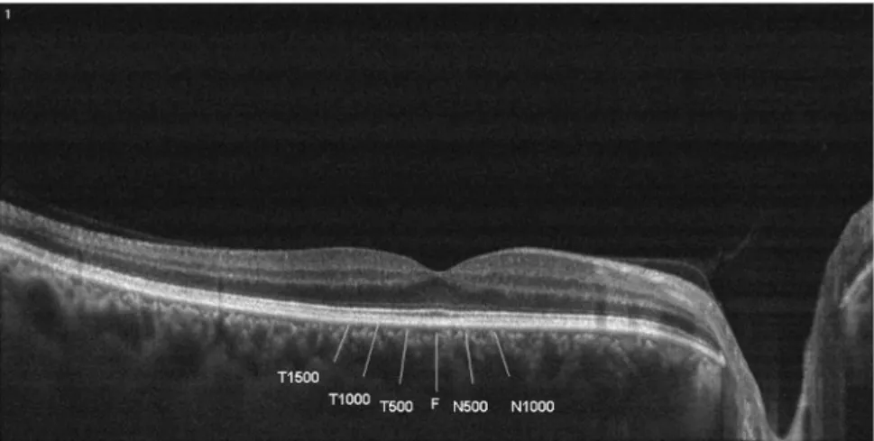
Choroidal thickness was measured perpendicularly from Bruch’s membrane equivalent to the choroid-sclera interface at the fovea and at 5 more points located at 500μm nasal to the fovea, 1000 μm nasal to the fovea, 500μm temporal to the fovea, 1000 μm temporal to the fovea, and 1500μm temporal to the fovea using
OptovueRTVue software’s ‘chorioretinal imaging mode’. Choroid-sclera interface was defined as the outermost dark-to-bright boundary (Figure 1). Choroidal thickness measurements were performed manually by two masked physicians (FCE and CS). If there was a difficulty to allocate the sclera-choroid interface in an EDI-OCT scan, the choroidal thickness measurement was excluded from statistical analysis. All basal OCT scans were performed at the same time of the day (in the morning) to avoid diurnalfluctuations. The two measurements were averaged; the differences between readings performed by the masked physicians were found to be within 10μm of the mean. If the measurements did not agree within 10μm, then they were repeated, and if the inconsistency persisted, a third masked physician (LA) took a
measurement. Of course, there was some difficulty on few images. But these scans were excluded from statistical analysis if differences between readings performed by the masked physicians were not found within 10μm. The interobserver reproducibility of the choroidal
measurements was evaluated by measuring the interclass correlation coefficient (ICC).
Statistical analysis was performed using the statistical package SPSS v 15.0 (SPSS Inc., Chicago, 2006).
Interobserver correlation (IC) was calculated for all choroidal thickness measurements. Baseline choroidal thickness measurements of the groups were compared by ANOVA. The categorical variables between the groups were analyzed using theχ2test. Po0.05 was considered as statistically significant.
Results
The mean age of the patients in group 1 and 2 was 63.7± 9.1 years (range 47–80 years). Patients in group 3 had a mean age of 61.9± 9.5 years (range 44–79 years), and those in group 4 had a mean age of 58.3± 9.4 years (range 41–76 years). Groups 1, 2, and 3 included 16 males and 14 females; group 4 included 17 males and 13 females. There was no statistically significant difference regarding age (P= 0.197) and gender (P = 0.937) between groups. Mean IOP at the time of examination was found to be 17.2± 4.3 mm Hg (range 14–21 mm Hg) in group 1, 16.8± 3.7 mm Hg (range 12–20 mm Hg) in group 2, 18.3± 3.6 mm Hg (range 15–22 mm Hg) in group 3 and 16.1± 3.2 mm Hg (range 13–19 mm Hg) in group 4. Mean axial length values of the patients was 23.10± 1.20 mm (range 22.12–23.91 mm)in group 1, 23.21 ± 0.9 mm (range 22.11–23.96 mm)in group 2, 23.01 ± 1.23 mm (range 21.78– 24.21 mm) in group 3, and 23.24± 1.04 mm (range 22.43– 24.64) in group 4. There was no statistically difference regarding IOP levels (P= 0.08) and axial length (P = 0.121) between groups.
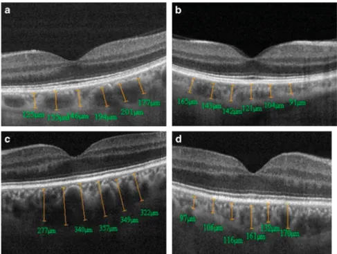
The mean subfoveal choroidal thickness was 237.35± 58.01μm in group 1, 330.75 ± 47.84 μm in group 2, 206.3± 86.75 μm in group 3, and 311.8 ± 51.42 in group 4. Significant differences in the mean subfoveal choroidal thickness were found between groups 1 and 2 (Po0.001), groups 1 and 4 (P= 0.004), groups 2 and 3 (Po0.001), and groups 3 and 4 (Po0.001). The differences in mean subfoveal choroidal thickness between groups 1 and 3 (P= 0.253) and groups 2 and 4 (P = 0.492) were not statistically significant. Moreover, as shown in Table 1, choroidal thicknesses of groups 1 and 3 were always thinner than those of groups 2 and 4 in all eyes at all six measurement points. Figure 2a–d illustrates the choroidal
thickness for an individual with bilateral and unilateral PEX syndrome group, respectively.
The interobserver ICC reproducibility for the mean choroidal thickness was 0.938 (95% CI, 0.931–0.984) and ICC was40.93 for all measurement points (Po0.001). Table 2 shows the comparison of the mean choroidal thickness measurements by two different observers between groups.
Discussion
PEX has been reported as a systemic vascular disease and is occasionally referred to as PEX vasculopathy.1,3–5 Elastin is the major part of the extracellular matrix of arterioles and previous studies have shown an association of vascular elastosis with pseudoexfoliative materials. Therefore, a possible association has been hypothesized between the presence of ocular pseudoexfoliation and vascular diseases.1–4In several studies it has been reported that cerebrovascular disease, arterial
Table 1 Mean± SD (in μm) of choroidal thickness measure-ments of groups 1, 2, 3, and 4
Group 1 Group 2 Group 3 Group 4 N2 252.45± 75.25 333.65 ± 67.78 207.05 ± 69.44 320.9 ± 76.61 N1 276.15± 76.88 335.05 ± 68.86 209.1 ± 75.48 311.75 ± 57.48 F 237.35± 58.01 330.75 ± 47.84 206.3 ± 86.75 311.8 ± 51.42 T1 259.35± 76.31 338.65 ± 48.09 199.5 ± 76.31 296.85 ± 45.22 T2 242± 76.56 321.95 ± 46.03 188.25 ± 76.12 285.3 ± 51.68 T3 242.4± 76.01 304.4 ± 49.74 182.55 ± 76.45 261.45 ± 56.55
N2: choroidal thickness at 1000μm nasal to the fovea; N1: choroidal thickness at 500μm nasal to the fovea; F: choroidal thickness at fovea; T1: choroidal thickness at 500μm temporal to the fovea; T2: choroidal thickness at 1000μm temporal to the fovea; T3: choroidal thickness at 1500μm temporal to the fovea.
Figure 1 T-1500: Choroidal thickness at 1500μm temporal to the fovea; T-1000: Choroidal thickness at 1000 μm temporal to the fovea; T-500: Choroidal thickness at 500μm temporal to the fovea; F: Choroidal thickness at fovea; N-500: Choroidal thickness at 500 μm nasal to the fovea; N-1000: Choroidal thickness at 1000μm nasal to the fovea.
hypertension, ischemic heart disease, and coronary artery disease appeared to be more frequent in patients with PEX syndrome.3,4Repo et al13revealed a high rate of iris
transluminance and pseudoexfoliation among patients with transient ischemic attacks. They hypothesized that hypoxia has an important contributory role in
pseudoexfoliation.
PEX is known as a risk factor for a wide spectrum of ocular complications, especially secondary open-angle glaucoma, and has also been suggested to be associated with retinal vein occlusion (RVO).13–15One possible explanation for the association between PEX and RVO might be a potential PEX vasculopathy of the retinal veins causing thrombosis due to damaged venous vascular endothelium.14Iris blood vessel abnormalities are
frequently observed in patients with PEX syndrome. A vasculopathy of iris blood vessels leading to degeneration of vessel walls and obliteration of blood vessels with iris hypoperfusion may be observed in PEX syndrome.15Moreover, reduced cerebral bloodflow in
PEX syndrome has been reported previously.8
A structurally and functionally normal choroidal vasculature is essential for retinal function. As a highly vascular ocular structure, the choroid is directly influenced by intraocular and perfusion pressure. Therefore, a precise clinical evaluation of choroidal morphology should be important for understanding the pathogenesis of many retinal and choroidal diseases.16,17
To date, SD-OCT featuring high resolution and scanning speeds has allowed the choroid to be assessed in vivo.12
Figure 2 Choroidal thickness measurements by enhanced depth imaging optical coherence tomography in right eye (a) and left eye (b) of a patient with bilateral PEX syndrome and in the unaffected eye (c) and affected eye (d) of a patient with unilateral PEX syndrome.
Table 2 Comparison of the mean subfoveal choroidal thickness measurements of the group 1, group 2, group 3, group 4 between two different observers
Group 1 Group 2 Group 3 Group 4
Observer 1 Observer 2 Observer 1 Observer 2 Observer 1 Observer 2 Observer 1 Observer 2 N2 256.15± 76.34 250.45 ± 72.46 335.66 ± 66.76 330.44 ± 67.18 210.34 ± 70.09 206.05 ± 69.89 324.9± 77.34 318.9± 76.18 N1 273.19± 74.28 278.23 ± 76.05 333.12 ± 67.98 336.23 ± 68.08 210.23 ± 76.23 206.1± 75.18 313.67 ± 55.78 309.65 ± 56.39 F 239.49± 61.34 234.31 ± 59.11 324.09 ± 51.45 335.12 ± 47.08 202.31 ± 80.67 208.3± 84.12 315.03 ± 53.23 308.81 ± 50.52 T1 254.43± 75.34 260.31 ± 76.48 335.45 ± 49.08 339.67 ± 48.29 197.15 ± 76.11 202.22 ± 74.09 293.77 ± 47.32 299.28 ± 45.67 T2 242.12± 75.58 245.87 ± 76.08 327.86 ± 45.44 320.95 ± 44.98 185.34 ± 74.13 191.22 ± 77.02 288.14 ± 50.45 285.02 ± 53.88 T3 241.45± 74.11 246.65 ± 77.09 301.19 ± 49.34 305± 50.23 184.53 ± 75.34 181.65 ± 74.12 258.67 ± 55.46 264.48 ± 56.02
N2: choroidal thickness at 1000μm nasal to the fovea; N1: choroidal thickness at 500 μm nasal to the fovea; F: choroidal thickness at fovea; T1: choroidal thickness at 500μm temporal to the fovea; T2: choroidal thickness at 1000 μm temporal to the fovea; T3: choroidal thickness at 1500 μm temporal to the fovea.
Spaide et al18described an‘enhanced deep imaging’ (EDI) technique to optimize the parameters of OCT allowing full thickness choroid visualization. Since then, choroidal thickness has been increasingly investigated in ocular diseases, as well as in healthy eyes.19–23
In our study, OptovueRTVue software’s ‘chorioretinal imaging mode’ was used to measure choroidal thickness. Branchini et al12stated that choroidal thickness might be
measured by RTVue using the‘chorioretinal imaging mode’. In this mode, it is possible to image the choroidal– scleral interface by adjusting the position of the
instrument relative to the patient’s eye, such that sensitivity to the choroid is maximized. Although choroidal thickness was measured manually in our study, the results showed high interobserver reproducibility.
Eyes with clinically visible PEX accumulation tend to show higher IOP levels than normal eyes, even when glaucoma may not be present.22Moreover, IOP level may influence choroidal thickness as it has been shown in the past.11In our study, to eliminate the effects of IOP level
over the choroidal thickness; participants with IOP level 422 mm Hg were not included in the study.
It has been reported that choroidal thickness increases in patients with Voght–Koyanagi–Harada disease20and
central serous chorioretinopathy21or decreases in patients
with pathologic myopia,10age-related macular
degeneration,16glaucoma,22and diabetic retinopathy.23
These studies speculated that abnormal choroidal thickness may be associated with abnormal choroidal circulation. Sogava et al24mentioned that there was no
correlation between the choroidal thickness and choroidal bloodflow in healthy young subjects. However, in two different studies, the authors reported that sildenafil citrate increases choroidal thickness because of the vasodilatory effect of sildenafil citrate on the choroidal circulation.25,26
Histopathological studies showed that PEX
accumulations in the ocular and peribulbar tissues have been detected not only in affected eyes, but also clinically unaffected eyes and therefore it was stated that the so-called unilateral PEX syndrome is clinically asymmetric rather than truly unilateral.27Our study showed that the
eyes of patients in the bilateral PEX group (group 3) and the affected eyes of the patients in the unilateral PEX group displayed choroidal thicknesses that were thinner than those of the eyes of normal subjects (group 4) and the unaffected eyes of the patients in the unilateral PEX group (group 2). Interestingly, the mean choroidal thickness in all six measurement points was found to be significantly thinner than the fellow eye, in the eyes with clinically apparent PEX syndrome.
PEX has been shown to affect smaller vessels rather than the major ones.2It accumulates in cells that have a
regulatory role for local microcirculation such as, vascular
endothelial cells, smooth muscle cells and pericytes.27 Therefore, a possible explanation for reduced choroidal thickness, may be related to impairment local choroidal microcirculation. Moreover, as a result of accumulation of the PEX material in the vessel walls, vascular alterations like increased permeability, obstruction, and loss of small vessels have been described in the PEX syndrome.13,15 Cursiefen et al14reported a histopathologic and
microscopic study of PEX in eyes with ischemic central retinal vein occlusion and did notfind any structural abnormalities or PEX deposits in the retinal vessels on electron microscopy. The same possibly might be true of the choroidal vasculature. There are no histopathologial studies related to choroidal vessels in the literature, however, independent from any vascular structural abnormalities or accumulation of the PEX material in the choroid vessels, tissue hypoxia due to reduced carotid, and choroidal bloodflow might be responsible for thinner choroidal thickness in PEX-affected eyes.
In 1997, Sibour et al9 studied choroid pulsatile blood
flow differences between the apparently healthy eye and the affected eye in patients with unilateral PEX syndrome. Interestingly, they found that the PEX-affected eye had a flow less than that of the other eye and a mean 14% ocular bloodflow reduction in the affected eye compared with its fellow. Moreover Scullica et al5found reduced carotid
bloodflow evaluated by ecodoppler in the PEX-affected eye compared with the fellow eye. In our study, reduced choroidal thickness values were detected in clinically PEX-affected eyes, which also correlates with both Sibour’s and Scullica’s studies. As a result, we speculated that choroidal bloodflow is not affected until the PEX material clinically appears in the eye. In addition, reduced carotid and choroidal bloodflow owing to ipsilateral vascular PEX material accumulation might be responsible for decreased choroidal thickness in the
PEX-affected eyes.
The limitation of our study is that, a larger cohort study would have allowed a more meaningful analysis on the relation between PEX syndrome and choroidal thickness. However, this can also be achieved in a further study based on the preliminaryfindings of this study. Moreover, the high interclass correlation coefficients between the two physicians (ICC40.93, for all measurement points) make the results more valuable.
In conclusion, patients with clinically PEX-affected eyes tend to have thinner choroids than both the fellow eye and the eyes of healthy individuals, which might be a result of the tissue hypoxia induced by reduced choroidal and carotid bloodflow associated with this disease. Further studies with larger groups, especially unilateral PEX syndrome, and longer follow-up periods are necessary to come to a more definite conclusion.
Summary
What was known before
K There are several studies related to both ocular and choroidal bloodflow in PEX syndrome. However, there have not been any reports to investigate choroidal thickness in patients with PEX syndrome. What this study adds
K Our study has concluded that choroidal thickness is significantly decreased in clinically affected eyes with PEX Syndrome that might support the tissue hypoxia associated with this disease.
Conflict of interest
The authors declare no conflict of interest. References
1 Schlötzer-Schrehardt U, Naumann GO. Ocular and systemic pseudoexfoliation syndrome. Am J Ophthalmol 2006; 141(5): 921–937.
2 Meyer E, Haim T, Zonis S, Gidoni O, Gitay H, Levanon D et al. Pseudoexfoliation: epidemiology, clinical and scanning electron microscopic study. Ophthalmologica 1984; 188(3): 141–147.
3 Mitchell P, Wang JJ, Smith W. Association of
pseudoexfoliation syndrome with increased vascular risk. Am J Ophthalmol 1997; 124(5): 685–687.
4 Mistlberger A, Gruchmann M, Hitzl W, Grabner G. Pulsatile ocular bloodflow in patients with pseudoexfoliation. Int Ophthalmol 2001; 23(4-6): 337–342.
5 Scullica L, Buceti R, Castagna I, Ferreri G, Trombetta JJ. Functional aspects of pseudoexfoliation: Physiopathological features. New Trends Ophthalmol 1993; 8: 163–168.
6 McCourt EA, Cadena BC, Barnett CJ, Ciardella AP, Mandava N, Kahook MY. Measurement of subfoveal choroidal thickness using spectral domain optical coherence tomography. Ophthalmic Surg Lasers Imaging 2010; 41(Suppl): S28–S33.
7 Sander B, Larsen M, Thrane L, Hougaard JL, Jørgensen TM. Enhanced optical coherence tomography imaging by multiple scan averaging. Br J Ophthalmol 2005; 89(2): 207–212.
8 Yüksel N, Anik Y, Kiliç A, Demirci A, Cağlar Y.
Cerebrovascular bloodflow velocities in pseudoexfoliation. Graefes Arch Clin Exp Ophthalmol 2006; 244(3): 316–321. 9 Sibour G, Finazzo C, Boles Carenini A. Monolateral
pseudoexfoliatio capsulae: a study of choroidal bloodflow. Acta Ophthalmol Scand Suppl 1997; 224: 13–14.
10 Fujiwara T, Imamura Y, Margolis R, Slakter JS, Spaide RF. Enhanced depth imaging optical coherence tomography of the choroid in highly myopic eyes. Am J Ophthalmol 2009; 148 (3): 445–450.
11 Usui S, Ikuno Y, Uematsu S, Morimoto Y, Yasuno Y, Otori Y. Changes in axial length and choroidal thickness after intraocular pressure reduction resulting from trabeculectomy. Clin Ophthalmol 2013; 7: 1155–1161. 12 Branchini L, Regatieri CV, Flores-Moreno I, Baumann B,
Fujimoto JG, Duker JS. Reproducibility of choroidal
thickness measurements across the three spectral domain optical coherence tomography systems. Ophthalmology 2012; 119(1): 119–123.
13 Repo LP, Teräsvirta ME, Koivisto KJ. Generalized transluminance of the iris and the frequency of the pseudoexfoliation syndrome in the eyes of transient ischemic attack patients. Ophthalmology 1993; 100(3): 352–355.
14 Cursiefen C, Hammer T, Küchle M, Naumann GO, Schlötzer-Schrehardt U. Pseudoexfoliation syndrome in eyes with ischemic central retinal vein occlusion. A histopathologic and electron microscopic study. Acta Ophthalmol Scand 2001; 79(5): 476–478.
15 Helbig H, Schlötzer-Schrehardt U, Noske W, Kellner U, Foerster MH, Naumann GO. Anterior-chamber hypoxia and iris vasculopathy in pseudoexfoliation syndrome. Ger J Ophthalmol 1994; 3(3): 148–153.
16 Manjunath V, Goren J, Fujimoto JG, Duker JS. Analysis of choroidal thickness in age-related macular degeneration using spectral-domain optical coherence tomography. Am J Ophthalmol 2011; 152(4): 663–668.
17 Polska E, Polak K, Luksch A, Fuchsjager-Mayrl G, Petternel V, Findl O et al. Twelve hour reproducibility of choroidal bloodflow parameters in healthy subjects. Br J Ophthalmol 2004; 88(4): 533–537.
18 Spaide RF, Koizumi H, Pozzoni MC. Enhanced depth imaging spectral-domain optical coherence tomography. Am J Ophthalmol 2008; 146(4): 496–500.
19 Margolis R, Spaide RF. A pilot study of enhanced depth imaging optical coherence tomography of the choroid in normal eyes. Am J Ophthalmol 2009; 147(5): 811–815. 20 Fong AH, Li KK, Wong D. Choroidal evaluation using
enhanced depth imaging spectral-domain optical coherence tomography in Vogt-Koyanagi-Harada disease. Retina 2011; 31(3): 502–509.
21 Imamura Y, Fujiwara T, Margolis R, Spaide RF. Enhanced depth imaging optical coherence tomography of the choroid in central serous chorioretinopathy. Retina 2009; 29(10): 1469–1473.
22 Kubota T, Jonas JB, Naumann GO. Decreased choroidal thickness in eyes with secondary angle closure glaucoma. An aetiological factor for deep retinal changes in glaucoma? Br J Ophthalmol 1993; 77(7): 430–432.
23 Esmaeelpour M, Považay B, Hermann B, Hofer B, Kajic V, Hale SL et al. Mapping choroidal and retinal thickness variation in type 2 diabetes using three-dimensional 1060-nm optical coherence tomography. Invest Ophthalmol Vis Sci 2011; 52(8): 5311–5316.
24 Sogawa K, Nagaoka T, Takahashi A, Tanano I, Tani T, Ishibazawa A et al. Relationship between choroidal thickness and choroidal circulation in healthy young subjects. Am J Ophthalmol 2012; 153(6): 1129–1132. 25 Vance SK, Imamura Y, Freund KB. The effects of sildenafil
citrate on choroidal thickness as determined by enhanced depth imaging optical coherence tomography. Retina 2011; 31(2): 332–335.
26 Kim DY, Silverman RH, Chan RV, Khanifar AA, Rondeau M, Lloyd H et al. Measurement of choroidal perfusion and thickness following systemic sildenafil (Viagra(®)). Acta
Ophthalmol 2013; 91(2): 183–188.
27 Hammer T, Schlötzer-Schrehardt U, Naumann GO. Unilateral or asymmetric pseudoexfoliation syndrome? An ultrastructual study. Arch Ophthalmol 2001; 119(7): 1023–1031.