141
Examination of IL-1β level as an inflammasome
marker in Alzheimer's disease
Onur Bulut1 , Anıl Tanburoğlu2 , Gülşah Bölük3 , Nurhak Demir4 , Erden Eren5 , Ufuk Vurgun5 , Şermin Genç5 , Görsev Yener1
1Department of Neurology, Dokuz Eylül University School of Medicine, İzmir, Turkey
2Clinic of Neurology, Başkent University, Adana Dr. Turgut Noyan Training and Research Center, Adana, Turkey 3Clinic of Neurology, Bilecik State Hospital, Bilecik, Turkey
4Clinic of Neurology, İstanbul Marmara University, Pendik Training and Research Hospital, İstanbul, Turkey 5Clinic of Neuroscience, Dokuz Eylül University, İzmir Biomedicine and Genome Center, İzmir, Turkey
Corresponding Author: Onur Bulut E-mail: [email protected] Submitted: 9 May 2017 Accepted: 20 May 2019
You may cite this article as: Bulut O, Tanburoğlu A, Bölük G, et al. Examination of IL-1β level as an inflammasome marker in Alzheimer's disease.
Neurol Sci Neurophysiol 2019; 36(3): 141-7.
Abstract
Objective: Interleukin (IL)-1β is believed to be responsible for the neurotoxicity of amyloid plaques in Alzheimer’s disease
(AD). In the present study, serum levels of IL-1β, and correlations with clinical features and neuropsychiatric test results were examined.
Methods: Thirty-eight patients with AD and 38 healthy control patients were included in the study. Serum IL-1β levels in
pa-tients with AD and control were analyzed using enzyme-linked immunosorbent assay method. The Mini-Mental Test Score (MMSE) and Geriatric Depression Scale (GDS) were administered to both the patient and control groups. Furthermore, the clin-ical dementia rating, detailed neuropsychologclin-ical test battery, and neuropsychiatric inventory were administered to the AD group. It was determined that the serum IL-1β measurements of the patient and control groups were not statistically different, and IL-1β measurements in the patient group were not correlated with the MMSE and GDS.
Results: The relationship of IL-1β measurements in the patient group with other clinical data was not significant. Among the
patients' neuropsychological tests, a moderately, significant negative correlation was found only between the clock drawing test and visual learning score and serum IL-1β levels.
Conclusion: Our study is in agreement with other studies in which no significant difference was found between patients with
AD and healthy controls in terms of serum IL-1β levels, but the moderately negative correlation obtained with the clock drawing test and visual learning score indicates a weak relation. This result may indicate that stronger relations will be determined in large-scale studies involving larger numbers of patients.
Keywords: Alzheimer's disease, cytokine, dementia, enzyme linked immunosorbent assay, interleukin-1β
INTRODUCTION
In Alzheimer disease (AD), which amyloid plaques are a distinctive feature, amyloid depositions that combine in a broad and indefinite manner form ‘diffuse plaques,’ and those with distinct neuritic elements form ‘neuritic plaques’ (1). Amyloid beta (Aβ) isoforms function as a chemoattractant in the core of amyloid plaques and lead to massive microglial activation (2). Microglial cells are thought to be the driving force in plaque formation and the evolution of plaques (3). Interleukin (IL)-1β, the key cytokine in the inflammatory response to Aβ, and the pathway it is in are important in the microglial synthesis of proinflammatory and neurotoxic factors, and it has critical importance in the inclusion of microglial cells to exogenous Aβ in the brain, along with caspase-1 and inflammasomes (4). IL-1 was found at 30 times greater concentrations in the glial cells of brain tissue sections of patients with AD (5).
Today it is known that IL-1 is classically composed of IL-1α and IL-1β, but 11 members of the family have been de-fined (6). IL-1β is an important mediator of the inflammatory response and plays a role in various cellular activities such as cell proliferation, differentiation, and apoptosis. Its relevant gene is found on chromosome 2 with the other 8 interleukin family members (7).
142
The purpose of this study was to investigate whether serum IL-1β levels were different in patients with AD compared with healthy controls, whether IL-1β was a peripheral biomarker candidate, whether serum IL-1β was affected by factors such as age and sex in patients with AD and healthy controls, and whether it was correlated with clinical measurements such as the Mini-Mental State Examination (MMSE) test scores, instru-mental daily life activities, and the Neuropsychiatric Inventory (NPI), and the results of neuropsychological tests in the pa-tient group. The correlation of serum IL-1β levels with neu-ropsychological test results other than the NPI has not been investigated until today, and our study is the first study in this respect in the literature.
METHODS
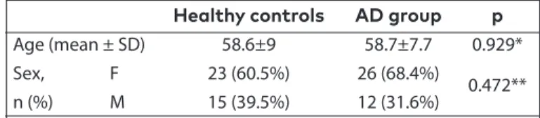
Our study was a case-control study. A total of 38 patients including 12 males and 26 females who had been diagnosed as having probable AD according to the 2011 National In-stitute of Neurological and Communicative Disorders and Stroke and the Alzheimer’s Disease and Related Disorders Association (NINCDS-ADRDA) criteria were included in the study. The control group was composed of the patient’s rel-atives who volunteered to participate in the study, who had no first-degree consanguinity and were healthy individuals with no neurologic disease and MMSE scores of 24 and above (n=38). The mean age of the patients was 58.7±7.7 years (minimum: 37 years, maximum: 75 years). Twelve (31.6%) were male, and 26 (68.4%) were female. The mean age of the controls was 58.6±9 years (minimum: 43 years, maximum: 84 years). Fifteen (39.5%) were male, and 23 (60.5%) were fe-male. No significant difference was found in the distribution of the age (p=0.929) and sex (p=0.472) of the patients and controls (Table 1).
The years of education were obtained by asking the patients and/or their relatives and controls during face-to-face inter-views. The cognitive status was assessed using the MMSE and Clinical Dementia Rating (CDR). Depression was determined using the Geriatric Depression Scale (GDS). Standardized neuropsychological tests were used for the neuropsycho-logical assessment. Neuropsychoneuropsycho-logical assessments were performed by collecting the data of the patients and controls via face-to-face interviews between December 24th, 2012, and August 28th,2015, by taking informed consent forms. Periph-eral blood samples (10 cc) were taken from the patients and controls.
The tests that were administered to patients were the MMSE, CDR, NPI, Auditory Verbal Learning Test (AVLT), Wechsler Memory Scale visual reproduction subtests, Semantic Fluen-cy Test, Lexical FluenFluen-cy Test, Clock Drawing Test, and Boston Naming Test.
Healthy controls were administered the MMSE and GDS. In our study, the Turkish version of the MMSE test for literate and illiterate patients was used (8). The Öktem Verbal Memory Processes test was applied for the assessment of verbal mem-ory, and instant learning and total learning were assessed (9). The Wechsler Memory Scale visual reproduction subtests was used in the assessment of visual memory, and visual learning score was assessed (10). The animal category was used for se-mantic fluency, and the KAS (reproducing words begin with letters K, A and S) test was used for lexical fluency (11, 12). The Schulman Method was used in the assessment of the Clock Drawing Test (13). The Boston Naming Test was used in the as-sessment of naming (14). The Turkish version of the GDS was used in the assessment of depression (15).
The main independent variable of the study was the IL-1β level. Apart from the independent variable (IL-1β) for the basic hypoth-esis, age, sex, education, MMSE and GDS scores of the cases and controls were determined and compared. Furthermore, other findings of the patient group related to AD (CDR, NPI, AVLT in-stant learning, AVLT total learning, Visual Learning Score, Seman-tic Fluency, Lexical Fluency, Clock Drawing, Boston Naming) were presented as descriptive statistics. Peripheral blood samples were taken from the patients and controls during the examina-tion. Serums were separated through centrifugation and stored at -80°C. Blood samples were taken from patients and controls and placed into a red-capped biochemical tube. The samples were placed into receiving tubes and centrifuged at 2000 g for 10 minutes, the serums were resolved and stored at -80°C until the day of the study. The analysis of the collected serum samples was performed using a Human IL-1 beta enzyme-linked immu-nosorbent assay (ELISA) kit (Catalog No: BMS224/2). Each serum sample was studied with kit standards. Standards were made in serial dilutions from the main stock (16 ng/mL) including Stan-dard 1 (8 ng/mL), StanStan-dard 2 (4 ng/mL), StanStan-dard 3 (2 ng/mL), Standard 4 (1 ng/mL), Standard 5 (0.5 ng/mL), Standard 6 (0.25 ng/mL), Standard 7 (0.125 ng/mL) and Standard 8 (0 ng/mL). Standard dilution was performed with deionized water.
Standards, and sera from the controls and patients were placed in wells in accordance with the plaque map, then, biotin conjugate was added, and they were allowed to incu-bate at room temperature for 2 hours. After being washed for three times, the Streptavidin-HRP solution from the kit was added to each well, and it was allowed to incubate at room temperature for 1 hour again. The TMB substrate solution was added to the wells, washed three times after incubation, and the plaque was allowed to incubate at room temperature for
Healthy controls AD group p
Age (mean ± SD) 58.6±9 58.7±7.7 0.929* Sex, F 23 (60.5%) 26 (68.4%)
n (%) M 15 (39.5%) 12 (31.6%) 0.472**
*t-test; ** Chi-square
AD: Alzheimer’s disease; SD: standart deviation; F: female; M: male
Table 1. Distribution of the patients and controls accord-ing to age and sex
143
10 minutes while protecting them against light. At the end of this period, the Stop solution needed to stop the reaction was added, and readings were performed. Readings were performed at 450 nm using an ELISA Multiplaque reader. The assay was studied on 1 ELISA plaque.
Statistical Analysis
The suitability of the obtained data for normal distribution was checked using the Kolmogorov-Smirnov and Shapiro-Wilk tests. Normally distributed data are presented as mean and deviation, and the t-test was applied in the comparison of two groups. The suitability of the data for the parametric distribution was tested again by firstly applying logarithmic transformation for non-normally distributed data, the comparison of two group distributions for non-parametrically distributed data was per-formed using the Mann-Whitney U test; these data are present-ed as mpresent-edian (minimum value-maximum value). The presence of a correlation between IL-1β and age and other clinical assess-ments was assessed using the Pearson rho coefficient under parametric conditions, and with the Spearman rho coefficient under non-parametric conditions. The obtained variables were compared using Chi-square analysis. In the AD-control separa-tion, the distinctiveness of IL-1 was performed using receiver op-erating curve (ROC) analysis. In all statistical evaluations, p<0.05 was accepted as the level of significance. The Statistical Package for the Social Sciences 22.0 program (SPSS IBM Corp.; Armonk, NY, USA) was used for data analysis.
RESULTS
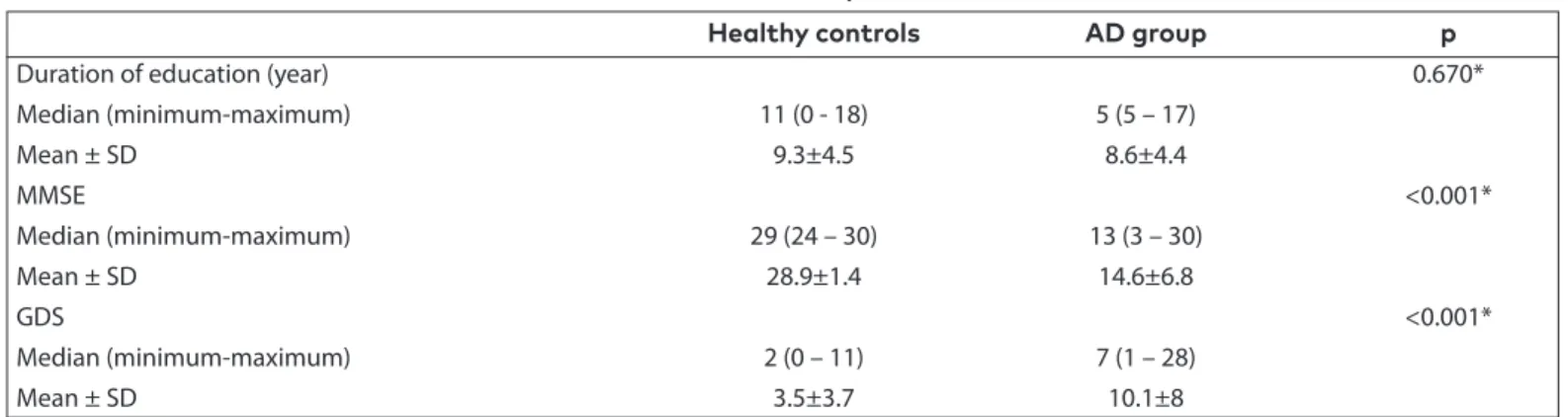
Thirty-eight patients and 38 healthy controls who had been diagnosed as having probable AD according to the NINCDS-ADRDA criteria were included in the study. For the control group, voluntary individuals who gave a positive answer to the verbal notification, lived in their own houses, and con-tinued daily life activities independently between December 24th, 2012, and August 28th,2015, were included in the order of application. The median duration of education of the patients was 5 years (minimum: 5, maximum: 17, mean: 8.6±4 years), and the median of the controls was 11 years (minimum: 0, maximum: 18, mean: 9.3±4.5 years). No significant difference
was found between the durations of education of the patients and controls (p=0.670, Table 2). The median MMSE score of the patients was 13 (minimum: 3, maximum: 30, mean: 14.6±6.8), and that of the controls was 29 (minimum: 24, maximum: 30, mean: 28.9±1.4). The MMSE scores of the patient group were found to be significantly lower compared with the control group (p<0.001, Table 2).
The patients’ median GDS value was 7 (minimum: 1, maxi-mum: 28, mean: 10.1±8), and for the controls it was 2 (mini-mum: 0, maxi(mini-mum: 11, mean: 3.5±3.7). The GDS score in the patient group was found to be significantly higher compared with the control group (p<0.001, Table 2).
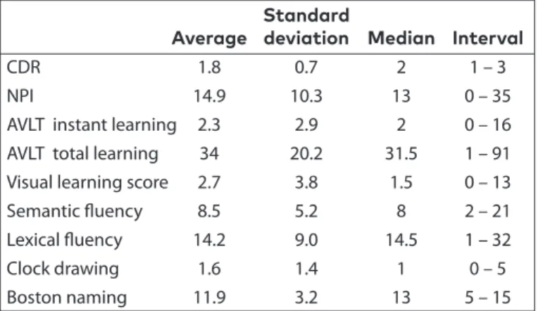
The CDR, NPI score and neuropsychological test scores in pa-tients with AD are presented in Table 3.
The fit of the IL-1β measurements to normal distribution was evaluated between the patient and control groups, and they were found not to be normally distributed. The necessary sta-tistical transformation was applied at logarithm base 10. Be-cause the interleukin distribution did not comply with normal distribution with these data too, the two groups were com-pared nonparametrically using the Mann-Whitney U test. It was observed that the median IL-1β measurement of the patient group was 1.07 (0-112.70), and was 0.84 (0-69.98) for the control group. It was found that the distribution of the IL-1β measurements of the patient and control groups was not statistically different (MWU p-value 0.658, Table 4).
Correlations tests were performed to examine the association of IL-1β measurement with other data.
A nonparametric correlation test was used to examine the correlations of IL-1β measurements with age. When the correlation between age and IL-1β was examined without considering the diagnosis (r= -0.1, p=0.392), no significant correlation was found between them. There was also no cor-relation between age and IL-1β levels when the corcor-relation
Healthy controls AD group p
Duration of education (year) 0.670*
Median (minimum-maximum) 11 (0 - 18) 5 (5 – 17) Mean ± SD 9.3±4.5 8.6±4.4 MMSE <0.001* Median (minimum-maximum) 29 (24 – 30) 13 (3 – 30) Mean ± SD 28.9±1.4 14.6±6.8 GDS <0.001* Median (minimum-maximum) 2 (0 – 11) 7 (1 – 28) Mean ± SD 3.5±3.7 10.1±8
* Comparison of two non-parametric independent groups, Mann-Whitney U test AD: Alzheimer’s disease; SD: standart deviation; GDS: geriatric depression scale
144
analysis between IL-1β and age was examined separately in the patient group (r= -0.151 p=0.374) and healthy controls (r= -0.80 p=0.633).
In the patient group, the correlation of IL-1β measurements with the MMSE was not significant (r= -0.080 p=0.646). It was determined that IL-1β was not correlated with the GDS (r= -0.080 p=0.711). In the patient group, the correlation of IL-1β measurements with the other clinical data was also found not to be significant (p>0.05).
A moderate negative significant correlation was found only with the Clock Drawing Test (r= -0.508, p=0.016) and the Visu-al Learning Score (r= -0.435, p=0.043) among the neuropsy-chological tests. No significant correlation was detected with the results of other neuropsychological tests (p>0.05, Table 5). In the control group, IL-1β measurements were found to be correlated with the MMSE (p=0.028 r= -0.357). IL-1β was not correlated with the GDS (p=0.790 r= -0.045) (Table 6).
DISCUSSION
Interleukin-1 levels in AD were first investigated in 1989; an increase in IL-1 immunoreactive neurons in the temporal lobes of brains with AD and Down’s syndrome and a signifi-cant increase in IL-1 immunoreactive products in brains with AD were determined in this study. IL-1 immunoreactive astro-cytes were observed around the plaques in the gray matter of those with AD (16).
Astrocytic activation is responsible for the formation of dys-trophic neurites in the amyloid deposits and the support of the glial-cytokine-mediated cascade underlying the neuro-pathologic changes in AD (17). It was observed that IL-1 in-creased the expression of adhesion molecules in glial cells via nuclear factor-kappa B (18). In a study performed on astrocy-tomas, it was shown that IL-1 increased the level of vascular cell adhesion molecule-1 on astrocytes and led to astrocyte activation, and that astrocyte activation was effective in the cross of other leukocytes from the blood brain barrier (19). The research data on circulating cytokines as a marker of pe-ripheral cytokine dysregulation in AD, in which inflammation in the central nervous system (CNS) has been demonstrated, are inconsistent; the probable cause of this is that the presence of the blood-brain barrier weakens the validity of biomarkers such as brain molecules through peripheral blood (20). The blood-brain barrier is not permeable to polar solutes and macromol-ecules due to its tight connections (21). Although the increase in permeability in the blood-brain barrier has been shown in AD, the fact that normal aging also increases the degradation and permeability in the blood brain barrier has been shown in a study with human hippocampal sections (22, 23). The inclusion of mostly early-onset cases in our study may have led to the lower levels of IL-1β in the AD group, which were not found to be higher compared with the controls when the effect of aging was taken into account. In studies in which there was a signif-icant difference in the patient group for IL-1β in the literature, the average age was between 70 and 80 years (24-26).
Standard
Average deviation Median Interval
CDR 1.8 0.7 2 1 – 3
NPI 14.9 10.3 13 0 – 35 AVLT instant learning 2.3 2.9 2 0 – 16 AVLT total learning 34 20.2 31.5 1 – 91 Visual learning score 2.7 3.8 1.5 0 – 13 Semantic fluency 8.5 5.2 8 2 – 21 Lexical fluency 14.2 9.0 14.5 1 – 32 Clock drawing 1.6 1.4 1 0 – 5 Boston naming 11.9 3.2 13 5 – 15
CDR: clinical dementia rating; NPI: neuropsychiatric inventory; AVLT: auditory verbal learning test
Table 3. The clinical dementia rating, behavioral scale (NPI) score, and neuropsychological test scores in pa-tients with AD
Healthy controls AD group p
IL-1β 0.84 1.07 0.658*
IL-1 β: interleukin (IL)-1β; *: Mann Whitney u test
Table 4. Comparison of the IL-1β values between the healthy controls and patient group
Rho p Age -0.151 0.374 MMSE -0.080 0.646 GDS -0.080 0.711 NPI 0.159 0.411 CDR 0.297 0.118
AVLT instant learning -0.115 0.554 AVLT total learning -0.097 0.638 Visual learning score -0.435 0.043 Semantic fluency -0.017 0.929 Lexical fluency -0.207 0.566 Clock drawing test -0.508 0.016 Boston naming test -0.259 0.212
MMSE: mini-mental state examination; GDS: global deterioration scale; NPI: neuropsychiatric inventory; CDR: clinical dementia rating; AVLT: auditory verbal learning test
Table 5. Nonparametric correlations of IL-1β in patient group
Rho p
Age -0.80 0.633
MMSE -0.357 0.028
GDS -0.045 0.790
MMSE: mini-mental state examination; GDS: global deterioration scale
Table 6. Nonparametric correlations of IL-1β in healthy control group
145
Cytokine release in AD is thought to be an early initiating fac-tor rather than the late outcomes of the disease (27). The in-flammatory response that increases as an effort of glial cells to clear the Aβ deposits at the early stages of the disease de-creases with the loss of normal neuronal circulation and the death of neurons at late stages, and a decrease occurs in the cytokine and chemokine release (28). In a study, IL-1β levels were lower in patients with mild cognitive impairment (MCI) than in patients with mild AD, and this result was interpreted as inflammation being an early event in AD (29). The fact that the patients in our study were at the clinical dementia stage could explain the low levels of IL-1β.
In the literature, IL-1β levels were examined in studies with various designs in terms of whether it distinguished patients from controls or different patient groups and in terms of whether it was correlated with some parameters in patients. Although it was mostly examined in the serum and plasma, there are also studies in which it was examined in cerebro-spinal fluid (CSF) (30). In some of these studies, significant differences were found between the groups. In a study con-ducted in 20 patients with mild-moderate AD and 20 healthy controls, it was determined that serum IL-1β levels increased in patients with AD compared with the controls, but this in-crease was only in those with early-onset AD in the subgroup analysis, there was a negative correlation between the IL-1β level and MMSE, and the IL-1β level was positively correlated with age in healthy individuals (31). In a study of 145 patients with AD and 51 control individuals without dementia, plasma IL-1β was detected only in 17 patients with AD and 1 healthy control, and the serum IL-1β levels were stated to be higher in those with in AD compared with the controls (24). In a study involving 60 patients with late-onset AD, 80 with vascular de-mentia (VaD), 40 patients with cerebrovascular disease with-out dementia, and 42 healthy control individuals, plasma IL-1β levels were found to be higher those with VaD, late-onset AD, and cerebrovascular disease without dementia compared with the controls (25). In another study involving 88 patients with mild-moderate AD, 74 with MCI, and 31 healthy controls, serum IL-1β levels were found to be significantly higher in patients with AD compared with the controls; the values of patients MCI were found between the two, and it was report-ed that its difference with the controls was not statistically significant. In that study, patients with MCI were divided into three groups as amnestic in the single-domain, non-amnesic, and amnestic in the multiple-domain. The serum IL-1β levels in multiple-domain MCI were found to be significantly higher compared with amnestic MCI, nonamnestic MCI, and controls, and close to the level in AD; in all cases, the serum IL-1β lev-els were found to be negatively correlated with the cognitive parameters (MMSE and episodic memory), and it was shown that the effect of age and sex on IL-1β was not significant (26). In another study involving 55 patients with AD and 37 healthy controls, which aimed to investigate correlations of inflam-matory cytokines in AD and cognitive functions and
depres-sion, serum IL-1β levels were found to be significantly higher in patients with AD with and without depression compared with the controls, and no correlation was found between the MMSE and IL-1β in patients with AD with and without depres-sion (32).
In a study with 29 patients with AD, 17 with VaD, 13 with MCI, and 23 healthy controls in Turkey in 2007, a correlation was shown between the MMSE and IL-1β levels in the AD group (33). In another study with 21 patients with early-onset AD, 53 with late-onset AD, 30 with MCI, 40 with Parkinson’s disease, and 53 healthy controls in Turkey in 2015, serum IL-1β levels were found to be significantly higher in the early-onset AD group compared with healthy controls of the same age. Sim-ilarly, in patients with early-onset AD and Parkinson’s disease, both had significantly higher levels compared with patients with MCI; higher in those with early-onset AD compared with late-onset AD (nonsignificant); and significantly higher in pa-tients with Parkinson’s disease compared with the controls, patients with MCI and late-onset AD of the same age; and also serum IL-1β was found to be low in those with high MMSE scores among patients with Parkinson’s disease (34).
There are also negative studies in which similar relationships were not found. In a study in which IL-1β levels and were investigated in blood taken from 8 patients with AD and 9 normal individuals or those with non-AD CNS disease twice 2-5 year intervals and with other cytokines in CSF, IL-1β was detected in CSF in 3 of 8 patients with AD and 4 of 9 control individuals; no significant difference was found between the patients with AD and controls, and no relationship was found between cognitive deterioration and CSF IL-1β levels in pa-tients with AD (30). In a study involving 44 papa-tients with AD, 7 with multi-infarct dementia, 10 with mixed dementia, 18 with undetermined dementia, 12 with cognitive impairment, and 43 healthy controls in which cytokine levels were investigated by stimulation with lipopolysaccharide, cytokine levels were found to be significantly higher in those with multi-infarct de-mentia compared with the controls, and it was concluded IL-1β levels were not affected by lipopolysaccharide stimulation (35). In another study involving 60 patients with AD, 60 with VaD, and 33 healthy control individuals, no significant differ-ence was found between the groups in terms of serum IL-1β levels, no correlation was found between serum IL-1β levels and MMSE and functional assessment scales in patients with AD, and a negative correlation was found with IL-1β MMSE only in VaD (36). In a study involving 72 patients with AD and 6 healthy controls in 2011, plasma IL-1β levels in the AD group were found to be within normal limits (37). In a study with 40 patients with AD, 20 with Parkinson’s disease, and 42 healthy controls in 1994, in which IL-1β levels were investigated in CSF and serum, no difference was found in CSF and serum IL-1β levels between the groups (38). In another study with 72 patients including 56 with AD, 16 with VaD, and 15 healthy control individuals in 2008, it was found that there was no
146
difference in the mean interleukin levels between those with possible AD and VaD, serum IL-1β levels were not correlated with age in patients with dementia, there was no relationship between interleukin levels and dementia severity, and blood IL-1β levels were higher in the early-onset patients, although it was found that there was no correlation between interleu-kin levels and GDS (39).
Thirty-eight patients with AD and 38 healthy controls were in-cluded in our study; there are studies with smaller and larger groups in the literature. IL-1β levels were assessed using ELISA in serum, and they have also been assessed using similar tech-niques in other studies.
Cytokine levels in peripheral circulation are affected by many factors such as sample collection, processing, and storage, and even before taking the patient’s sample, the time sam-pling, nutritional status of the patient, acute stress, storage method and duration, and choice of anticoagulant may affect the circulating cytokine levels (40). That the time of sampling was not constant, the patients’ fasting status was not known, and drug uses were not established may have led to the fact that cytokine levels were not found as significant.
Our study is among studies in which no significant difference was found between patients with AD and healthy controls in terms of the serum IL-1β levels. Moreover, the fact that IL-1β measurements in the patient group were not correlated with the MMSE is also consistent with the literature.
Our study is the first to determine that IL-1β is not correlated with the GDS; the correlation of IL-1β with the GDS has not been examined previously in the literature. Correlations were analyzed with detailed neuropsychological assessment re-sults for the first time in our study.
In this study, it was observed the IL-1β levels in serum were not sufficient to distinguish patients with AD from controls. However, a moderate, negative, significant correlation was found in the Clock Drawing Test, one of the neuropsycholog-ical tests, and the Visual Learning Score, and no significant correlation was found with other neuropsychological test results.
As a result of our study, the most probable reason for the fact that no difference was found between the groups is the small size of the sample. The weak correlations obtained in our study may indicate that stronger correlations will be obtained in studies in which more patients and controls are included.
Ethics Committee Approval: Ethics committee approval was
received for this study from the Dokuz Eylül University In-stitute of Health Sciences Neurological Sciences support-ed by TÜBİTAK (date: 5.4.2012, protocol no: 32-SBKAEK).
Informed Consent: Written informed consent was obtained
from patients and healthy controls who participated in this study.
Peer-review: Externally peer-reviewed.
Author Contributions: Concept – G.Y., Ş.G., O.B.; Design –
G.Y., Ş.G., O.B.; Supervision – G.Y., Ş.G., O.B.; Resources – E.E., U.V., Ş.G.; Materials – O.B., A.T., G.B., N.D., E.E., U.V., Ş.G., G.Y.; Data Collection and/or Processing – G.Y., Ş.G., A.T., O.B., G.B., N.D.; Analysis and/or Interpretation – G.Y., O.B.; Literature Search – G.Y., Ş.G., O.B.; Writing Manu-script – G.Y., O.B., N.D.; Critical Review – G.Y., O.B.
Conflict of Interest: The authors have no conflicts of interest
to declare.
Financial Disclosure: The authors declared that this study
has received no financial support. REFERENCES
1. Dickson DW. The pathogenesis of senile plaques. J Neuropathol Exp Neurol 1997; 56: 321-339. [CrossRef]
2. Akiyama H, Barger S, Barnum S, et al. Inflammation and Alzhei-mer's disease. Neurobiol Aging 2000; 21: 383-421. [CrossRef] 3. Wegiel J, Wang KC, Imaki H, et al. The role of microglial cells and
astrocytes in fibrillar plaque evolution in transgenic APP(SW) mice. Neurobiol Aging 2001; 22: 49-61. [CrossRef]
4. Halle A, Hornung V, Petzold GC, et al. The NALP3 inflammasome is involved in the innate immune response to amyloid-beta. Nat Immunol 2008; 9: 857-865. [CrossRef]
5. Griffin WS, Stanley LC, Ling C, et al. Brain interleukin 1 and S-100 immunoreactivity are elevated in Down syndrome and Alzheimer disease. Proc Natl Acad Sci U S A 1989; 86: 7611-7615. [CrossRef] 6. Dinarello CA. Interleukin-1 in the pathogenesis and treatment of
inflammatory diseases. Blood 2011; 117: 3720-3732. [CrossRef] 7. Webb AC, Collins KL, Auron PE, et al. Interleukin-1 gene (IL1)
as-signed to long arm of human chromosome 2. Lymphokine Res 1986; 5: 77-85.
8. Güngen C, Ertan T, Eker E, Yaşar R, Engin F. Reliability and vality of the standardized Mini Mental State Examination in the di-agnosis of mild dementia in Turkish population. Turk Psikiyatri Derg 2002; 13: 273-281.
9. Öktem Tanör Ö. Öktem Sözel Bellek Süreçleri Testi (Öktem-VMPT) El Kitabı. Türk Psikologlar Derneği Yayınları, 2011, No: 34, Ankara. 10. Wechsler D. Manual for the Wechsler Memory Scale-Revised,
The Psychological Corporation, 1987, San Antonio, TX.
11. Tumaç A. The effect of age and education on performance in some frontal damage-susceptible tests in normal subjects. İs-tanbul Üniversitesi, Sosyal Bilimler Enstitüsü, Unpublished mas-ter's thesis, 1997.
12. Öktem Tanör Ö. Nöropsikolojik Değerlendirme In: Tanrıdağ O, Davranış Nörolojisi, Nobel Tıp Kitabevleri, 2016, İstanbul. 13. Shulman KI, Gold DP, Cohen CA, Zucchero CA. Clock-drawing
and dementia in the community: A longitudinal study. Int J Geri-atr PsychiGeri-atry 1993; 8: 487-496. [CrossRef]
14. Fernández-Blázquez MA, Ruiz-Sánchez de León JM, López-Pina JA, Llanero-Luque M, Montenegro-Peña M, Montejo-Carrasco P. A new shortened version of the Boston Naming Test for those aged over 65: an approach from item response theory. Rev Neu-rol 2012; 55: 399-407. [CrossRef]
147
15. Ertan T, Eker E. Reliability, validity, and factor structure of thegeriatric depression scale in Turkish elderly: are there different factor structures for different cultures? Int Psychogeriatr 2000; 12: 163-172. [CrossRef]
16. Griffin WS, Stanley LC, Ling C, et al. Brain interleukin 1 and S-100 immunoreactivity are elevated in Down syndrome and Alzheimer disease. Proc Natl Acad Sci U S A 1989; 86: 7611-7615. [CrossRef] 17. Mrak RE, Sheng JG, Griffin WS. Correlation of astrocytic S100
beta expression with dystrophic neurites in amyloid plaques of Alzheimer's disease. J Neuropathol Exp Neurol 1996; 55: 273-279. [CrossRef]
18. Moynagh PN, Williams DC, O'Neill LA. Activation of NF-kappa B and induction of vascular cell adhesion molecule-1 and intracel-lular adhesion molecule-1 expression in human glial cells by IL-1. Modulation by antioxidants. J Immunol 1994; 153: 2681-2690. 19. Rosenman SJ, Shrikant P, Dubb L, Benveniste EN, Ransohoff RM.
Cytokine-induced expression of vascular cell adhesion mole-cule-1 (VCAM-1) by astrocytes and astrocytoma cell lines. J Im-munol 1995; 154: 1888-1899.
20. Lee KS, Chung JH, Choi TK, Suh SY, Oh BH, Hong CH. Peripheral cytokines and chemokines in Alzheimer's disease. Dement Geri-atr Cogn Disord 2009; 28: 281-287. [CrossRef]
21. Abbott NJ, Patabendige AA, Dolman DE, Yusof SR, Begley DJ. Structure and function of the blood-brain barrier. Neurobiol Dis 2010; 37: 13-25. [CrossRef]
22. Marques F, Sousa JC, Sousa N, Palha JA. Blood-brain-barriers in aging and in Alzheimer's disease. Mol Neurodegener 2013; 8: 38. [CrossRef]
23. Montagne A, Barnes SR, Sweeney MD, et al. Blood-brain barrier breakdown in the aging human hippocampus. Neuron 2015; 85: 296-302. [CrossRef]
24. Licastro F, Pedrini S, Caputo L, et al. Increased plasma levels of interleukin-1, interleukin-6 and alpha-1-antichymotrypsin in patients with Alzheimer's disease: peripheral inflammation or signals from the brain? J Neuroimmunol 2000; 103: 97-102. [CrossRef]
25. Zuliani G, Ranzini M, Guerra G, et al. Plasma cytokines profile in older subjects with late onset Alzheimer's disease or vascular dementia. J Psychiatr Res 2007; 41: 686-693. [CrossRef]
26. Forlenza OV, Diniz BS, Talib LL, et al. Increased serum IL-1beta level in Alzheimer's disease and mild cognitive impairment. De-ment Geriatr Cogn Disord 2009; 28: 507-512. [CrossRef] 27. Tarkowski E, Andreasen N, Tarkowski A, Blennow K. Intrathecal
inflammation precedes development of Alzheimer's disease. J Neurol Neurosurg Psychiatry 2003; 74: 1200-1205. [CrossRef] 28. Galimberti D, Schoonenboom N, Scheltens P, et al. Intrathecal
chemokine synthesis in mild cognitive impairment and Alzhei-mer disease. Arch Neurol 2006; 63: 538-543. [CrossRef]
29. Magaki S, Mueller C, Dickson C, Kirsch W. Increased production of inflammatory cytokines in mild cognitive impairment. Exp Gerontol 2007; 42: 233-240. [CrossRef]
30. Lanzrein AS, Johnston CM, Perry VH, Jobst KA, King EM, Smith AD. Longitudinal study of inflammatory factors in serum, ce-rebrospinal fluid, and brain tissue in Alzheimer disease: inter-leukin-1beta, interleukin-6, interleukin-1 receptor antagonist, tumor necrosis factor-alpha, the soluble tumor necrosis factor receptors I and II, and alpha1-antichymotrypsin. Alzheimer Dis Assoc Disord 1998; 12: 215-227. [CrossRef]
31. Alvarez XA, Franco A, Fernández-Novoa L, Cacabelos R. Blood levels of histamine, IL-1 beta, and TNF-alpha in patients with mild to moderate Alzheimer disease. Mol Chem Neuropathol 1996; 29: 237-252. [CrossRef]
32. Khemka VK, Ganguly A, Bagchi D, et al. Raised serum proinflam-matory cytokines in Alzheimer's disease with depression. Aging Dis 2014; 5: 170-176. [CrossRef]
33. Oztürk C, Ozge A, Yalin OO, et al. The diagnostic role of serum inflammatory and soluble proteins on dementia subtypes: cor-relation with cognitive and functional decline. Behav Neurol 2007; 18: 207-215. [CrossRef]
34. Dursun E, Gezen-Ak D, Hanağası H, et al. The interleukin 1 alpha, interleukin 1 beta, interleukin 6 and alpha-2-macroglobulin se-rum levels in patients with early or late onset Alzheimer's dis-ease, mild cognitive impairment or Parkinson's disease. J Neuro-immunol 2015; 283: 50-57. [CrossRef]
35. De Luigi A, Fragiacomo C, Lucca U, Quadri P, Tettamanti M, Gra-zia De Simoni M. Inflammatory markers in Alzheimer's disease and multi-infarct dementia. Mech Ageing Dev 2001; 122: 1985-1995. [CrossRef]
36. Yasutake C, Kuroda K, Yanagawa T, Okamura T, Yoneda H. Serum BDNF, TNF-alpha and IL-1beta levels in dementia patients: com-parison between Alzheimer's disease and vascular dementia. Eur Arch Psychiatry Clin Neurosci 2006; 256: 402-406. [CrossRef] 37. Corsi MM, Licastro F, Porcellini E, et al. Reduced plasma levels of
P-selectin and L-selectin in a pilot study from Alzheimer disease: relationship with neuro-degeneration. Biogerontology 2011; 12: 451-454. [CrossRef]
38. Pirttila T, Mehta PD, Frey H, Wisniewski HM. Alpha 1-antichymo-trypsin and IL-1 beta are not increased in CSF or serum in Alzhei-mer's disease. Neurobiol Aging 1994; 15: 313-317. [CrossRef] 39. Angelopoulos P, Agouridaki H, Vaiopoulos H, et al. Cytokines in
Alzheimer's disease and vascular dementia. Int J Neurosci 2008; 118: 1659-1672. [CrossRef]
40. Zhou X, Fragala MS, McElhaney JE, Kuchel GA. Conceptual and methodological issues relevant to cytokine and inflammatory marker measurements in clinical research. Curr Opin Clin Nutr Metab Care 2010; 13: 541-547. [CrossRef]