Haliç University, Printed in Turkey.
Characterization of
Bacillus
species by numerical analysis of their
SDS-PAGE protein profiles
Ismet Berber
Department of Biology, Faculty of Art and Science, Yüzüncü Y›l University, 65080, Van, Turkey
Received 1 December 2003; Accepted 26 January 2004Abstract
In the present study nine reference Bacillusstrains were characterized by whole-cell protein profiles using sodium dodecyl sulfate-polyacrylamide gel electrophoresis (SDS-PAGE). A numerical classification of the protein profiles revealed two distinct clusters at 47% similarity level. Cluster 1 comprised four strains belongs to B. subtilisand B. megateriumspecies at similarity levels changed between 67 and 85%, while cluster 2 consisted five strains belongs to various Bacillusspecies at similarity levels changed between 50 and 70%. The strains of the cluster 2 were clearly separated from that of cluster 1 by numerical analysis. Our results indicates that SDS-PAGE method combined with computerized analysis of cellular protein profiles provide an effective approach to investigate of taxonomic relationships within Bacillusspecies.
Key words:Characterization, Bacillus,SDS-PAGE, numerical taxonomy
Bacillus
türlerinin SDS-PAGE protein profillerinin nümerik analizle karakterizasyonu
Özet
Bu çal›flmada, dokuz referans Bacillus›rk› sodyum dodesil sülfat poliakrilamid jel elektroforezi kullan›larak elde edilen toplam hücresel protein profillerine göre karakterize edildi. Protein profil sonuçlar› esas al›narak yap›lan nümerik s›n›fland›rma %47 benzerlik düzeyinde iki ayr› grup (cluster) oluflturdu. Grup 1 benzerlik düzeyi %67-85 aras›nda de¤iflen B. subtilisve B. megaterium türlerine ait dört ›rk›; grup 2 benzerlik seviyesi % 50-70 aras›nda de¤iflen farkl› Bacillus türlerine ait befl ›rk› içermektedir. Nümerik analizde grup 1’e ait ›rklar grup 2’den aç›kça ayr›lmaktayd›. Bizim sonuçlar›m›z, SDS-PAGE yöntemiyle elde edilen hücresel protein profillerinin bilgisayar analizleriyle birlefltirilmesinin Bacillus türlerinin taksonomik iliflkilerinin incelenmesinde etkili bir yaklafl›m sa¤lad›¤›n› gösterdi.
Anahtar sözcükler:Karakterizasyon, Bacillus,SDS-PAGE, nümerik taksonomi
Introduction
The genus Bacillus are generally defined as gram-positive, aerobic or facultative anaerobic, motile, peritrichous flagella and endospore-forming rod-shaped microorganisms (Claus and Berkeley, 1986). This diversity was apparent even with classical
phenotypic characterization based primarily on morphology, nutrition, growth characteristics; and various substrate utilization and physiological assessments (Slepecky and Hemphill, 1992). Although physiological reactions are generally used to determine the species of the genus, inconsistencies in test results can make identification difficult (Ash et al., 1991).
Description of the genus has been improved by using information obtained from DNA base composition and DNA-DNA hybridization studies which was listed in
Bergey’s Manual of Systematic Bacteriology40 (Claus and Berkeley, 1986). However, in the literature there are newly identified species which were shown to be genetically and phenotypically distinct from other
Bacillus species and have not been described in
Bergey’s Manual(Slepecky and Hemphill, 1992). A number of different methods have been used for typing Bacillus species follow: Serotyping, bacteriophage typing, bacteriocin activities, antibiogram and biotyping, plasmid typing, analysis of cellular fatty acid content, native-PAGE, small-subunit-ribosomal RNA sequencing and genome analysis (Ash et al., 1991; Berber and Cokmus, 2001). Although these methods have been used for identification of Bacillus
species, characterization of these microorganisms is still not well defined (Ivanova et al., 1999).
A second level information for a cell, other than sequencing of bacterial genome, can be obtained from cellular protein profiles. Different types of electrophoresis were used to explore the profiles. The protein profiles produced by SDS-PAGE of whole cell extract have been found correlates closely with DNA-DNA hybridization results suggests it could be appropiate to use SDS-PAGE for rapid bacterial identification (Vauterin et al., 1990; Niemi et al., 1993; Berber et al., 2003).
Combination of polyacrylamide gel electrophoresis (PAGE) of proteins with computerized analysis of profiles provided an effective approach to investigate the taxonomic relationships among many bacterial species (Kersters, 1985; Costas, 1992). This paper describes PAGE results of nine reference Bacillus
species. The aim of the study was to evaluate the usefulness of the technique as a taxonomic tool in this genus.
Materials and methods
Bacteria and growth conditions
The test bacteria used in our study have been provided from Prof Dr. Cumhur Cokmus (Department of Biology, Faculty of Sciences, Ankara University Ankara TURKEY). All cultures were grown at 30°C for 24 h on NYSM (Difco, DETRO‹T)) agar and propagated at least twice before use.
P reparation of whole-cell proteins
For each culture, a loopful of overnight growth on NYSM agar plate was suspended in 15 ml NYSM broth, and incubated on rotated incubator for 48 h (at 30°C, 150 rpm). Samples were then transferred into 1.3 ml eppendorf tubes, centrifuged for 3 minutes at 12,100 rpm, and the collected cells were washed three times with distilled water. The washed cells were stirred after adding 25 µl SDS-samples buffer on (0.06 M Tris-HCl, 2.5% Glycerol, 0.5% SDS, 1.25% ß-mercaptoethanol) and the proteins were denatured by boiling the tubes for 5 minutes (Laemmli, 1970).
SDS-PAGE
Solubilized proteins were subjected to SDS-PAGE in gel slabs of 1 mm thickness (3.5cm, 4% stacking and 16.5cm, 12.5% resolving gels) as described by Laemmli (1970). Electrophoresis was performed with a discontinuous buffer system in a BRL gel apparatus model V16-2BRL Gaithersburg MD, USA. The gel was run at 30 mA until the bromophenol blue marker had reached to the bottom of the gel. Gels were then stained with Coomassie Brillant Blue R-250.
Data analysis
Gels were examined by naked eyes directly and the protein profiles were recorded as binary data, that is, 1 or 0. The resultant data were typed into the MINITAB (Version 13.1) program. This is a program for data input and analysis of binary data and is run on IBM computer. The similarity and relationship between the protein traces of test strains were expressed in a dendrogram derived by using the Pearson product-moment correlation coefficient and unweighted pair group method with arithmetic averages algorithm.
Results
Figure 1 shows the whole-cell protein profiles of
Bacillusspecies obtained by sodium dodecyl sulphate polyacrylamide gel electrophoresis. There were considerable differences in protein profiles of Bacillus
species at 20.000-66.000 kDa region. Most of Bacillus
species, except B. megaterium DSM 32 and B. megaterium ATCC 1842, contain the protein band (marked by 1) at the top of each lane. B. subtilisand B.
subtilisDSM 10 strains had similar protein patterns for the bands marked by 10 and 12 (lines A and B). However, B. subtilisDSM 10 strain distinguished from other strain with the presence of a band (marked by 16). B. megaterium DSM 32 (Fig 1, line C), B. megaterium ATCC 1842 (Fig 1, line D) and B. megaterium(Fig 1, line F) had crucial differences in protein bands, although they belong to the strains of same specie. These strains were separated from each other in the presence or absent of some protein bands marked by 3, 4, 6, 11, 13 and 15. B. licheniformisand
B. thuringiensis var. israelensis obviously differed from other Bacillus species (Fig 1, lines E and H). Moreover, some variations were observed between protein profiles of two species (B. sphaericus MRS 400 and B. cereus 7064) and other Bacillus species (Fig 1, lines G and I). Protein pattern of B. cereus7064 strain was slightly similar to B. sphaericusMRS 400 strain, although, strain was distinguished by the presence of four darkly protein bands (marked as 5, 8, 9, 14).
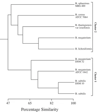
A dendrogram produced after numerical analysis of the whole-cell protein profiles using the Pearson product-moment correlation coefficient and unweighted pair group method with arithmetic averages algorithm (UPGMA) is shown in Figure 2. Numerical analysis revealed clearly two distinct clusters at a similarity level of 47% as shown in the dendrogram (Fig 2). Cluster 1 comprised four strains
at the similarity levels changed between 67 and 85% belongs to B. subtilisand B. megateriumspecies. Two members of the cluster 1 (B. subtilis and B. subtilis
DSM 10), formed the cluster at over 85 % of similarity level, had a characteristic two pairs of banding pattern at the 20.000-29.000 kDa in the molecular weight region (Fig 1). Cluster 2 included five strains related to different Bacillus species. The similarity level of members of the cluster 2 changed between 50 and 70%. The strains of the cluster 2 were clearly separated from cluster 1 by numerical analysis.
Discussion
Gram-positive, rod-shaped, aerobic or facultative anaerobic spore-forming bacteria have been assigned to the genus Bacillus. The genus Bacillus is phenotypically heterogeneous with its members exhibiting an extremely wide range of nutritional requirements, growth conditions, metabolic diversity and DNA base composition (Claus and Berkeley, 1986). In addition, the results of 16S rRNA sequence analysis reconfirm the insufficient defined genera on
Figure 1: SDS-PAGE of whole-cell proteins of Bacillus species. Lines: A, B. subtilis; B, B. subtilis DSM 10; C, B. megaterium DSM 32; D, B. megaterium ATCC 1842; E, B. licheniformis; F, B. megaterium; G, B. sphaericus MRS 400; H, B. thuringiensis var. israelensis; I, B. cereus ATCC 7064; M, Molecular weight marker (x103kDa).
Percentage Similarity B. sphaerious MRS 400 B. cereus ATCC 7064 B. thuringiensis var israelensis B. megaterium B. licheniformis B. megaterium DSM 32 B. megaterium ATCC 1842 B. subtilis DSM 10 B. subtilis Cluster 1 Cluster 2 47 65 82 100
Figure 2: Electrophoretic protein patterns and dendrogram based on unweighted pair group method with arithmetic averages algorithm (UPGMA) of the protein patterns of whole-cell of Bacillus species.
the basis of phenotypic criteria (Woese, 1987). Protein electrophoresis has been of great value for delineation of numerous bacterial taxa (Vauterin et al., 1990; Costas, 1992). Each of the different electrophoretic techniques has its own discrimination level and field of application. It is also widely acknowledged that the electrophoretic separation of cellular proteins is a sensitive technique which mainly provides information on the similarity of the strains at and below the species level. In addition, it is also generally accepted that the objective comparison of electrophoretic protein patterns provides a reliable measure of genomic inter-relationship.
Our results have showed that electrophoretic methods can provide valuable information which may be used in identification of Bacillus strains. These results are in good agreement with previous researches (Lewis et al., 1987; Cokmus and Yousten, 1994; Zheng and Slavik, 1999; Berber and Cokmus, 2001). It is known that protein profiles of whole-cell and extracellular proteins are good enough to distinguish most of bacterial genera at species level (Elliott and Facklam, 1993; Sacilik et al., 1998; Berber et al., 2003). Some researchers (Costas et al., 1993; Cokmus and Yousten, 1994; Atalan et al., 2000) also differentiated the strains of P roteusspecies, strains of
Bacillus sphaericus and strains of Streptomyces
species by whole-cell proteins using SDS-PAGE at the subspecies level.
The conventional tests based on the phenotypic characteristics can clearly lead to misclassification in some bacterial taxa. Recently, it has been reported that the elecrophoretic technique, as a practical method, is necessary for integrated use of phenotypic characters in identification of bacterial genera at all level (Murray et al., 1990). In our results, numerical analysis of one-dimensional SDS-PAGE of the protein patterns of whole-cell of Bacillus species provides a useful approach towards clarifying relationship within the
Bacillus species. As each species cluster had characteristically distinctive protein band patterns, we suggest that simple visual comparison of the principal bands provides a rapid means of identifying isolates from various sources, by combination of cellular protein patterns. We conclude that numerical analysis of SDS-PAGE of whole-cell proteins is an extremely useful in taxonomic assesment in studing Bacillus
species.
Acknowledgement
I would like to thank Prof. Dr. Cumhur COKMUS (Department of Biology, Faculty of Sciences, Ankara University, Tando¤an 06100 Ankara-TURKEY) for bacterial strains.
References
Atalan E, Manfio GP, Ward AC, Kroppestedt RM. and Goodfellow M. Biosystematic studies on novel Streptomycetes from soil. Antonie Van Leeuwenhoek. 77: 337-353, 2000.
Ash E, Farrow JAE, Wallbanks S and Collins MD. Phylogenetic heterogeneity of the genus Bacillus revealed by comparative analysis of small-subunit-ribosomal RNA sequences. Letters in Applied Microbiolgy. 13: 2002-2006, 1991.
Berber I and Cokmus C. Characterization of Bacillus sphaericus strains by Native-PAGE. Bull of Pure and Appl Sci. 20 (1): 17-21, 2001.
Berber I, Cokmus C and Atalan E. Characterization of Staphylococcus species by SDS-PAGE of Whole-Cell and Extracellular Proteins. Microbiology. 72 (1): 42-47, 2003.
Claus D and Berkeley CW. The genus Bacillus. In: Bergey’s Manual of Systematic Bacteriology. Vol 2. Sneath PHA (Ed). Williams, Wilkins, Baltimore. 34: 1105-1139, 1986. Cokmus C and Yousten AA. Characterization of Bacillus
sphaericus strains by SDS-PAGE. J Invertebr Pathol. 64: 267-268, 1987.
Costas M, Holmes B, Frith KA, Riddle C and Hawkey PM. Identification and typing of P roteus penneri and P roteus vulgaris biogroups 2 and 3, from clinical sources, by computerized analysis of electrophoretic protein patterns. J of Applied Bacteriology. 75: 489-498, 1993. Costas M. Classification, identification and typing of
bacteria by the analysis of their one-dimensional polyacrylamide gel electrophoretic protein patterns. In: Advances in Electrophoresis. Vol 5. Chrambach A, Dunn NJ and Radola BJ (Ed). 351-408, 1992.
Elliott JA and Facklam RR. Identification of Leuconostoc spp. by Analysis of Soluble Whole-Cell Protein Patterns. J Clin Microbiol. 31 (5): 1030-1033, 1993.
Ivanova PE, Vysotskii MV, Svetashev VI, Nedashkovskaya OI, Gorshkova NM, Mikhailov VV, Yumoto N, Shigeri Y, Taguchi T and Yoshikawa S. Characterization of Bacillus strains of marine origin. International Microbiol. 2: 267-271, 1999.
Kersters K. Numerical methods in the classification of bacteria by protein electrophoresis. In: Computer Assisted Bacterial Systematic. Goodfellow M, Jones D and Priest FG (Ed). London: Academic Pres. 337-368, 1985.
Laemmli UK. Cleavage of structural proteins during the assembly of the head of bacteriophage T4. Nature (London). 227: 680-685, 1970.
Lewis LO, Yousten AA and Murray RGE. Characterization the surface protein layers of the mosquito pathogenic strains of Bacillus sphaericus. J Bacteriol. 169 (1): 72-79, 1987.
Murray RGE, Brenner DJ, Colwell RR, Devos P, Goodfellow M, Grimont PAD, Pfennig, N, Stackebrant E and Zavarzin GA. Report of the ad hoc commite on approches to taxonomy within the preteobacteria. International Journal of Systematic Bacteriology. 40: 213-215, 1990.
Niemi RM, Niemela SI, Bamford DH, Hantula J, Hyvarinen T, Forsten T and Raateland A. Presumptive fecal Streptococci in environmental samples characterized by one-dimensional sodium dodecyl sulfate-polyacrilamide gel electrophoresis. App and Env Microbiol. 59 (7): 2190-2196, 1993.
Sacilik SC, Osmanagaoglu O, Berber I and Cokmus C. A new method for characterization of gram positive cocci by SDS-PAGE of extracellular proteins. 8thInternational
Symposium on the Genetics of Industrial Microorganisms. Jerusalem, Israel. 65, 1998.
Slepecky RA and Hemphill HE. The Genus Bacillus Nonmedical in “The Prokaryotes”. Vol II. Balows A, Trüper HG, Drowkin M, Tno WH, Schleifer KH (Ed). Springer-Verlag, New-York, Berlin, Heidelberg, London, Paris, Tokyo, Hong-Kong, Barcelona, Budapest. 48, 1697-1745, 1992.
Vauterin L, Vantomme R, Pot B, Hoste B, Swings J and Kersters K. Taxonomic analysis of Xhantomonas campestris pv. begonidae and X. campestris pv. pelargonii by means of phythopathological, phenotypic, protein electrophoretic and DNA hybridization methods. Systematic and Applied Bacteriol. 13: 166-167, 1990. Woese CR. Bacterial Evolution. Microbiological Reviews.
51: 221-271, 1987.
Zheng G and Slavik MF. Isolation, partial purification and characterization of a bacteriocin produced by a newly isolated Bacillus subtilis strain. Letters in Applied Microbiology. 28: 363-367, 1999.