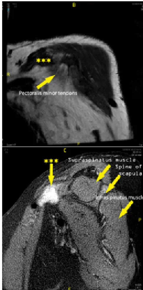
Başlık: Isolated spontaneous pectoralis minor tendon rupture in a patient with chronic renal failure Yazar(lar):ÖRÜCÜ, Merve; KUTLAY, Șehim; GÜNEȘ, Seçilay; GÖK, HaydarCilt: 69 Sayı: 2 Sayfa: 117-120 DOI: 10.1501/Tipfak_0000000929 Yayın Tarihi: 2016 PDF
Tam metin
Şekil
Benzer Belgeler
In conclusion, a diagnosis of isoniazid-induced cerebellitis should be considered when cerebellar signs develop in patients undergoing hemodialysis and treated with isoniazid..
A relatively forceful injection, ex- ceeding the capacity of the isolated septal artery (assumed to take a right anterior oblique image of the RCA), resulted in mul-
To the best of our knowledge, intramyocardial rupture of an isolated septal coronary artery was first defined in a patient with myocar- dial noncompaction and concurrent
Here, we report the case of a patient with par- oxysmal AF and end-stage renal disease (ESRD) requiring hemo- dialysis, in whom apixaban successfully and safely resolved a LAA
In conclusion, in the chronic renal failure patients, having peripheral catheters with fever and high inflammatory markers, infective endocarditis should be suspected
TPA addition induces the phosphorylation of JNKs and ERKs, but not p38, protein in HL-60 cells, and incubation of HL-60 cells with JNKs inhibitor SP600125, but not ERKs inhibitor,
The levels of nitrite, an oxidative product of nitric oxide, were increased within lipopolysaccharide-treated macrophages in a concentration-dependent manner (P < 0.01).
Fig-1: (a) Block representation of 4:2 compressor(b) Normal4:2 compressor Proposed 4:2 compressors.. In the paper four compressor that, double quality reconfigurable estimated
