A. O. Vet. Fak. Derx. 37 (2): 269-285, 1990
PATHOLOGICAL FlNDINGS IN CALVES WITH HYDRANENCEPHALY'
Rıfkı Hazıroğlu2
Hidranensefalili bU1.llğllarda patolojik bulgular'
Özet: Bu çaltşma Iıidranensefalili 22 buzağı üzerinde
gerçekleştiril-miştir. Buzağııann
ı
3'ü Alanya, 9'u İçel İli ve çevresinden, 1985 yı/ında seyreden epizooti .wasll1da sağlanmıştır. Hidranensefalili buzalIı/ara ve bunlartll bazılarmm annelerine aitı
8 kan serum u örneğinin PirbrightAraştırma Enstitüsü'nde yapılan serolojik muayenesinde, hepsinin deği-şen derecelerde Akabane virusuna karşı nötralizan antikorlar içerdiği saptanmıştır. Bu serumların 8'inde ise Mavidil'e karşı EL/SA pozitif . sonuç almmıştır. Aynca Etlik Hayvan Hastaltkıarı Araştırma Enstitü-sü'nce çaltşmada kullamlan buzağılarm ikisinin kanmdan Mavidil vi-rusu izole edilmiştir. lzolasyonu yapı/an virus/ar Pirbright Araştırma Enstitüsü'nde serotip 4 olarak identifiye edi/miştir. Buzağı/arda körlük dışıııda belirgin hir klinik bulguya rastlanmamıştır. Makroskobik ola-rak, beyinde hemisferlerin değişen derecelerde serebrospinal sıvı ile dolu olduğu gözlenmiştir. Beyin stem'i ise normal görünümdedir. Corpus striatum, thalamus, hippocampus, tectum mesencephaly ve cerebellum' da basmca bağlt değişiklikler dikkati çekmiştir. Epijiz ayrılması ve Iıipoplazisine birer buzağıda rastlanmıştır. Ventriküler sistemde 3. be-yin kamarasmda kireç birikinti/erinin oluşturduğu tıkanma sadece bir ol-guda gözlenmiştir. Çok belirgin cavum septi pellucidi, septum pellucidum' da de/ekt ve kısmi corpus cal/osum yokluğu diğer makroskobik bulgulardandır. Mikroskobik olarak, kısmen kalm olan rezidüel hemis-fer dokusu normal histolojik tabakalanmayı göstermesine rağmen ince olan doku sklerotik görünümdedir. Bazı alanlarda ise sadece leptome-ninksler yer almaktadır. Rezidüel hemisfer dokuları genelolarak ince bir substantia alba katmdan oluşmuştur ve bu katta görülen gliozis
ne-i This report is summarized from author's doctorate thesis written in Turkish. 2 Associate professor, Department of Pathology, Faculty of Veterinary Medicine, University of Ankara, 061 Lo Ankara.
deniyle ana kaviteye doğru Çıkmtılar yapmaktadır. ince olan rezidüel henzisfer dokusunda görülen sklerotik görünüm, benzer şekilde hemen hemen her olguda, medul/a oblongata'da promontorium gliosum calami scriptorii'de de gözlenmiştir. Aynca subependimal rozetler ile subepen-mal gliozis, fokal gliozis, indijeransiye nöyronal nodül/er, subkorti. kal kavitasyonlar ve haf(f perivaskiiler infiltrasyonlar diğer bulgular-dandır. Mye/inasyon normaldir. Tüm olgularda görülen bu yangısal olmayan değişiklikler ya111sırabir buzağıda nonpurulent ensefalitis sap-tanmıştır.
Summary: This study was based 011 hydranencephalic 22 calves. These calves were obtained Ji'om the provinces of Antalya and İçel during an epizootic period in
ı
985. The serum neutralizing antibodies against Akabane virus were detected in eighteen blood sera tak en from hydranencepha/ic calves and their mothers by Pirbright Animal Virus Resem'dı Institute (A VRI) in England. Pozitive results for bluetongue virus were also determined in eight of eighteen sera using ELISA. In addition, the bluetongue virus was isolated Ji'om the' blood of two hydran-encepha/ic calves by Etlik Animal Disease Research Institute (ADRI) in Turkey. These isolates were identifieel as bluetongue virus serotype 4 by Pirbright AVRI. None the calves showed any remarkable clinical symptoms except b/indness. Macroscopical/y, hemispheres were fil/ed with various amounts of cerebrospinal fluid (CSF). The brain stems were normal in appearance. Changes due to compression were noticed in the thalamus. corpus striatum, hippocampus, tectum mesencephaly and cerebel/um. The displacement and hypoplasia of the pineal body were encountered in two different calves. Obstructioll of the third ventricle due to precipitation of calcium salts was observed in one ca/f. The other macroscopical findings were cystic septum pellucidum, defect of the septum pellucidum and partial absence of the corpus cal/osum. Microscop-ically, narrow residual hemispheric tissues were scierotic in appearance, although the relatively large tissues had normal histologic strııctures and some areas of residual tissues were composed of only leptomeninges. Generally, residual hemispheric tissues had narrow substantia a/ba pro-truding to the membranous sacs due to gliosis. Sclerotic appearances were also observed in the promontorium gliosum calami scriptorU of the medulla oblongata in almost all of the cases. The other microscopical findings were subependymal rosettes, gliosis, subcOl.tical cavitations andmild perivascular cujfings. Myelination was normal. These changes were found in all cases except one, that case had nonpurulent encephalitis.
PATHOLOCICAL FI:'IiDl:\CS IN CALVES WITH HYDRANENCEPHALY 271
Introduction
Intracranİal malformations and especially hydranencephaly have
been reported in relation to prenatal infections with viruses of several
families in calves. e.g. Reoviridae-bluetongue virus (15, 17, 29),
Chuzan virus (7); Bunyaviridae-Akabane virus (4, i i, 12), Aino
virus (4, 22); Togaviridae-bovine viral diarrhea virus (2).
In the spring of i980, the first severe outbreak of arthrogryposis
and hydranenccphaly (AG / HE) in calves was reported in the westem
part of Turkey (33). Similar congenital deformities have been recorded in field outbreaks among the calves in Japan (13, 14, 24), Australia (5,8,31) and Israel
(ıo,
16,24), and it is generally accepted that these can be caused by the Akabane virus (4, ii-i4, 20, 23). As well ,!-S this, there are indications that the bluetongue virus (BTV) may cause con-genital defects in the central nervous system (CNS) both in the field and laboratory (15, 17,26,27,29). Secondly, another outbreakcharac-terized only hydranencephaly was observed in calves in the southem
part of Turkey in 1985/ 86.
This paper presents the results of a study of the pathology of 22
affected calves from the southem part of Turkey in 1985.
Materİals and Methods
A total of 22 hydranencephalic calves were supplied from the
provinces of Antalya and İçel between May 1985-July 1985: 13 from
the region around Antalya and 9 from the region around İçel. Table i
shows calves' age, brain weight, breed, sex and origin.
Calves no 2iand 22 were conveyed to the University of Ankara,
Faculty of Veterinary Medicine, Department of Clinical Sciences for
clinical examination, electroencephalography (EEG) and
electrocar-diography (ECG) on July 12, 1985.
Blood sera samples relating to the first 1i calves and some of
theİr mothers were sent to Pirbright AVRI, for examination of
neutra-lizing antibodies against the Akabane and bluetongue viruses.
Following a clinical examination of the live animals apart from
numbers 1, 2, 3 and 4, they were necropsied. The brain and spinal cord
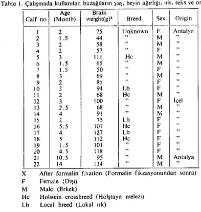
Table 1. Age, brain weight, breed, sex and origin of the ealves used in this study. Tablo 1. Çalışmada kullanılan buzağıların )aş. beyin ağırlığı. ırk, seks ve orijinkri.
Age Brain
Calf no (Month) weight(g)X Breed Sex Origin
--_.- --- ----.- --- --- ---i 2 75 t!nknClwn F Antaly~ 2 1.5 44 " M ,. 3 2 58 ,. M " 4 2 57 " F " 5 3 ii i He M " <> 1.5 65 " M " 7 1.5 50
..
F ,. 8 3 69..
M " 9 2 8, " F " LO 3 94 Lb F " ıı 2 68 He M " 12 3 100..
F içel 13 2.5 68..
M " 14 4 91 " to.'İ..
i; 2. 75 Lb F..
16 5.5 107 He F ,. 17 4 127 Lb F..
IR 'i 112 l-Ie F " 19 1.5 101 " F " .'-0 4.5 i LS " F "i
2122 10.5ı4 13495 "" MM Antalya..
X After formalin fixation (Formalin fikzasyonundan sonra) F Female (Dişi)
M Malc (Erkek)
He Holstein erossbreed (Holştayn melezi) Lb Loeal breed (Lokal ırk)
range of other organs and tissues were taken from the calves. Following
fixation with 10
%
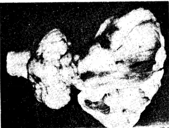
buffered formalin the brains were weighed andsliced transversally (Fig. 1) and gross lesion s recorded. Tissue samples were taken from the brain and cord, and other viscera. These were processed for histological examination by conventional methods.
Sec-tions were stained with haemotoxylin and eosin (HE) and selected
sections from the CNS were also stained with Klüver and Barrera's
luxol fast blue (LFB) for myelin-Gomori's method for reticulum fibers and Holzer's method for neuroglial fibers.
Results
Clinical findings: The major defect was a lack of intelligence, and blindness. In a small series tested, the eye preservation reflexes werc absent, but pupillary light reflcxes were presenL They were dummies,
PATHOLOGICAL FINDINGS IN CALVES WITH HYDRANENCEPHALY 273
f
Q /) " d cfgh ıj tl lmno
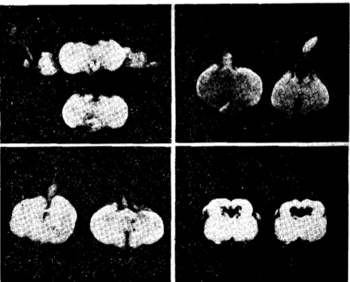
Fig. I. The coronal sections taken from affected brains for microscopic examination (Şekil ı.Mikroskobik inceleme için lczyonlu beyinlerden alınan koronal kesitleri gösteren
şema).
uncoordinated in gait, unable to stand properly and ınoved erratically when stimulated. The calves showed cud chewing behaviour as if fodder existed inside their mouths and exhibited licking action, Pulsation, temperature and respiration were normal and they had hearing abilities.
Other C1inic observations were salivation, vomitus, nervous
symp-tom s such as convulsion and tremor, lacrimation, strabismus, domed
cranium and torticollis in different calves. In the calves transported from Antalya to Ankara, there were insignificant activities of neurons
in EEG and ECG was normal. Pirbright A VRl's results are shown
table 2.
Gross pathology of the central nervous system: The cerebral hemispheres or major part s of them were represented by a fluctuant flu-id-filled sac bounded by a thin delicate membrane which was attaehed
to the meninges and which frequently collapsed as the calvarium was
removed. The fluid was watery and c1ear or blood-stained. Occasionally
the hemispheric lesions were asymmetrical and occasionally
symmet-rica!' Cystic cavities were found in the residual heınispheric tissues and
midbrain. They were located in areas bilateral or unilateral to the
mesencephalic cana!. Although the brain stern appeared unchanged
from the exterior, cross sections showed solitary and multiple cystic eavities of various sizes in the ncrve tissue. Particularly large cavities,
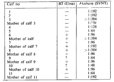
Table 2. Results of ELI SA test and SVNT carried out on blood sera of calves for BT and Akabane viruses. respectively.
Tablo L. Mavidil ve Akabane virusları için buzağıların kan serumlarında yapılan ElISA ve SVNT (serum \ irus nötrali,asyon testi) testlerı s8;il1çlarl.
Calf no RT (Eltsa) Akabaile (SVNT)
--- ---
--_
.... ----ı -- 1:192 7- - 1:192 3 - >i:384 Mother of calf 3 - ı:"5(. 4 - I: 128 5 -- 1:64 6 T, ı:96 Muther of calf + >1 :384 7 ~ 1:96 Mother of calf 7 + 1:192 8 + >1:384 Mother of calf 8 - 1:96 9 ? 1:64 Mother of calf 9 1:96 LO ? 1:48 Mother of calf LO + 1:96 II-
1:64 Mother of calf IL - 1:192hemispheric tissues (Fig. 2). The inner surface of the cavity was
usuaIly moist, with a slight aceumulation of CSF.
After formalin fixation brain weight (Table i) ranged from 44
to 134 g (normal 180-250 g). The spinal cord appeared normal in size
and did not have any macroseopic lesions.
Most of the cases had smaIl nodular masses of 1-2 mm in diameter on the leptomeninges, eorpus cal10sum and nucleus caudatus. Parti al absence and defect of the eorpus eal10sum were observed in some braİns. Total absenee and defects were also observed in the septum peIlueidum.
Moreover, cystic septum peIlucidum cal1ed double septum or 5 th
ventricle or cavum septi pel1ucidi anatomical1y was found in one case (Fig. 3). In some cases, the sept um peIlucidum was intact, but extremely
thin. There were deformities in the shape of the nucleus caudatus,
thalamus, meseneephalon and eerebeIlum. These deformities could
be due to the compression of the hemispheres which were full
of fluid. Another marked finding was band-shaped residual brain tis-sues between eorpus callosum-residual hemispheres, nucleus caudatus-residual hemispheres, and residual hemispheres-residual hemispheres (Fig. 4). The hippoeampus was great1y redueed unilateral1y or
bilate-raIly and in some of the cases a granular surfaee was observed.
PATHOLOGICAL FINDINGS IN CALVES WITH HYDRAI'\ENCEI'H:\LY 275
Fig. 2. The cystic cayitations of the residual hemispheric tissue sectioned fro:ıı the lobus frontalis. ease no 19.
(Şekil 2. Lobus frontalis'ten arta kalan he-misferik dokudan yapılan kesitteki kistik kayitasyonlar. Olgu no ı9).
Fig. 3. Cyslic septum pellucidum. Case no 5.
(Şekil -:. Kistik septum pellucidunı. Olgu no 5)
Fig. 4. Hydl'anencephalic brain, slight cerebcllar hypoplasia, and band-shaped n,sidual nerye tissue. Case no 7.
(Şekil 4. Hidranensefalik beyin ,hafif serebellar hipoplazi ye band şeklindeki kalıntı sinir dokusu. Olgu no 7)
of the nearabsence of the frontal temporal and parietal lobes. As a
result, the corpus striatum, thalamus and hippocampus were exposed.
The other macroscopical findings were obstruction of the 3 rd
yentric1e (Fig. 5), displacement and hypoplasia of the pineal body
(Figs. 6, 7), dilatation of the meseneephalic canal (Fig. 8) and a
I'ig. 5. Obstruction of the third ventricle rlue to calcium saIts. Case no 2.
(Şekil 5. Kalsiyum tuzlarınııı neden oldugu 3. ventrikiildeki tıkanma.
Olgu no 2)
Fig. 6. Displacement of theııineal body. Case no 20, 16.
(Seki! 6. Epifizis sel'ebrinin normal konumundan ayrılması, Olgu no 20, 16)
Fig. 7. Hypoplasia of the pineal body. Case no 20, 18.
(Şekil 7. Epifizis screbrinin hipoplazisİ. Olgu no 20, 18)
Fig. 8. Dilatation and cystic cavities around the mesencephalic cana!. Casc no 2.
(Şekil 8. Meıensefalik kanal çevresinde kistik kavitasyonlar ve genişleme. Olgu no 2)
A Siight cerebeHar hypoplasİa (Fig. 4). hernİation and İrregularİty
of the folium were encountered in so me calves.
Gross pathology of other fissues and organs: There were no
consİst-ent assocİated lesions outside the braİn. The wounds of the back and
legs, subcutaneous haemorrhages of the head and legs, subcutaneous
jelatinous accul11ulatİons and hepatisation of the cranİal lo bes İn the lungs were observed İn calves nos 13,14, LS and 16, respectİvely. Apart
from pneumotic lesİons İn the lungs, these must be traumatİc due to
blindness. Therc were no pathological changes İn the eyes and optic
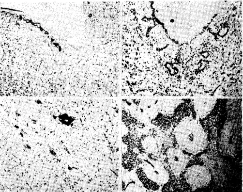
PATHOLOCICAL FI:\Dl:\"CS 1:"J" CALVES WITH I-IYORAl\ENCEPHALY 277 Histology of the central nervous system: Hydranencephaly was characterized histologicaııy by a cerebral cortex so thin that at low magnification it f itted into one microscopic field. Jts internal surface
with epenoymal ceııs was frequently incomplete or absent. Abundant
ınesodermal fibers associated with numeroııs capiııaries ""ere observed in the residual hemispheric tissues (Fig. 9), corpus callosum, septum
peııucidum, hippocampus, thalaınus, pincal body, hypophysis,
mesen-cephalon and promontorium gliosum calami scriptorii of the medulla
oblongata. These gaye a sclerotic appearance to the tissue (Fig. 10).
The thin residual hemispheric tissues were sclerotic in appearance
(Fig. i i), although the relatively large tissues had anormal histologic strııctllfe and so me areas of residual tissues were composed of only leptomeninges.
There were alsa gIial infiltrations, bat h nodular and diffuse, in the vicinity of the ependymal lining. Quite of ten these glial nodules were seen to protrude into the ventricles or mesencephalic cana\.
Cystic cavitations, subependymal rosettes, mineralisations,
mi-crohaemorrhages and spongiotic areas were observed in various parts
of the CNS, and subcortical cavitations. substantia grisea- substantia
alba-substantia grisea or mixed structure (Fig. 12) were seen in the
residual cerebral cortex in some cases. Ependymal dysplasia located
in the vicinity of the central cana! was also found.
The cystic cavities were a space formed in the nerve tissue without any special type of wall, in which both nerve cells and nerve fibers
dissappeared completely. Neuroglial ceıı infiltrated into the cavity
and of ten mixed with fragments of nerve fibers or the cavity was totally
empty under the microscope (Fig. 13). The nerve tissue around the
cavity was destroyed and there was disruption and rarefaction of
nerve fibers or was pack ed with CSF under pressure.
The cavitation was of perivascıılar origin, and was probably
inflıı-enced by the excess pressııre of the CSF. The Wirchow-Robin spaces
became extremely dilated. The CSF invaded the nerve tissue adjacent
to the perivascular space, which then disrupted and dissappeared. A
cavity involving blood vessels eventuaıı formed in or near this space. These types of cystic cavities were especially found in the
l11esenceph-alan and rhombencephalon.
it is known that ependymal roset formatian in congenital
L
Fig. 9. Abundant argentaffin fibers in e!ose assoeiation with numerous blood vessels in
the residur.! hemispheric tissue. Case no 15, Gomori's retieuluın stain.
(Şekil 9. Arta kalan hemisferik dokuda yer alan çok sayıdaki kan damarları ile yakın
iliş-kili argentaffin fıbriller. Olgu no ı5, Gomori'nin retikulum boyası)
Fig. LO. Sclerotic appearanee in the residual hemispherie tissue. Case no ı1, HE
(Şekil LO. Arta kalan hemisferik dokudaki sklerotik görünüm. Olgu no lL, HE)
Fig. ı ı. Complete sclerotic appearance in the narrow residua! hemislıpheric tissue.
Case no 7, Gomori's reticulum stain.
(Şekil ı ı.Arta kalan dar hemisferik dokudaki tam sklerotik görünüm. Olgu no 7,
Gomori'nin reıikulum boyası)
Fig. 12. Mixed substantia alba and grisca in the eerebra: eortex. Case no 2, Klüver Barrera's
myelin stain.
(Şekil 12. Serebral kortekste birbirıne girmiş substantia alba ve grisea. Olgu no 2, Klüver
Barrera'nın myelin boyası)
Subependymal rosettes were alsa observed in all calves (Figs. 14, 15) except calf no 22. As well as this, same ependymal rosettes were found in the deep part of the nerve tissue. These may depend on pressure of the CSF.
A few smail foci of mineralisation, which probably deveIoped dur-ing the growth of the brain, occurred in the residual hemispheric tissues,
I'ATHOLOGICAL FJ:,\J)J~GS IN CALVES VdTlf HVDRANENCEPH"LV 279
Fig. 13. Subeartical eavitations and a few mineralisation foei in the eerebrai cortex. Case no 19, HE.
(Şekil 13. Serebral kortekste subkortikal kavitasyonlar ve az sayıdaki mineralizasyon odakları. Olgu no 19, HE)
thalamus (Figs. 13, 16). pİneal body and hypophysis. The obstruction
of the 3 rd ventricle due to cakium salts was observed in one calf.
Furthermore, this type of cakium precipitate was free in the bases of the hemispheric sacs of so me of the affected brains.
Deformities in the shape of the cerebellum, probably duc to CSF
compression were encountered and they showed no remarkable
pa-thological changes except that case no 7 and case no 2 had hypoplastic changes including depletition of the extemal granular layer, Purkinje cell ectopia, and reduction in the width of the granular or molecular layer. In these affected cases, the cortical architecture was disturbed,
foliation was rudimentary and islands embedded within the substance
were surrounded by Purkinje and granular cells (microgyria) (Fig. 17).
Two calves (Calves no i1 and 12), which werc not able to walk
during their life, exhibited microhaemorrhages in the lateral and
ventral hom of the medulla spinalis.
Myelination was normal in the sections stained by the LFB
meth-od. A marked systematic increase in glial fibers was frequently seen
in the periphery of the dilated perivascular spaees, the wall of the ven-tricles and cavities in sections stained with Holzer's method.
Inflammatory changes were not round in the CNS of the calves
Fig. 14. Partial absence of the ependymal layer in the 3 rd ventridc and sııbependyma! rosettes. ease no G, HE.
(Şekil 14, Ü,:üncü ventrikülü döşeyen ependim hücrelerinin kısmi yokluğu ve subependi-mal rozetler. Olgu no 6, HE)
Fig. ıs. Close magnification of the subependymal roselles. Case no ıs, HE (Şekil ıs. Subependimal rozetlerin daha büyük görünümü. Olgu no ıs, HE) Fig. 16. Mineralisation in the thalamus. ease no 14, HE
(Şekil ]6. Talamusta mineralizasyon. Olgu no 14, HE)
Fig. 17. Coronal sections of the cerebellum. The Purkinje and granular cells as islands are displaced into the molecular iayer. ease no 7, HE
(Şekil 17. Serebellumun koronal kesiti. Moleküler tabakaya doğru yer değiştirmiş Purkinje ve granular hücrenin oluşturduğu adacıklar. Olgu no 7, HE)
There were no lesions in the eyes and optic nerves histologically İn any of the cases examiııcd for this.
Discussion and Conclusion
In Turkey, bluctongue diseasc (BTD) in sheep was first clinically diagnosed İn the south of the country, in 1944 (1). Af ter a 30-year break,
PATHOLOGICAL f1NDI:"iGS IN CALVES \\IITJ-! J-!YORANENCEI'HAT.Y 281
spread to other areas in the same region in i978 and
ı
979 (I, 30, 35).In that period avirus isolated frol11 a sheep was identified as BTV
serotype 4 (35).
In March 1980, the first outbreak of AG / HE in calves in the south-westem part of Turkey was reported (33). In May 1980 serological
in-vestigations carried out on blood collected in westem Turkey showed
antibodies to Akabane virus in adult cattle and calves and in a
colostrum-deprived calf. However, the Akabane virus was not isolated.
In Octabel' 1980 BTV serotype 4 was isolated from a newbom calf
with AG / HE (35).
Almost five years la ter, in May 1985, we observed another Oııt-break in calves around Antalya and İçel in the southem part of Turkey (Fig. 18) which manifested itself only by hydranencephaly. Alsa, some ofthese calves and some oftheir mothers had antibodies to the Akabane virus and same were positive against BTV. In view of the fact that these calves had taken calastrum, it is difficıılt to evaluate whether it is active
Fig. U~.Provinces involved İn the outbreak in calves betwcen i985 and i986. and location map of Turkey.
immunity in the ealf or it is obtained from the mother. In addition, BTV was isolated from ealves nos 5 and
ı
7 by Etlik ADRl and identifiedas serotype 4 by Pirbright AYRI.
B1uetongue and Akabane viruses are transmitted by biting midges
of the genus Culieoides (6, 9,
ı
8, 30) or mosquitoes (19, 28). Central nervous system anomalies have been observed in ealves bom to damsexposed by BTY, Akabane virus and other viruses -e.g., boyine viral
diarrhea, Aino, Chuzan- in the first trimester of pregnaney (II,
ı
5, 23, 26). In the last outlıreak, similarly, pregnant eows could have been infected by the virus or viruses at the end of summer 1984 by ınidges. However, there İs no dat:! on Culieoides movements or on meteorolo-gical eonditions, but it is possible tnat during suitablc weather eondi-tions, biting midges may migrate from other mediterranean countries and this beeame active in the southem part of Turkey. Also, there is serological evidence of Akabane activity in Cyprus, Jsrael, NorthemSyria and Jordan (Fig. 18) (P.S. Mellor, personel communication).
Clinical and macroscopical findings in hydranencephalic calves
were almost identical with the literature (8, i i, J5, 21, 24, 29, 34) ex-cept the obstruction of the 3 rd ventricle, displacement and hypoplasia
of the pineal body and cystic septum pellucidum. Cystic septum
pellucidum has been cncountered in newbom lambs which their dams
infected with pestiviruses (3), but have not been recorded in calves. For the first time, these macroseopical findings are being reported in ealves with hydranencephaJy.
AIso, histologieal findings reported were similar to those of our cases (8, iJ, 15, 21, 24,29,34). The sclerotic structure was recognized in various parts ofCNS of the calves. it is noted that sclerotic appearance of the superior and inferior collieuli is a constant and interesting feature
of hereditary encephalomyopathy in Hereford calves (32), whereas
it was not considered to be hereditary in our cases. Since most of
our cases were third or fourth calves and the first or second calves bom to the same mother suffered no congenital deformities or CNS anoma-lies we ruled out all eongenital defects. As well as this, the breeds of our cases were diffcrenL The CSF may have caused reactivity of the glial tissue resulting in dense fibrillary gliosis and this may give a sclerotic view to the tissue. In this condition, it can be seen in every
cascs exposed by the pressure of the CSF, but it has not been
Pi\THOL.OCICi\L FINDli'\CS IN Ci\LVES WITH HYDRi\NENCEPHALY 283
Marked nerve cell loss with fibrillary gliosis in the ventral hom of the spinal cord and in the nucleus fasciculi lateralis, nucIeus nervi hypoglossi and nucleus dorsalis nervi vagi of the medulla oblongata
have been noted in calves with AG / HE (21). Our cases were
only hydranencephalic and there were no marked lesions except
haemorrhages in the spinal cords and in those of the nucIei of the
medulla oblongata.
In this complicated situation, we believe that the etiology of these
outbreaks is stili ohscure. BTV and Akabane virus together or
sepa-rately may be the causative agents of these outbreaks. Epidemiologic, serologic and virologic studies are needed to elucidate this condition in Turkey. A project is in progress for this purpose.
Referenees
I. Anon(i980). Epizootiology, diagnosis aııd colltrol of blueıoııgul! in Turkey. Bull. Off. int. Epiz., 92: 567-574.
2. Badman, R.T., Mitchell, G., Jones, R.T., Westbury, H.A. (1981). Association of bovine viral diarrhoea virus iııfectioıı lo hydraneııcephaly and other central Ilervous system lesiolis iııperinatal calı'es. Aust. Yet. J., 57: 306-307.
3. Barlow, R.M. (1980). Morphogenesis of hydmnencephaly and other intracmnial malfor-m~ıioııs in progeny of pregnallt ell'es infected ıvith pestiviruses. J. Comp. Path., 90; 87-98.
4. Coverdale, O.R. (1978). Congeııital abnormalities iıı calves associated with Akabane virus aııd Aiııo virus. Aus. Vet. J., 54: 151-152.
5. Della-Porta, A.J., Murray, M.D., Cybinski, D.H. (i976). Congenital bovine epizootic arıhrogryposis aııd hydraııeııcephaly in Austmlia. Aust. Yet. J., 52: 496-501. 6. Doherty, R.L., CarIey, J.G., Standfast, H.A., Dyce, A.L., Snowdon, W.A. (l9i2).
Virus strains isolated from arthropods during aıı epizooıic of bovine ephemeral fever iii Queeııslond. Aust. Yet. J., 48: 81-86.
7. Goto, Y., Miura, Y., Kono, Y. (1988). Epidemiolol!İcal survey of an epidemic of congenital abnormaliıies vith hydraneııcephaly-cerebeflar hypoplasia syndrome occl/ring in 1985/ 80 aııd seroepidemiological investigatioııs on Chuzan virus, a putative causal agent of the disease, in Japan. Jpn. J. Yet. Sci., 50: 405-413.
8. Hartley, W.J., DeSaram, W.G., Della-Porta, A.J., Snowdon, W.A., Shepherd, N.C.
(ı977). Paıhology of coııgenital bOl'ine epizootic arthrogryposis aııd hydranencepha/y aııd ils relationship to Akabaııe virus. Aust. Yet. J., 53: 319-325.
9. Jennings, M., Boorman, J.P.T'j Ergün, H. (1983). Cu/icoides from westem Turkey in
relation to bluetongue disease of sheep aııd cal/le. Rev. Elev. Med. vet. Pays. trop., 36: 67-70.
10. Kalmar, Eo, Peleg, B.Ao, Savir, Do(i975). Arthrogryposis-Hydraneııcephaly syııdrome
iııııewbom caııle, sheep aııd goats-serological survey for antibodies agaiııst the Akabaııe virus. Refuah Vet., 32: 47-54.
11. Konno, So, Moriwaki, Mo, Nakagawa, Mo (1982). Akabane diseaseiııcattle: Coııgeniıal abııorıııalities caused by viral infeclioıı. Spontaııeous disease. Vet. PathoL, 19: 246-266. 12. Konno, So, Nakagawa, Mo (1982). Akabaııe disease iıı caııle: Congeııital abnormalities
caused by viral iııfectioıı. Experimeıııal disease. Vet. PathoL, 19: 267-2'/9.
13. Kurogi, Ho, Inaba, Y., Goto, Yo, M1ura, Y., Takaha~hi Ho, Sato, Ko, Omori, T., Matu-moto, M. (1975). (1975). Serologic evidence for etiologic role of Akabaııe virusiıı epizo-otic abortion-arthrogryposis-hydranencephaly in catlle in Japaıı, 1972-1974. Areh. ViroL. iJ.7: 71-83.
14. Kurogi, H., Inaba, Y., Taka!ıashi, E., Sato, K., Omori, T., Miura, Y., Goto, Y.• Fujiwa-ra, Yo, Hatano, Y., Kodama, K., Fukuyama, S., Sasaki, N., Matumoto, Mo (1976). Epizo otic coııgeııital arthrogryposis-hydraııcnceplıııly syndrome in catıle: lsolatioıı of Akabaııe virus from affected fetuses. Arch. ViroL, 51: 67-74.
15. MacLahlan, N.J., Osburn, B.I. (l9~3). Blueıoııgue virus-induced hydranenceplıaly iıı
caııle. Vet. PathoL, 20: 563-573.
16. Markusfield, O., Mayer, Eo (1971). Aıı arthrogryposis aııd Iıydranencephaly syndrome
iııcalvesiııIsrael, 1969/ 1970- Epidemiological aııd clinical aspecls. Refuah Vet., 28: 51-6\.
17. McKercher, D.G., Saito, J.K., Singh, KoV. (1970). Serologic evidence of aıı etiologic role for bluetongue virus iııhydranencephaly 0/calves. J. Am. Vet. Med. Assoc., 156: 1044-1047.
18. Mellor. PoS., Pitzolis, G. (1979). Obsen'atioııs on breeding sites and light-trap collec-tioııs of Culicoides duriııg aıı (Juıbreak0/blueıoııgue iııCyprus. Bull. EntomoJ. Res., 69: 229-234.
19. Metselaar, Do, Robin J. (1976). Akalıane rirus isolaıed iıı Kenya. Vet. Rec., 99: 86. 20. Miura, Y., Hayashi, S., Ishihara, To, Inaba, Y., Omori, To, Matumoto Mo (1974). Neu-tralizing antibody against Akl.1baııevirus in precolostral sera from calves with congeııital arthrogryposis-hydraneııcephaly syııdrome. Arch. Ges. Virusforsch., 46: 377-380. 21. Moriguchi, R., Izawa, H., Soekawa, M. (1976). A patlıological study 011calves witlı
arthyrogryposis and hydraııencephaly. Zentralbl. Veteriniiermed (B) 23: 190-199. 22. Moriwaki, M., Maiura, Y., Hayashi, S., Ishitani, R. (i977). Histopathological/indings
of calres iıı/ecıed experimentaııY with Aino virus. NatL Inst. Anim. Health Q, 17:
95-106.
23. Narita, Mo, Inui, So, Hashiguchi, Yo (1979). The pathogenesis of coııgeniıal encephalo-pathies in sheep experimentaııY induced by Akabane rirus. J. Comp. Path., 89: 229-240. 24. Nobel, T.A., Klopfcr, Uo, Neumann, F. (1971). Pathologyolan arthrogryposis-hydruıı-encephaly syndrome in domeslic rumiııall/siııIsrae11969/ 70. Refuah Vet., 28: 144-151. 25. Omori, To, Inaba, Y., Kurogi, Ho, Miura, Y., Nobuto, K. Ohashi, Y., Matsumoto, Mo
(1974). Viral abortion arthyrogryposis-Iıydranencephaly syndrome in catlle iıı Japan, 1972-1974. BulL Off. int. Epiz., 81: 447-458.
PATHOLOGICAL flNDINGS IN CALVES WITH HYDRANENCEPHALY 285
26. Osbum, 8.1., Johnson, R.T .• Silverstein, A.M., Prendergast, R.A., Jochim, M.M., Levy S.E. (1971). Experiıııeıııal viral-indueed eongenital eneep/ıa/opathies 1/ The pat-/ıagenesis 0/bluerongııe meCİne virus in/eelion in /erallaıııbs. Lab. Jnvest., 25: 206-210. 27. O~burn, B.I., Silverstein, A.M., Prendergast, R.A., Johnson, R.I., ParshaII CJJr. (1971).
Experiıııeıııal ı,iral-indueed congeııital eneephalopat/ıies iPathology 0/hydraneneephaly and porencephaly caused by bll/etongl/e vaccine virus. Lab. 'Invest., '25: 197-205. 28. Oya, A., Okuno, T., Ogata, T., Kobayashi, I., Matsuyama, T. (1961). Akabane virus.
a newarbor rims isolared iıı Japaıı. Jpn. J. Med. Sci. Bial., 14: 101-108.
79. Richards, W.P.C, Crenshaw, G.L., BushnelI, R.B. (1971). Hydraneneephaly 0/ealves associated wiıh natural bluerongue virus in/ection. Carneıı Vet., 61: 336-348. 30. Sellers, R.F., Pedgley, D.E. (1985). Possible windborne spread to western Turkey 0/
blueron!!ue virusiıı1977 and 0/Akabane virusiıı1979. J. Hyg. Camb., 95: 149-158. 31. Shepherd, N.C, Gee, CD., Jessep, T., Timmins, G., CarrolI, S.N., Bonncr, B.R. (1978)
Congeniral bOl'ine epizooric arrhrogryposis and hydranencephaly. Aust. Vet. J., 54: 171-177.
32. Urman, H.K., Gracc, O.D. (1964). Heredirary eneephalomyopathy ..1 hydrocephalus syııdrome iıı ııewbom calres. Corneıı Vet., 54: 229-249.
33. Urman. H.K., Milli, O.H., Mert, N., Berkin, Ş., Kahraman, M.M., Yüce, H., Avvuran, H. (1979). Türkiye'di! buzağılarda koııjeniral epizoorik artrugryposis ve hydraııeneepha/ie olayları. A.Ü. Vet. Fak. Derg., 26: 287-295.
34. Whittem, J.H. (1957). Congenira/ abnorma/ities in eahes: artrngryposis and hydranen-cephal)'. J. Path. Bact., 73: 375-387.
35. Yonguç, A.D., Taylor, W.P., Csontos, L- Worral, E. (1982). Bluetongue İlı western Turkey. Vet. Rec., lll: 144-146.