Short Communication / Kısa Bilimsel Çalışma
Pathological and electron microscopical observations on naturally
occurring contagious ecthyma outbreak in two wild goats
(Capra aegagrus aegragus)
Özlem ÖZMEN
1, Hüseyin DOLU
11Mehmet Akif Ersoy University, Faculty of Veterinary Medicine, Department of Pathology, Burdur, Turkey.
Summary: Numerous young wild goats (Capra aegagrus aegragus) were reported to suffer from a contagious and fatal disease
in a wildlife protection area in Antalya, Turkey, in January, 2017. Because of the large and partly inaccessible mountain area, total number of the dead goats were not known. However, approximately 15 goats were estimated to die from the disease; two of them were submitted for necropsy. At gross examination, lesions mainly localized on lips and numerous proliferative papules and pustules, necrotic scabs and swelling, as well as edema, hyperemia and ulcers, were observed at the commissure of the lips. Necropsy of both goats was performed and tissue samples were examined histopathologically and electron microscopically for the presence of viral agents. Histopathological examination revealed necrotic and proliferative cheilitis and dermatitis with eosinophilic intracytoplasmic inclusion bodies in keratinocytes. Electron microscopy revealed typical parapoxvirus particles. According to gross, histopathological and ultrastructural findings the disease was diagnosed as contagious ecthyma.
Keywords: Contagious Ecthyma, electron microscopy, pathology, wild goats (Capra aegagrus aegragus).
Doğal olarak şekillenmiş bulaşıcı ektima salgınında iki yaban keçisinde (capra aegagrus aegragus)
patolojik ve elektron mikroskobik gözlemler
Özet: Ocak 2017’de, Antalya'nın yaban hayatı koruma alanında, çok sayıda yaban keçisinde (Capra aegagrus aegragus) bulaşıcı
ve ölümcül bir hastalık tespit edildi. Hastalık büyük ve dağlık bir arazide şekillendiği için ölü sayısı tam olarak saptanamamakla birlikte yaklaşık 15 yaban keçisi yavrusunun öldüğü tahmin edilmekteydi ve bunlardan ikisine teşhis amaçlı nekropsi yapıldı. Makroskobik muayenede lezyonlar çoğunlukla dudaklarda lokalize olmuştu ve dudakların kommisuralarında çok sayıda proliferatif papül, püstül, nekrotik kabuklanmalar, şişkinlikler ile beraber ödem, hiperemi ve ülserler görüldü. Her iki keçi yavrusuna da nekropsi yapılarak histopatolojik incelemeler ve elektron mikroskobik olarak viral ajanların tespiti için doku örnekleri toplandı. Histopatolojik incelemelerde keratinositlerde eozinofilik intrasitoplazmik inklüzyon cisimciklerinin de bulunduğu nekrotik ve proliferatif şelitis ve dermatitis tespit edildi. Elektron mikroskobik olarak tipik parapoksvirüs partikülleri ortaya konuldu. Hastalık makroskobik, histopatolojik ve ultrastrüktürel bulgulara göre bulaşıcı ektima olarak teşhis edildi.
Anahtar sözcükler: Bulaşıcı ektima, elektron mikroskopi, patoloji, yaban keçisi (Capra aegagrus aegragus).
Contagious ecthyma (CE) is an acute, contagious, debilitating and economically important zoonotic viral skin disease of sheep, goats and wild ruminants with a worldwide distribution (8,10,11). The disease is caused by a double-stranded DNA virus which is a prototype of Parapoxvirus genus of Chordopoxvirinae subfamily within the Poxviridae family (1).
Contagious ecthyma is characterized by papules, vesicles and pustules localized on lips, nose, ears, eyelids, and in some cases on the feet or perineal region. Lesions can also occur inside the mouth, particularly in young animals. Erythema, macule, vesicle formation, pustules and then scabs and scars occur in the skin of affected
ruminants (7,8,10,11). In later stages, infection is confined to the squamous epithelium and may involve the oral cavity, eyelids, teats, and coronary band, subsequently predisposing affected animals to secondary infections (12). Because of the painful lesions that often occur in the mouth and on muzzle, the disease may result in anorexia or even starvation. Young animals may refuse to nurse, and lesions on the udder of the dam can cause her to abandon her own offspring and foot lesions can cause transient lameness. The disease usually resolves spontaneously. It has high morbidity and low mortality, but can cause significant debilitation due to the inability of affected animals to suckle or graze (7-12).
Although, contagious ecthyma is known to occur in wild ruminants, there is no study about the pathological finding of the disease in wild goats. The aim of this study was to examine the histopathological and ultrastructural features of naturally occurring contagious ecthyma in wild goats.
Approximately 15 young wild goats were estimated to die from the illness and two of them were presented from Akseki, Antalya to department of pathology for diagnosis. Necropsy was performed, and tissue sections were collected. For histopathological examination tissue specimens were fixed in 10% buffered formalin, embedded in paraffin, sectioned at 5 μm, stained with hematoxylin and eosin (HE) and submitted to microscopic examination after routine procedures. Stained sections were examined under light microscopy (Olympus CX41, Olympus Corporation, Tokyo, Japan). Morphometric evaluation and microphotography were performed using the Database Manual Cell Sens Life Science Imaging Software System (Olympus Corporation).
Tissue samples taken from the labial lesions were processed also for transmission electron microscopy (TEM). In short, samples were fixed in 2.2% gluteraldehyde and postfixed in 1% osmium tetroxide (OsO4) prepared in 0.1 M phosphate buffer solution. The
samples were then dehydrated in graded alcohol series and embedded in Araldite CY212 (Agar Scientific, Stansted, UK). Ultrathin sections were taken from plastic blocks and stained with uranyl acetate/lead citrate. These sections were investigated and photographed by a Zeiss 906E TEM (Zeiss Electron Microscopy, Thornwood, NY).
Contagious ecthyma occurred in a wild goat herd in national wildlife park in Akseki, Antalya. Although the total number of the dead animals were not known because of the large and mountainous area of the national park, approximately 15 young wild goats were estimated to die from the illness. The staff of the national park collected seven dead goats and two of them were presented for diagnosis. At necropsy, severe hyperemia, edema, papules and necrotic lesions, and ulcers on and around the lips were observed (Figs. 1-2). Secondary bacterial pneumonia in lungs were observed in one goat. Both goats had signs of starvation and severe cachexia possibly due to mouth lesions.
Histologically, lip lesions were similar in both animals. Histopathological examination revealed severe hyperemia, edema, multifocal epidermal hyperplasia with prominent rete ridges, hyperkeratosis, acanthosis and necrotic crusts. Papules, vesicles, and pustules, together with severe inflammatory reaction (mostly composed of neutrophil leukocytes) were observed in lesioned areas. Spongiosis characterized by swollen keratinocytes and ballooning degeneration was a common finding in epidermis. The nuclei of the cells were generally karyopyknotic and karyolytic in some cells. Rarely
keratinocytes contained one or more round to oval, brightly eosinophilic intracytoplasmic inclusion bodies (Figs. 3-4).
The epidermal layer of the lips exhibited diffuse erosion and multifocal ulcers. Generally epidermal cells were replaced by thick necrotic crusts that were secondarily infected. Sections taken from lesioned lip areas were composed of degenerated keratin, loss of keratinocytes, hyperemia, edema, fibrin clusters, necrotic cellular debris, degenerated neutrophil leukocytes, histiocytes, and lymphocytes.
Ultrastructural examination of the lip tissues of both animals revealed degenerate keratinocytes containing numerous ovoid shape, typical 200-300 nm wide cytoplasmic parapoxvirus virions (Fig.5). Numerous keratinocytes become karyopyknotic and karyolitic, while some of them contained inclusion bodies.
Although contagious ecthyma can be seen in wild ruminants, there is little knowledge about the pathology of the disease in wild goats. This study describes for the first time the gross, microscopic and ultrastructural findings in naturally occurring contagious ecthyma in wild goats. Contagious ecthyma infection of domestic sheep and goats is generally neglected worldwide due to the low morbidity rates. This study showed that contagious ecthyma can be more severe and cause high mortality especially in young wild goats. However, it should be kept in mind that harsh living conditions in natural settings may also have a role in high mortality rates.
Contagious ecthyma is a disease of worldwide distribution, however, it is seldom reported in the literature due to its low morbidity and negligible economic consequences (15). The causing virus, which is found in skin lesions and scabs, is thought to enter the skin through cuts and abrasions. The virus is very resistant to inactivation in the environment (9). Although contagious ecthyma is an endemic disease in domestic ruminants in Turkey, there is no report about its occurrence in wild animals and this is the first report indicating the presence of contagious ecthyma in wild ruminants in Turkey.
The clinical disease is characterized by proliferative, crusted, and sometimes pustular lesions around the lips, muzzle and occasionally the udder, feet and vulva (5,13,14). Gross lesions in domestic ruminants are characterized by multifocal to coalescing proliferative and ulcerative dermatitis which is localized to the squamous epithelium at mucocutaneous junctions, particularly around the mouth and nares (4,6,9,10). Although examination of the clinical findings in these flocks was impossible, gross lesions were similar to those that appear in domestic ruminants. Hard winter conditions might have increased the mortality rates in wild animals in our study. Similarly, increased mortality rates in winter months in chamois were also reported previously (13).
Figure 1. Multifocal-coalescing necrotizing and proliferative cheilitis and dermatitis in a wild goat. Şekil 1. Bir yaban keçisinde multifokal-nekrotik ve proliferatif şelitis ve dermatitis tablosu.
Figure 2. Erosive ulcerative cheilitis with necrotic crusts in another wild goat.
Şekil 2. Bir başka yabani keçide nekrotik kabuklanma ile eroziv ülseratif şelitis tablosu.
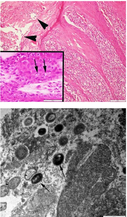
Figure 3. Severe, proliferative, and necrotizing lesions at the muco-cutaneous junction of the lip. Epithelial hyperplasia (arrows) and necrotic crusts (stars). HE staining, Bar=500 μm.
Şekil 3. Dudağın mukokutaneöz birleşme yerinde şiddetli, proliferative ve nekrotik lezyonlar. Epiteliyal hiperplazi (oklar) ve nekrotik kabuklar (yıldızlar), HE, Bar=500 μm.
Figure 4. Epithelial hyperplasia, spongiosis, acanthosis, pustule formations (arrow heads) and brightly eosinophilic intracytoplasmic inclusion bodies (arrows) (inset) in keratinocytes. HE staining, Bar=100 μm, Bar=50 μm (inset).
Şekil 4. Epiteliyal hiperplazi, spongiozis, akantozis, püstül oluşumu (ok başları) ve keratinositlerde parlak eozinofilik intrasitoplazmik inklüzyon cisimcikleri (oklar) (büyütülmüş resim). HE, Bar=100 μm, Bar=50 μm (büyütülmüş resim).
Figure 5. Degenerated keratinocyte with numerous free parapoxvirus particles (arrows) within the cytoplasm. Viruses show typical characteristics of an inner core surrounded by an intermediate coat further bounded by an envelope. Transmission electron microscope, lead citrate and uranyl acetate. Bar=1 μm.
Şekil 5. Sitoplazmasında çok sayıda serbest parapoxvirus partikülleri (oklar) içeren dejenere keratinosit. Orta kısımda yarıklanma gösteren ve bir zarfla sınırlanmış tabaka ile çevrili karakteristik viruslar. TEM, kurşun sitrat ve uranil asetat. Bar=1 μm.
Severe to moderate lesions such as enlargement of the lymph nodes, arthritis, and pneumonia resulting from sore mouth has been reported in CE cases in domestic ruminants (3). Similarly, secondary pneumonia was observed in a goat in this study. The young ages and insufficient food intake may be possible cause of secondary problem in CE.
Histologic lesions of contagious ecthyma are characterized by marked epidermal hyperplasia, ballooning degeneration, and eosinophilic intracytoplasmic inclusion bodies within keratinocytes that are only briefly detectable at the vesicular stage (3-5). There are frequently superimposed bacterial infections in the affected skin (9). Similar findings were observed at histopathological examination in our study; typical and characteristic
intracytoplasmic inclusion bodies were seen in keratinocytes.
Ultrastructurally, the cytoplasmic inclusions contain numerous 200-300 nm wide brick-shaped virions typical of parapoxviruses with the characteristic inner core surrounded by an intermediate coat further bounded by an envelope (2). In this study, although gross and histopathological findings were characteristic for contagious ecthyma, the diagnosis was confirmed by electron microscopic demonstration of the viral agents; typical parapoxviruses were seen in keratinocytes.
This paper reports contagious ecthyma infection in wild goats in Turkey for the first time. The possible transmission of echtyma virus in wild goats may be from affected domestic goats grazing on the same mountain pastures.
References
1. Chan KW, Yang CH, Lin JW, et al. (2009): Phylogenetic
analysis of parapoxviruses and the C-terminal heterogeneity of viral ATPase proteins. Gene, 432, 44-53.
2. Cheville NF, Lehmkuhl H (2009): Cytopathology of viral
diseases. 318–327. In: NF Cheville, H Lehmkuhl (Eds),
Ultrastructural Pathology: The Comparative Cellular Basis of Disease, 2nd edition, Wiley-Blackwell, Danvers. 3. Gelberg HB (2012): Alimentary system and the peritoneum,
omentum, mesentery, and peritoneal cavity. 326-327. In: JF
Zachary, MD McGavin (Eds), Pathologic Basis of Veterinary Disease, 5th edition, Elsevier, St. Louis. 4. Ginn PE, Mansell JEKL, Rakich PM (2007): Skin and
appendages. 664–666. In: MG Maxie (Ed), Jubb, Kennedy,
and Palmer’s Pathology of Domestic Animals, 5th edition, vol. 1, Elsevier, Philadelphia.
5. Haig DM, Mercer AA (1998): Ovine diseases: orf. Vet Res, 29, 311-326.
6. Hargis AM, Ginn PE (2012): The integument. 1023. In: JF Zachary, MD McGavin (Eds), Pathologic Basis of Veterinary Disease, 5th edition, Elsevier, St. Louis. 7. Hosamani M, Scagliarini A, Bhanuprakash V, et al.
(2009): Orf: an update on current research and future
perspectives. Expert Rev Anti Infect Ther, 7, 879-893.
8. Li W, Ning Z, Hao W, et al. (2012): Isolation and
phylogenetic analysis of orf virus from the sheep herd outbreak in northeast China. BMC Vet Res, 8, 229.
9. Murphy FA, Gibbs EPJ, Horzinek MC, et al. (1999):
Poxviridae. 289-291. In: Veterinary Virology, 3rd edition, Academic Press, San Diego, California.
10. Nandi S, De UK, Chowdhury S (2011): Current status of
contagious ecthyma or orf disease in goat and sheep-A global perspective. Small Rum Res, 96,73-82.
11. Navarre CB, Lowder MQ, Pugh DG (2002):
Oral-esophageal diseases. 66-67. In: DG Pugh (Ed), Sheep and
Goat Medicine, 1st edition, W.B. Saunders Company, Philadelphia.
12. Savory LJ, Stacker SA, Fleming SB, et al. (2000): Viral
vascular endothelial growth factor plays a critical role in orf virus infection. J Virol, 74, 10699-10706.
13. Scagliarini A, Vaccari F, Turrini F, et al. (2011):
Parapoxvirus infections of red deer in, Italy. Emerg Infect
Dis, 17, 684-687.
14. Smith KJ Skelton HG, James WD, et al. (1991).
Parapoxvirus infections acquired after exposure to wildlife.
Dermatology, 127, 79-82.
15. Zhao K, Song D, He W, et al. (2010): Identification and
phylogenetic analysis of an orf virus isolated from an outbreak in sheep in the Jilin province of China. Vet
Microbiol, 142, 408-415.
Geliş tarihi: 15.05.2017 / Kabul tarihi: 26.10.2017
Address for correspondence:
Prof. Dr. Özlem ÖZMEN Mehmet Akif Ersoy University, Faculty of Veterinary Medicine, Department of Pathology,
15030, Istiklal Yerleskesi, Burdur, Turkey e-mail: [email protected] Phone: +90 248 2132170