* Correspondence: [email protected]
J Exp Clin Med 2021; 38(3): 383-386
doi: 10.52142/omujecm.38.3.34
1. Introduction
Pulmonary thromboembolism (PTE) is a disease that describes the clinical findings resulting from the occlusion of the main pulmonary artery and its branches. Although it is frequently diagnosed and the ratio of mortality is around 10%, its un-diagnosis can result by mortality. To diagnose PTE, the clinician should suspect first. In PTE cases, chest pain, syncope and shortness of breath are the main complaints. As these complaints can be also the symptoms of cardiovascular diseases, the electrocardiography (ECG) must be performed at an early stage. Although it is known that in PTE there are some ECG changes, the sensitivity and specificity are low. Sinus tachycardia, right bundle branch block and S1Q3T3 pattern can frequently be observed as ECG findings for PTE (Siddiqa et al., 2020; Yeh and Chang, 2008; Sinha et al., 2005). Whereas ST elevations are highly suggestive of acute coronary syndrome (ACS), ST elevations associated with PTE have infrequently been reported in literature. With this case report, we aim to take attention to the issue by presenting a PTE case with an ST elevation.
2. Case report
A 52-year-old man presented to the emergency department with complaints of pain in the right side of his chest and back, followed by a convulsion. There were no chronic illnesses in patient’s story. The patient’s vital findings were: Arterial blood
pressure: 110/60 mmHg; Pulse rate: 60/min; sPO2: 98% and body temperature: 36.7oC. The ECG performed due to the chest pain demonstrated normal sinus rhythm (NSR), heart rate (HR) 60/min, ST elevation in leads V1-V2, DI, AVL, negative T-waves in leads V3-6 (Fig. 1). During the examination in the emergency department, the patient had a convulsion similar to that he had at home. However, neither at home nor at the hospital he didn’t have urinary incontinence or the postictal phase afterwards. His convulsion recovered without any medication. The patient described a chest pain after this convulsion-like activity. Therefore, his second ECG was performed. (Fig. 2). By the second ECG, NSR, heart rate of 58/min, ST elevations in leads DI, AVL, V1-4 and ST depression in leads D2, D3 and AVF were detected. The head computer tomography (CT), taken due to convulsion, was interpreted as normal. No pathological finding was observed in the posterior-anterior chest radiography. pH:7,33 pO2:67 mmHg, pCO2: 30 mmHg, lactate:3.5 mmol/L were detected in the patient’s blood gas. Cardiology consultation was requested for the patient after no pathological finding was detected in complete blood count and biochemistry tests. Echocardiography (ECHO) performed by a cardiologist showed normal left ejection fraction and mild right ventricular dilatation. The highly sensitive troponin test result was 7 (normal reference range 0-14 pg/ml).
Journal of Experimental and Clinical Medicine https://dergipark.org.tr/omujecm
Case Report
A case of pulmonary thromboembolism with ST elevation and literature review
Ethem ACAR1,* , Ahmet DEMİR1 , Birdal YILDIRIM1 , Ahmet DEMİR1 , Aysel GÖKÇEK21Department of Emergency Medicine, Faculty of Medicine, Muğla Sıtkı Koçman University, Muğla, Turkey 2Department of Cardiology, Faculty of Medicine, Muğla Sıtkı Koçman University, Muğla, Turkey
Received: 14.07.2020 • Accepted/Published Online: 03.02.2021 • Final Version: 23.04.2021
Abstract
Electrocardiography (ECG) is an auxiliary test applied for differential diagnosis of Pulmonary Thromboembolism (PTE). However, its specificity is low, but it is more often used for differential diagnosis. Although there are some changes on ECG in PTE, ST elevation is not a finding that we expect to see. With this case report, we aimed to draw attention to the issue with the diagnosis of PTE in a patient with ST elevation in ECG. 52-year-old male patient presented to the emergency department with a complaint of convulsion after chest and back pain. During the examination in the emergency department, he had a convulsion and woke up on his own without entering a postictal phase. After that, he had a chest pain. For this reason, ECG was performed to the patient and revealed ST elevation in leads DI, AVL, V1-4. The results of the patient’s EEG and head computer tomography (CT) were normal, so coronary CT angiography was performed. And it was observed that the coronary arteries were patent but embolism was detected in his cross-sectional pulmonary arteries. Thus, he was treated with the diagnosis of PTE. By the obtained results in this case that ST elevation may be a finding of PE, although it is rare.
Acar et al. / J Exp Clin Med
384
Fig. 1. Patient's 1st ECG done in the emergency department. Patient’s electrocardiography (ECG) performed for chest pain showed normal sinus rhythm (NSR), heart rate (HR) 60/min, ST elevation in V1-2 leads, t negative in DI, DII, aVL and V3-6 leads
Fig. 2. 2nd ECG done after the patient's convulsion-like activity. Patient’s 2nd electrocardiography (ECG) done after the patient's convulsion-like activity: ST elevation was detected in DI, AVL, V1, V2, V3, V4 leads, St depression was detected in DII-III, AVF leads
Fig. 3. Coronary Computer tomography (CT) Angiography images of the patient. The patient's CT coronary showed no lesions in the coronary arteries
Fig. 4. PTE compatible image seen on coronary Angio computer tomography (CT) of the patient. The patient's coronary CT showed no lesions in the coronary arteries. However, embolism was observed in the right main pulmonary artery and the branch
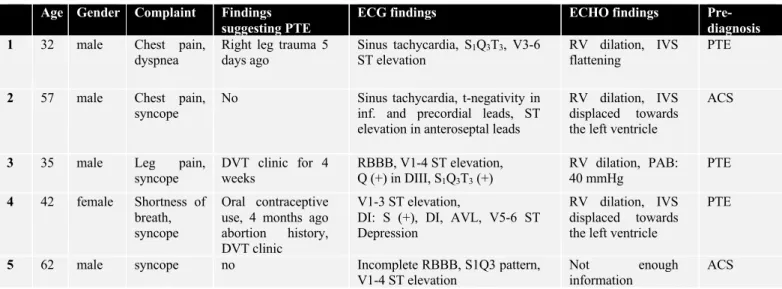
Table 1. The characteristics of the patients with have ST elevation associated with PTE according to the literature
The patient was admitted to the coronary intensive care unit with the pre-diagnosis of ACS. Coronary angiography or thrombolytic therapy was not initiated in the first place because
the neurological event was not ruled out in the story and ECHO findings were not fully associated with ACS. Coronary CT angiography was performed during his follow-up in the intensive care unit. No significant lesions in the coronary Age Gender Complaint Findings
suggesting PTE
ECG findings ECHO findings
Pre-diagnosis 1 32 male Chest pain,
dyspnea
Right leg trauma 5 days ago Sinus tachycardia, S1Q3T3, V3-6 ST elevation RV dilation, IVS flattening PTE
2 57 male Chest pain,
syncope No Sinus tachycardia, t-negativity in inf. and precordial leads, ST elevation in anteroseptal leads
RV dilation, IVS displaced towards the left ventricle
ACS
3 35 male Leg pain, syncope DVT clinic for 4 weeks RBBB, V1-4 ST elevation, Q (+) in DIII, S1Q3T3 (+) RV dilation, PAB: 40 mmHg PTE 4 42 female Shortness of breath, syncope Oral contraceptive use, 4 months ago abortion history, DVT clinic
V1-3 ST elevation,
DI: S (+), DI, AVL, V5-6 ST Depression
RV dilation, IVS displaced towards the left ventricle
PTE
5 62 male syncope no Incomplete RBBB, S1Q3 pattern,
V1-4 ST elevation
Not enough
information
Acar et al. / J Exp Clin Med
385 arteries were detected in the coronary CT. However, an
embolism was observed in the right main pulmonary artery and its branch when the sections were examined (Figs. 3 and 4). Therefore, electroencephalogram (EEG) and diffusion cranial magnetic resonance imaging were planned to rule out epileptic seizures for the patient whose treatment had been initiated. No pathology was detected in these examinations. The patient diagnosed with PTE and medicated with Coumadin, was discharged with full recovery on the 7th day of his hospitalization as no additional problems were developed. 3. Discussion
Relationships between PTE and ECG changes have been discussed in several literatures and have been reported that sinus tachycardia, right bundle branch block (RBBB) and S1Q3T3 were frequently observed as ECG findings of PTE (Siddiqa et al., 2020; Yeh and Chang, 2008; Sinha et al., 2005; Boey et al., 2015). The coexistence of PTE and ST elevation is not a common finding (Thomson et al., 2019). In the literature, a small number of cases on PTE associated with ST elevation (Table 1) were found.
When these cases were examined, it was observed that there were other findings suggesting PTE on ECG or clinical findings in addition to ST elevation. For example, Yeh and Chang stated that their 32-year-old male patient had suffered a right leg trauma five days ago before applying to hospital and reported that on the patient’s ECG sinus tachycardia and S1Q3T3 pattern in addition to ST elevation in leads V3-6 and RV dilatation in ECHO. Therefore, CT was ordered with the suspect of PTE and confirmed the diagnosis (Yeh and Chang, 2008). In the 57-year-old male case report, Wilson and Schaller (2008) stated that their patient had presented with syncope to the Emergency Department. They described that sinus tachycardia in addition to ST elevation in leads V1-4 on the ECG and RV dilatation in the ECHO were observed. However, t any pathology in PCI was detected, and in order to exclude PTE, the ventilation / perfusion scintigraphy was performed and diagnosed. Lin et al. (2009) stated that their case was a patient scheduled for Doppler ultrasonography (Doppler USG) due to leg pain, and the patient applied to the emergency department after having syncope. Deep vein thrombosis on Doppler USG, RV dilatation on ECHO and PAP: 40 mmHg were detected. On the ECG o, S1Q3T3 pattern and right bundle branch block in addition to ST elevation in leads V1-4 were detected. With these clinical findings CT with a pre-diagnosis of PTE was planned and diagnosis was confirmed. The researchers, presenting a 42-year-old female patient with a history of oral contraceptive use, described that their patient applied to the emergency department with complaints of leg pain and shortness of breath (Livaditis et al., 2004). They stated that V1-3 ST elevation on the patient's ECG, S in D1, RV dilatation and leftward displacement of the interventricular septum (IVS) in ECHO were found (Livaditis et al., 2004). In their case of a 62-year-old male without a chronic disease, Falterman et al. (2001) reported that their patient applied to the
emergency department due to syncope at home and related head trauma. They stated that the ECG of their patient had S1Q3 pattern, incomplete right bundle branch block and V1-4 ST elevation, first suspected of ACS, but later the patient died from a cardiac arrest during PCI, and PTE was detected at autopsy. It was observed that in three of the five cases mentioned above, there were serious risk factors for PTE or ECG changes. Although there were ST elevations in those cases, pulmonary CT angiography was performed primarily for PTE diagnosis. On the other hand, there is no such story in our case. The patient with the complaint of chest pain and shortness of breath, made us think of ACS clinic, except for the convulsion history which left us in doubt. In fact, we thought a loss of consciousness described as a convulsion was syncope. Because the blood gas taken after the loss of consciousness did not have acidosis and the patient did not have a postictal period. However, we still had to exclude epileptic seizures as differential diagnosis.
ECG changes in PTE are thought to observed due to the enlargement in the right structures of heart and the increase in pulmonary artery pressure. From this point on, we can expect to see ST depression in the precordial leads; however, ST elevation is much less common and the cause of ST elevation has not been clearly demonstrated (Ambesh et al., 2019; Cheng, 2005; Van Mieghem et al., 2004). Nevertheless, it is suggested that ST elevation may result from excessive right ventricular dilatation or ischemia, and in this case, it may cause pseudo-infarction findings in the anterior leads (Senthilkumaran et al., 2020; Wang et al., 2003).
In this case, patient also had a mild dilatation of the right ventricle and we can explain the ST elevation with this. In our case the patient wasn’t treated with PCI because the ECHO findings didn’t suggest STEMI and the patient’s relative insisted on convulsion. However, coronary CT angiography was performed on the patient. The coronary arteries were found to be normal and the pulmonary vascular embolism was detected.
As a result, although it is rare, ST elevation might be a sign of PTE. Therefore, PTE should to be ruled out before diagnosing the patient with ACS, especially if clinical findings suggest PTE. Conflict of interest None to declare. Acknowledgments None to declare. References
1. Ambesh, P., Kapoor, A., Kumar, S., Jain, S.K., 2019. The dilemma of the "ischemic-looking" electrocardiogram: Pulmonary embolism or acute coronary syndrome? Ann. Card. Anaesth. 22, 89-91.
2. Boey, E., Teo, S.G., Poh, K.K., 2015. Electrocardiographic findings in pulmonary embolism. Singapore. Med. J. 56, 533-537.
Acar et al. / J Exp Clin Med
386
3. Cheng, T.O., 2005. Mechanism of ST-elevation in acute pulmonary embolism. Int. J. Cardiol. 103, 221-223.
4. Falterman, T.J., Martinez, T.J., Daberkow, D., Weiss, L.D., 2001. Pulmonary embolism with ST segment elevation in leads V1 to V4: case report and review of the literature regarding electrocardiographic changes in acute pulmonary embolism. J. Emerg. Med. 21, 255-261.
5. Lin, J.F., Li, Y.C., Yang, P.L.,2009. A case of massive pulmonary embolism with ST elevation in leads V1-4. Circulation journal: official journal of the Japanese. Circulation Society. 73, 1157-1159.
6. Livaditis, I.G., Paraschos, M., Dimopoulos, K., 2004. Massive pulmonary embolism with ST elevation in leads V1-V3 and successful thrombolysis with tenecteplase. Heart. 90, e41. 7. Senthilkumaran, S., Karthikeyan, N., Meenakshisundaram, R.,
Florence, B., Thirumalaikolundusubramanian, P., 2020. Is ECG an aid to differentiate pulmonary embolism from ACS? Ann. Card. Anaesth. 23, 543
8. Siddiqa, A., Haider, A., Jog, A., Yue, B., Krim, N.R., 2020. Pulmonary Embolism Presenting as ST-Elevation Myocardial Infarction: A Diagnostic Trap. Am. J. Case Rep. 2020; 21, e927923-1–e927923-6.
9. Sinha, N., Yalamanchili, K., Sukhija, R., Aronow, W.S., Fleisher, A.G., Maguire, G.P., Lehrman, S.G., 2005. Role of the 12-lead electrocardiogram in diagnosing pulmonary embolism. Cardiology in review. 13, 46-49.
10. Thomson, D., Kourounis, G., Trenear, R., Messow, C.M., Hrobar, P., Mackay, A., Isles, C., 2019. ECG in suspected pulmonary embolism. Postgrad. Med. J. 95, 12-17.
11. Van Mieghem, C., Sabbe, M., Knockaert, D., 2004. The clinical value of the ECG in noncardiac conditions. Chest. 125, 1561-1576.
12. Wang, K., Asinger, R.W., Marriott, H. J. L., 2003. ST-Segment Elevation in Conditions Other Than Acute Myocardial Infarction. N. Engl. J. Med. 349, 2128-2135.
13. Wilson, G.T., Schaller, F.A., 2008. Pulmonary embolism mimicking anteroseptal acute myocardial infarction. J. Am. Osteopath. Assoc.108, 344-349.
14. Yeh, K.H., Chang, H.C., 2008. Massive pulmonary embolism with anterolateral ST-segment elevation: electrocardiogram limitations and the role of echocardiogram. The American journal of emergency medicine. 26, 632 e1-3.