Original Article
Corresponding Author Gizem Turachttps://orcid.org/0000-0001-8219-2015 Kocaeli University, Center for Stem Cell and Gene Therapies Research and Practice, 41380, Izmit, Kocaeli, Turkey
Tel: +90-262-3038817 Fax: +90-262-3591278 E-mail: [email protected] Received: February 8, 2018 Revised March 14, 2018 Accepted: March 20, 2018
The Effect of Recombinant Tyrosine
Hydroxylase Expression on the
Neurogenic Differentiation Potency of
Mesenchymal Stem Cells
Gizem Turac1,2, Gokhan Duruksu1,2, Erdal Karaoz3,4
1Center for Stem Cell and Gene Therapies Research and Practice, Kocaeli University, İzmit, Kocaeli, Turkey 2Department of Stem Cell, Institute of Health Sciences, Kocaeli University, İzmit, Kocaeli, Turkey
3Liv Hospital, Center for Regenerative Medicine and Stem Cell Research and Manufacturing, İstanbul, Turkey 4Department of Histology & Embryology, İstinye University, Faculty of Medicine, İstanbul, Turkey
Objective: Tyrosine hydroxylase (TH) is a rate-limiting enzyme in dopamine synthesis, making the enhancement of its activity a target for ensuring sufficient dopamine levels. Rat bone marrow mesenchymal stem cells (rBM-MSCs) are known to synthesize TH after dif-ferentiating into neuronal cells through chemical induction, but the effect of its ectopic ex-pression on these cells has not yet been determined. This study investigated the effects of ectopic recombinant TH expression on the stemness characteristics of rBM-MSCs.
Methods: After cloning, a cell line with stable TH expression was maintained, and the pro-liferation, the gene expression profile, and differentiation potential of rBM-MSCs were ana-lyzed. Analysis of the cells showed an increment in the proliferation rate that could be re-versed by the neutralization of TH.
Results: The constitutive expression of TH in rBM-MSCs was successfully implemented, without significantly affecting their osteogenic and adipogenic differentiation potential. TH expression improved the expression of other neuronal markers, such as glial fibrillary acidic protein, β-tubulin, nestin, and c-Fos, confirming the neurogenic differentiation capacity of the stem cells. The expression of brain-derived neurotrophic factor (BDNF) and ciliary neurotrophic factor (CNTF) significantly increased after the chemical induction of neuro-genic differentiation.
Conclusion: In this study, the expression of recombinant TH improved the neuroprotective effect of MSCs by upregulating the expression of BDNF and CNTF. Although the neuronal markers were upregulated, the expression of recombinant TH alone in rBM-MSCs was not sufficient for MSCs to differentiate into neurogenic cell lines.
Keywords: Tyrosine hydroxylase, Differentiation, Gene expression, Neuroprotection, Mesenchymal stem cell
INTRODUCTION
Tyrosine hydroxylase (TH) is a member of the aromatic ami-no acid hydroxylase family and is the rate-limiting enzyme in the biosynthesis pathway of the catecholamines. For that rea-son, TH expression plays a key role in some neurological
disor-ders, such as Parkinson disease (PD).1 To compensate the
de-creased level of dopamine, many strategies were developed to sustain the required level, such as transplanting fetal mesence-phalic dopamine cells,2,3 genetically modified cells expressing
TH,4-6 and direct transfer of TH-producing gene cassettes to
endogenous striatal cells.7-9
This is an Open Access article distributed under the terms of the Creative Commons Attribution Non-Commercial License (http://creativecom-mons.org/licenses/by-nc/4.0/) which permits unrestricted non-commercial use, distribution, and reproduction in any medium, provided the original work is properly cited.
Copyright © 2018 by the Korean Spinal Neurosurgery Society
Mesenchymal stem cells (MSCs) have the potential to differ-entiate into various cell types including neuronal cells. Howev-er, the neuronal differentiation potential of these cells is closely related to the cell origin, and it was previously demonstrated that dental pulp-MSCs possess higher differentiation capacity into neuronal cell lineages than bone-marrow derived MSCs.10
The improvement of the differentiation characteristics of MSCs into dopaminergic could be achieved by gene transformation.11
Genetic modification of MSCs was shown to provide an attrac-tive approach to produce therapeutically important proteins, like brain-derived neurotrophic factor (BDNF).12
In this study, rat bone marrow-derived MSCs (rBM-MSCs) were modified by transfection of rat TH gene. The extracellular production of TH was aimed to analyze the effect of the en-zyme on the differentiation potential of stem cells into neuronal cell lineages. The changes in cell proliferation and other stem cell characters after modification were also evaluated in this context.
MATERIALS AND METHODS
1. Isolation and Culture of rBM-MSCsThe bone marrow of Wistar Albino rat (n=5) was used to isolate MSCs. The methods used in this study were approved by Kocaeli University Ethics Committee for Animal Experi-ments (KOU HADYEK 6/4-2011). Isolation and culture of rBM-MSCs were performed as previously described.13 Under
sterile conditions, both rat femur and tibiae were excised, and cells were separated by density centrifugation by Ficoll-his-topaque (1.077 g/mL), and the cell pellet was resuspended in L-dulbecco's modified eagle's medium (L-DMEM) (Gibco, Invit-rogen, Paisley, UK). The cells were seeded in plastic tissue cul-ture flasks and incubated at 37°C in humidified air with 5% CO2. After the cells reached 70%–80% confluence, were
sub-cultured using 0.25% trypsin ethylenediaminetetraacetic acid (EDTA) solution (Gibco).The culture media was refreshed once every 3 days.
2. Flow Cytometry Analysis
The isolated cells were characterized with respect to follow-ing antigens in cytometer: CD29, CD45 CD90, CD54, CD106, major histocompatibility complex (MHC) Class I and MHC Class II, as previously described.14 All antibodies were supplied
by BD Biosciences (San Diego, CA, USA). Flow cytometry was performed using a FACSCalibur (BD Biosciences), and data were analyzed with Cell Quest software (BD Biosciences).
3. Differentiation of TH+ rBM-MSCs
Adipogenic and osteogenic differentiation were performed according to the protocol mentioned previously.14 To induce
adipogenic differentiation, cells (3,000 cells/cm2) were cultured
in L-DMEM (Gibco) supplemented with 10% fetal bovine se-rum (FBS) (Gibco), 0.5 mM isobutyl-methylxanthine (IBMX) (Sigma, St. Louis, MO, USA), 1 µM dexamethasone (Sigma), 10-μg/mL insulin (Gibco), 200 µM indomethacin (Sigma), and 1% Pen/Strep (Gibco) for 3 weeks. The presence of intracellular lipid droplets was confirmed by staining with 0.5 % Oil red O (Sigma). For osteogenic differentiation, cells (3,000 cells/cm2)
were cultured in L-DMEM supplemented with 0.1 µM dexa-methasone, 0.05 µM ascorbate-2-phosphate (Wako Chemicals, Richmond, VA, USA), 10 mM β-glycerophosphate (Sigma), 1% Pen/Strep and 10% FBS. After 4 weeks, osteogenic differentia-tion was assessed via staining with 2% alizarin red (pH 4.1–4.3; Fluka, Buchs, Switzerland).
For neurogenic differentiation, cells on collagen (type-I) coated coverslips were cultivated until 70% confluency. Cells were further cultured in differentiation medium (L-DMEM supplemented with 0.5 mM IBMX), epidermal growth factor (Biological Industries, Kibbutz Beit Haemek, Israel), basic fi-broblast growth factor (Biological Industries), neural stem cell proliferation supplements (StemCell Technology, British Co-lumbia, Canada), and 1% Pen/Strep.
4. Isolation of TH Gene From Rat Brain Tissue
The tissue was obtained from Wistar albino rat (4 months) by excision of the brain cortex. The tissue was transferred in RNA Later Solution (Qiagen, Hilden, Germany). Total RNA was iso-lated by the High Pure RNA Isolation Kit (Roche, Mannheim, Germany), according to the manufacturer’s instructions. The concentration and purity were detected by measurements at 260 nm and 280 nm. Complementary DNA (cDNA) synthesis was performed by Transcriptor High Fidelity cDNA Synthesis Kit (Roche).
5. Cloning of TH Gene
The second strand DNA synthesis and subsequent TH gene amplification were performed by Phusion DNA polymerase (Thermo, Braunschweig, Germany). The reaction mixture in-cluded 1X polymerase chain reaction (PCR) buffer, 0.2 mM de-oxynucleoside triphosphate, 0.5 µM of each primer, 1.25 mM MgCl2, and 2 µL of cDNA from the first strand reaction. An
initial 5-minute denaturation step at 94°C was followed by cDNA amplification cycles including denaturation at 94°C,
an-nealing at an appropriate temperature at 60°C and elongation at 72°C. The gene separated in agarose gel (Roche, 1%) was puri-fied by Agarose GelExtract Mini Kit (Prime5, Hamburg, Ger-many), restriction digested with HindIII and BamHI (Thermo, Fermentas, Vilnius, Lithuania) and ligated by T4 DNA Ligase (Thermo, Fermentas) into pFLAG-3 (Sigma) downstream of cytomegalovirus (CMV) promoter. Following subcloning and isolation by EndoFree plasmid isolation maxi kit (Qiagen), the plasmid was transfected by electroporation (Neon Transfection System, Invitrogen, Carlsbad, CA, USA) with respect to the in-structions provided by the manufacturer. The transfection pa-rameters for rBM-MSCs were 990 V/40 ms/2 pulses. After 48 hours of culture in L-DMEM with 10% FBS, the transformed cells were selected with respect to their resistance against 400 µg/mL G418 (Roche) under standard culture conditions. 6. Immune Staining
Samples were fixed in ice-cold methanol for 10 minutes and treated with 0.025% Triton X-100 (Merck, Darmstadt, Germa-ny) for permeabilization. Cells were incubated with 1.5% block-ing serum solution (Santa Cruz Biotechnology, Heidelberg, Germany) for 30 minutes at 37°C, and incubated overnight at 4°C with the primary antibodies. Samples were incubated with appropriate secondary antibodies for 25 minutes and covered with mounting medium containing DAPI (4´,6-diamidino-2-phenylindole) (Santa Cruz Biotechnology). The mounted cells were immediately examined under a fluorescence micro-scope (Leica DMI 4000B, Wetzlar, Germany).
7. Real-Time PCR Analysis
The gene expression levels were detected by LightCycler 480 DNA SYBR Green I Master (Roche) with gene-specific primers on LightCycler 480 real-time PCR instrument, according to the manufacturer’s protocol. Melting curve analysis for determining the dissociation of PCR products was performed from 60°C to 95°C to confirm the presence of a single peak. The second deriv-ative maximum method was performed to calculate Cp values.
To profile the gene expression in MSCs before and after gene transfer, real-time (RT)2 Profiler PCR Array for rat stem cells
(PARN-405F, SABioscience, Qiagen, Germany) was used ac-cording to the manufacturer’s protocol.
8. Western Blot Hybridization
Sodium dodecyl sulfate-polyacrylamide gel electrophoresis (SDS-PAGE) was performed according to Laemmli.15
SDS-PAGE was resolved in 11% polyacrylamide resolving gel with a
4% stacking gel. Protein samples were denatured by mixing with sample buffer (2% SDS, 10% glycerol, 0.1 M Tris–HCl at pH 6.8, 1% β-mercaptoethanol, traces bromophenol blue) and heated in boiling for 3 minutes. Gels were run in a Mini-PROTEAN Tetra Cell system (Bio-Rad, Herts, UK). The gels were run at 100 V. For Western blotting, proteins were transferred onto nitrocellu-lose membrane by i-Blot instrument (Invitrogen, Carlsbad, CA, USA) according to the manufacturer’s instructions. The mem-branes were blocked in phophate buffered saline supplemented with 3% skim milk and were incubated with antibodies against TH (Santa Cruz Biotechnology, sc-73152) or FLAG-Protein Ex-pression System (Sigma, F3165) at 4°C overnight. Following in-cubation with the specific secondary anti-mouse peroxidase-conjugated anti-IgG antibody (Santa Cruz Biotechnology, sc-2005). The bands were visualized by enhanced chemilumines-cence (ECL Plus; GE Healthcare, Uppsala, Sweden).
9. ELISA for TH
The level of rat TH in media was detected by enzyme-linked immunosorbent assay (ELISA) (USCN Life Science Inc., Wu-han, China) according to the manufacturer’s recommendation. The minimum detectable dose of TH was 59 pg/mL. All these experiments were repeated at least three times. The differences in the absorbance were measured by a ultraviolet-visible spec-trophotometer (VersaMax microplate reader, Molecular De-vices, Sunnyvale, CA).
10. Cell Proliferation and Cell Cycle Analysis
The water soluble tetrazolium-1 (WST-1) assay was used to estimate the proliferation rate of the cells in culture media. The cells were seed in 96-well plate, and cultured for 48 hours, be-fore reaching 70% confluence. The culture media was removed and the basal media supplemented with WST-1 (10%; Roche) was added to the well. After incubation at 37°C for 1 hour in dark, the color formation was and quantified at 450 nm with a microplate reader.
CycleTEST PLUS DNA Reagent Kit (BD Biosciences) was used to perform the DNA ploidy analysis in different rBM-MSCs following the manufacturer’s protocol. The cells were de-tached, and later the DNA content was labeled with propidium iodide. The analyses were performed by flow cytometer. Pe-ripheral blood mononuclear cells with known DNA amount were used as a control.
11. Neutralization Assay for TH
(Santa Cruz Biotechnology, sc-73152) was added in the culture medium at the amount of 0.2 µg/mL. The cells were cultured in this media for 8 days, and the medium was refreshed once ev-ery 2 days.
12. Statistical Analysis
All experiments were performed in triplicates. Data were re-ported as means±standard deviation. The statistical analyses were performed by SPSS ver. 10.0 (SPSS Inc., Chicago, IL, USA). Data were analyzed using 1-way analysis of variance or paired t-test. Differences between the experimental and control groups were declared statistically significant when p<0.05.
RESULTS
1. Expression of rat TH Gene in rBM-MSC
TH gene of 1,497-kb length was isolated from rat brain tissue
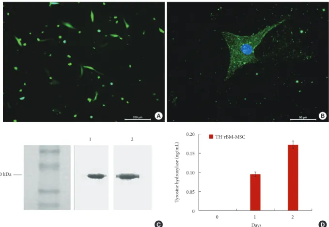
and ligated first to pUC19 for sequence analysis. The sequence of the fragment was checked by comparing it with TH gene se-quence in the database (GeneBank Acc. No.: NM 012740) for any mutation. The fragment was later ligated into a pFLAG-3 expression vector, and the construct was transferred into rBM-MSCs in passage 3 (P3). The presence of gene integration was confirmed by green fluorescent protein (GFP) expression in the cell (Fig. 1A). The expression of FLAG-Protein Expression Sys-tem fragment, which should be coexpressed with TH gene as fusion protein under the control of CMV promoter, was detect-ed in the cells (Fig. 1B). After the selection with G418 for 3 consequent passages, the ratio of GFP positive cells expressing was estimated by flow cytometry to be 92.09%±5.88%. This indicates that more than 90% of the cells are coexpressing TH gene with GFP (Supplementary Fig. 1). Western blot analysis for TH and FLAG protein fragments showed 2 fragments of nearly 60 kDa (Fig. 1C). The size of FLAG peptide did not
sig-Fig. 1. (A) The expression of the transgenic gene in rBM-MSCs. Following transfection, most of the cells showed GFP expres-sion after 3 days (scale bar: 200 µm). (B) The immunostaining against FLAG peptide (blue color) was performed to show the coexpressed TH gene (scale bar: 50 µm). (C) Western blot analysis against TH (1) and FLAG (2) showed the protein expression in size of about 60 kDa. (D) TH level was determined highest on the second day in the culture medium of TH+ rBM-MSCs.
rBM-MSC, rat bone marrow mesenchymal stem cells; GFP, green fluorescent protein; FLAG, protein expression system; TH, ty-rosine hydroxylase. A B 0 1 2 Days 0.20 0.15 0.10 0.05 0 Ty rosin e h ydr oxy la se (n g/mL) TH+rBM-MSC D C 1 2 60 kDa
nificantly affect the total size of TH protein on the blotting membrane. The time course of TH gene expression was shown that the expression altered during the culture (Fig. 1D). TH was not detected in the cell lines without TH gene transfection (wild-type and empty vector transfections). After one day of culture, the weak protein expression was measured in the trans-fected cells. The level of the expression was increased in the second day but sharply dropped down in the following day. The highest level of TH expression was found 0.17±0.01 ng/mL on day 2.
2. Effect of TH Gene Expression on rBM-MSC
After gene transfection, significant alteration in cell morphol-ogy was not observed. The flow cytometer analysis for both, rBM-MSCs and TH+ rBM-MSCs, demonstrate no significant
change in the surface markers (data not shown). However, the proliferation rate of transfected cells increased. The cell cycle status was evaluated to estimate any change (Fig. 2A). The analysis showed that the ratio of TH+ rBM-MSCs in S phase
was increased slightly to 18.67% from 14.77%, and the cells in G2/M phase and G1 phase was changed from 8% to 6.08% and
from 77.23% to 75.26%, respectively. The expression of neuro-nal markers was aneuro-nalyzed in rBM-MSCs and TH+ rBM-MSCs.
For this purpose, the immune staining was performed on Nes-tin, c-FOS, GFAP, TUBB, NF, and TUBB3 expressions (Fig. 3). Nestin, GFAP, and TUBB3 staining were found positive. How-ever, immune staining of cells against c-FOS, NF, or TUBB was negative. Epithelial marker (E-cadherin), pancreatic endoderm marker (Pdx1) and chondrogenic markers (Col2a1a, BMP3, and Acan) were downregulated in TH+ rBM-MSCs compared
to the cells before transformation (Fig. 2B). On the other hand, neural markers (N-Cadherin, Jag1, and Tubb3) and osteoblast marker (Col1a1) were increased. Dll3, which can inhibit the primary neurogenesis, was decreased, but Wnt1, which is a sig-naling molecule important in neural development, was also de-creased.
3. Alteration of Differentiation Potential After Gene Transfer The differentiation potential of cells into osteogenic, adipo-genic and neuroadipo-genic cell lineages was further analyzed by his-tochemical staining, and impact of TH gene expression on stemness character of cells was demonstrated. rBM-MSCs
Fig. 2. (A) The cell cycle status of rBM- and TH+ rBM-MSCs. Cell cycle distribution of transfected and untransfected cell lines
was measured by flow cytometry after propidium iodide staining. All experiments were repeated at least 3 times. (B) Changes in expression of differentiation markers in TH+ MSCs with respect to untransformed cells. TH, tyrosine hydroxylase;
rBM-MSC, rat bone marrow mesenchymal stem cells. G1 N-Cad herin Tubb 3 E-Cad herin Coll2a1a
Cyclin E1CXCL12 Jag1 Dll3 Wnt1 Acan BMP3 Pdx1 Coll1a1
S G2/M 80 75 70 12 8 4 0 -4 -8 4 2 0 -2 -4 -6 -8 30 20 10 0 20 10 0 Fo ld ch ange Fo ld ch ange rBM-MSC TH+ rBM-MSC A B
showed superior osteogenic differentiation capacity, and this feature of cells continued after gene transfer. After 28 days in osteogenic differentiation medium, mineral deposits were ob-served to form in TH+ rBM-MSCs (Fig. 4B). Later these
depos-its were stained with Alizarin Red S. There is no significant dif-ference in the staining patterns with this dye in the cell lines be-fore and after gene transfection (Fig. 4C). In the adipogenic dif-ferentiation medium, rBM- and TH+ rBM-MSCs were
differen-tiated to the same degree. The oil droplets formed were demon-strated by Red Oil O staining (Fig. 4E). Neurogenic differentia-tion capacity of TH+ rBM-MSC was analyzed after the
incuba-tion in neurogenic differentiaincuba-tion medium for 20 hours, 30 hours, 60 hours, and 10 days. Stem cells exposed to neural dif-ferentiation medium underwent dramatic morphological changes reminiscent of neuritis (Fig. 5C). The morphological changes were significant in the cells after 20 hours of incuba-tion, but the expression of neural cell line related markers did not appear before 30 hours of incubation. TH and coexpressed FLAG peptide expression were visible before the differentiation process, but significant increase in TH was detected after 30 hours of induction (Fig. 5D-O). This sudden increase in the staining could be related to the induction of endogenous TH
expression. The neurogenesis markers, Nestin and GFAP, were detected throughout the induction period, but Tubb3 and c-Fos expressions were only visible after 30 hours, disappeared after 60 hours of induction and not visible after 10 days (Fig. 6). The Tubb3 and c-Fos were expressed at the same time when endog-enous TH expression was at the highest point. Similarly, the ex-pression of neurotrophic factors, BDNF, and CNTF, were also observed in the same time period (Fig. 7A-H). The gene ex-pression analysis of neurogenic markers in differentiated/un-differentiated TH+ rBM-MSCs supported our findings with
re-spect to increased levels of TH, c-Fos, and Sox9 (Fig. 7J). TH gene showed 4 times higher expression level in differentiated TH+ rBM-MSCs than in undifferentiated TH+ rBM-MSCs. The
neurogenic markers, c-Fos, and Sox9, expressions were in-creased almost 6 and 3 fold, respectively.
4. Neutralization of the TH Effect in Transfected Cells The proliferation rate of TH+ rBM-MSCs increased 2 fold
af-ter TH gene transfection. To prove that this alaf-teration is due to the TH ectopic expression, the activity of TH was suppressed by the antibody (anti-TH). The culture of cells in the complete medium supplemented with anti-TH decreased the rate of
pro-Fig. 3. Immunophenotype analysis of TH+ rBM-MSCs for neurogenic markers. Representative staining patterns are shown for
Nestin (B, D), c-Fos (C, D), GFAP (F, H), β Tubulin (G, H), NF (J, L), TUBB3 (K, L). Nuclei were labeled with DAPI (A, E, I; blue). Only Nestin, GFAP, and TUBB3 staining were found positive. Scale bars represent 50 µm. TH, tyrosine hydroxylase; rBM-MSC, rat bone marrow mesenchymal stem cells; DAPI, 4´,6-diamidino-2-phenylindole.
A E I B F J C G K D H L
liferation significantly by 40% compared to unneutralized cells. But proliferation rate was still higher with respect to untrans-formed cells (Fig. 7I). This data supports the results of cell cycle analysis, in which the S phase of the cells was elevated (Fig. 2A). Besides, the expression of proliferation stimulating chemokine, CXCL12, was increased almost by 8 fold (Fig. 2B).
DISCUSSION
PD is the second most common neurodegenerative disease of the aging central nervous system after Alzheimer dementia.16
Stem cell-based therapies aim to provide a long-lasting symp-tomatic relief for PD patients. Stem cells are defined as imma-ture cells with a capacity of self-renewal and depending on their origin, can differentiate into specialized cell types or retain the potential to differentiate into somatic cells, including dopami-nergic neurons.17 MSCs are highly promising candidates for PD
treatment approaches. The application of MSCs might be effec-tive in a PD therapeutic strategy.18,19 Intrastriatal grafting of
MSCs into adult intact rats resulted in a significant elevation of TH expression and dopamine levels in the striatum.20 In other
reports showed the functional recovery in PD rodent models following transplantation of MSCs that are indicative of a neu-roprotective role in vivo.21,22
In this study, TH gene was transferred to rBM-MSCs, and stable gene expression was obtained in the cell line by selection with G418. The continuous expression caused the alteration in cellular physiology of TH+ rBM-MSCs becoming much smaller
in size. This modification might be explained by the increment of the proliferation rate, which was slightly increased after gene transfer. The expression level of TH was estimated as 0.17 ng/mL in the culture on the second day. This level is significantly lower compared with the neuronal differentiated MSC, in which TH level was estimated as 0.53 ng/mL.19 On the other hand, the TH
expression also affected the other properties of cells, like prolif-eration and differentiation potential.
Other studies involving the transfer of TH gene into other stem cells focus on their therapeutic effect in neurodegenera-tive diseases.11,23,24 However, the impact of TH expression in
stem cells on cell characteristics was sparsely investigated. In the study by Lu et al.,11 TH gene was delivered into MSCs by
adeno-associated virus, and the cells were transplanted into the
Fig. 4. Differentiation of TH+ rBM-MSCs into osteogenic (A–C) and adipogenic (D–E) lineages. After osteogenic differentiation
cells were stained with Alizarin Red-S (B) and control (A) (scale bar: A, B, 200 µm). (C) There is no difference in the staining pattern between rBM- and TH+ rBM-MSC. After induction to differentiate into adipogenic cell lineage, TH+ rBM-MSC were
stained with Oil Red O (E) and control (D) (scale bar: 50 µm). In the adipogenic differentiation medium, and TH+ rBM-MSCs were differentiated to the same degree. (E) The oil droplets formed were demonstrated by Red Oil O staining. TH, tyro-sine hydroxylase; rBM-MSC, rat bone marrow mesenchymal stem cells.
A D B E C rBM-MSC TH+ rBM-MSC
striatum of PD rat for functional analysis. Following injection, dopamine level was significantly improved with respect to the control rats, but the dose and timing of gene expression in the brain were found to be not regulated, as it was observed in nor-mal physiology state. These results propose that the expression of TH in these cells might not induce to differentiate into dopa-minergic or neuronal cells, as proposed by other studies.25
Simi-larly, the genetically engineered rBM-MSCs with TH gene was demonstrated to contribute to regeneration process, but rather differentiation they migrated in brain tissues and increased the survival rate of surrounding cells.24 Studies regarding in vivo
differentiation of rBM-MSCs into neuronal cells have shown unsatisfactory results in parallel.26 Their supportive effects
ob-served following transplantation mainly occurred indepen-dently of tissue integration, and these secreted stimulating fac-tors by the transplanted cells might be the source.27,28 These
se-creted factors contributing to the regeneration process of stem cells are the neurotrophic factors: BDNF and CNTF. The ex-pression of these proteins was increased notably in the neuro-genic differentiation medium. Although these markers were not markers for mature neural cell lines, they are important for the regeneration process after neurodegenerative diseases.
Fig. 5. Differentiation of TH+ rBM-MSCs into neuron-like cells. After 30 hours of induction in the neuronal differentiation
me-dium, cells displayed neuron-like morphologies (arrows) (B, C). (A) There was no neuronal differentiation in control culture. Scale bar: A, 200 µm; B, 50 µm; C, 20 µm. Immune fluorescence analysis of neural differentiated TH+ rBM-MSCs for distinct
time courses of 20 hours (D–G), 30 hours (H–K), and 10 days (L–O). The expressions of FLAG (D, E, H, I, L, M) and TH (F, G, J, K, N, O) were detected in differentiated (E, I, M, F, J, N) and undifferentiated (D, H, L, F, J, N) TH+ rBM-MSCs. Nuclei were
shown by DAPI in blue staining. Scale bar: D–O, 50 µm. TH, tyrosine hydroxylase; rBM-MSC, rat bone marrow mesenchymal stem cells; FLAG, protein expression system; DAPI, 4´,6-diamidino-2-phenylindole.
B C A D H L E I M F J N G K O
These are known to have function through activation of sepa-rate and specific receptors, and subsequent signaling pathways. The protective effect was reported to correlate with the endoge-nous up-regulation of these neurotrophic factors, which induce the activation of ERK1/2 and AKT pathways and the down-stream effect on the transcription factor CREB.29 These factors
would affect the other cells in the same microenvironment as neuroprotective against apoptosis. The BDNF expression in TH+ rBM-MSCs was observed even before the neurogenic
in-duction, but the CNTF appeared after 30 hours and went down before 60 hours of culture in differentiation medium. The cells might also respond similarly in the neural microenvironment by increasing the secretion of these factors the following trans-plantation into the brain tissue of animal disease model. In the comparable in vivo study related to the spinal cord injury, this paracrine effect was also observed.30
Nestin, GFAP, and TUBB3 expressions were detected in TH+
cells without any chemical induction. The presence of these markers might provide the suitable conditions for high efficient neurogenic differentiation. Other markers, for example, Tubb
and c-Fos, were expressed only after 30 hours under neurogen-ic differentiation condition when the neuron-like structures were observed to form. TH+rBM-MSCs could differentiate into
neuron-like cells with the morphology similar to neurons in the first days, but the number of viable cells was drastically de-creased after 10 days, indicating a possible elimination of cells from culture. In 10 days neurogenic differentiated group, the cells were still viable, although the number of cells was de-creased considerably. To maintain the viability, the components of neurogenic differentiation media might be improved better to sustain their expressions.
Under the normal culture conditions, we did not observe any morphological difference between rBM-MSCs and TH+
rBM-MSCs. However, the cells formed reticular-like structures in the culture. Increased N-Cadherin expression, which is present in neuronal cells,31 and decreased expression of the epithelial
marker, E-Cadherin, was observed. Despite the stem cell-spe-cific surface markers were not significantly changed, the up-regulation of N-Cadherin expression could indicate that TH+
rBM-MSCs are close to neuron-like cell character after TH
Fig. 6. Immune fluorescence analysis of neural differentiated TH+ rBM-MSCs for the time courses of 20 hours (A–D), 30 hours
(E–H), and 10 days (I–L). The expressions of GFAP, βTubulin, Nestin, and c-Fos were detected in differentiated (B, F, J, D, H, L) and undifferentiated (A, E, I, C, G, K) TH+ rBM-MSCs. Nuclei were shown by DAPI in blue staining. Scale bar: 50 µm. TH,
ty-rosine hydroxylase; rBM-MSC, rat bone marrow mesenchymal stem cells; DAPI, 4´,6-diamidino-2-phenylindole.
A E I B F J C G K D H L
gene transfection.
In the neurogenic differentiation, some pathways regulate these processes. It was shown that activation of Wnt/Shh path-way blocks the neurogenic differentiation.32 In the present
study, down-regulation by 4 times was observed in Wnt1 gene expression in TH+ rBM-MSCs, which could suggest that the
Wnt pathway might weakly influence the inhibition of neuro-genic differentiation. Besides the constitutive recombinant ex-pression, the endogenous TH expression also observed after neurogenic induction to differentiate. Therefore, the level of
TH expression was further improved during neurogenic
differ-entiation by repression of Wnt pathway. On the other hand, Wnt signaling was reported to increase the proliferation and to
impair differentiation of stem cells.33 This contradiction in TH+
rBM-MSCs could be explained by the acceleration of cell cycle by TH gene through affecting intracellular mechanisms. This effect of TH gene could be reversed by neutralization of gene product. We found that the ratio of cell in S phase increases while that of cells in G1 phase decreased in TH+ rBM-MSCs
culture, which indicates the promoting effect of TH gene trans-fection on cell proliferation. Furthermore, down-regulation of Cyclin E gene expression, which regulates the G1/S phase tran-sition, might have an effect on growth. Under normal condi-tions, the expression of this protein is related to the increase the number of cells in G1 phase and decreases those in S phase.34 In
fact, decrease G1 phase and increase S phase could be
attribut-Fig. 7. Expression of neurotrophic factors, BDNF, and CNTF, in undifferentiated (A–D) and in neurogenic differentiated TH+
rBM-MSCs for 30 hours (E–H). Nuclei were shown in blue by DAPI staining. (I) Cell proliferation analysis for neutralized TH+
rBM-MSCs, untreated TH+ rBM-MSCs, and rBM-MSCs. (J) Expressions of neurogenic markers in neurogenic differentiated/
undifferentiated TH+ rBM-MSCs were estimated by real-time polymerase chain reaction. TH showed 4 times higher expression
level in neurogenic differentiated TH+ rBM-MSCs. Scale Bars: 50 µm. BDNF, brain-derived neurotrophic factor; CNTF, ciliary
neurotrophic factor; TH, tyrosine hydroxylase; rBM-MSC, rat bone marrow mesenchymal stem cells; DAPI, 4´,6-diamidino-2-phenylindole. A E B F C G D H 0 1 2 Pax6 c-Fos GFAP Tubb 3
Sox9 Nestin Rex1 TH
Days Neurogenic markers 120 100 80 60 40 20 0 40 30 20 10 0 C ell n um ber (%) Fo ld ch ange Neutralized TH+ rBM-MSC rBM-MSC J I TH+ rBM-MSC Neurogenic differentiation (TH+ rBM-MSC)
ed to up-regulation of CXCL12 by 8 times. Accelerated G1/S transition in cell cycle might increase the proliferation rate by the interaction of CyclinD1 and CXCL12.35 Eventually,
up-reg-ulation of Jag1 gene expression, which involves in G1/S phase transition by interaction with Cyclin D1, and down-regulation of Dll3 gene expression promotes the activation of Notch path-way.36 The differentiation potential of TH+ rBM-MSCs toward
neurogenic cell lineages was higher compared to the naive rBM-MSCs. However, down-regulation of Coll2a1, Aggrecan and BMP3 point out the decrease in chondrogenic differentia-tion potential. Similarly, Pdx1 gene down-reguladifferentia-tion indicates the decrease in endodermal differentiation potential.
CONCLUSION
In conclusion, recombinant TH gene expression was not suf-ficient to transform the rBM-MSCs into neural cell lineages, but it still has the capacity to function as dopaminergic cells. After induction of neurogenic differentiation, the expression of endogenous TH gene was induced in TH+ rBM-MSCs, and the
regulation of TH synthesis could be maintained, which is im-portant for the regulation of the dopamine synthesis. The TH expression converted the rBM-MSCs into dopamine-secreting-cell like dopamine-secreting-cells with the differentiation potential into neural dopamine-secreting-cell lineages. The TH+ rBM-MSCs had been shown previously that
they could be used in the treatment of neurodegenerative dis-eases, like PD, but the terminal differentiation was not ob-served. Ectopic TH expression in rBM-MSCs improved the early neuronal differentiation markers, but rather differentia-tion into funcdifferentia-tional cells, the cells have the potential to be used as a neuroprotector.
CONFLICT OF INTEREST
The authors have nothing to disclose.ACKNOWLEDGMENTS
This study was supported by a grant of the Scientific and Re-search Council of Turkey (TÜBİTAK, Grant No.111M443). We thank Gülay Erman for her help in flow cytometry analysis and Özlem Sağlam for her technical assistance in Western Blot analysis.
SUPPLEMENTARY MATERIAL
Supplementary Fig. 1 can be found via https://doi.org/10. 14245/ns.1836010.005.
REFERENCES
1. Kessler MA, Yang M, Gollomp KL, et al. The human tyro-sine hydroxylase gene promoter. Brain Res Mol Brain Res 2003;112:8-23.
2. Herman JP, Abrous ND. Dopaminergic neural grafts after fifteen years: results and perspectives. Prog Neurobiol 1994; 44:1-35.
3. Olanow CW, Kordower JH, Freeman TB. Fetal nigral trans-plantation as a therapy for Parkinson’s disease. Trends Neu-rosci 1996;19:102-9.
4. Wolff JA, Fisher LJ, Xu L, et al. Grafting fibroblasts geneti-cally modified to produce L-dopa in a rat model of Parkin-son disease. Proc Natl Acad Sci U S A 1989;86:9011-4. 5. Horellou P, Brundin P, Kalén P, et al. In vivo release of dopa
and dopamine from genetically engineered cells grafted to the denervated rat striatum. Neuron 1990;5:393-402. 6. Jiao S, Cheng L, Wolff JA, et al. Particle
bombardment-me-diated gene transfer and expression in rat brain tissues. Bio-technology (N Y) 1993;11:497-502.
7. During MJ, Naegele JR, O’Malley KL, et al. Long-term be-havioral recovery in parkinsonian rats by an HSV vector ex-pressing tyrosine hydroxylase. Science 1994;266:1399-403. 8. Horellou P, Vigne E, Castel MN, et al. Direct intracerebral
gene transfer of an adenoviral vector expressing tyrosine hy-droxylase in a rat model of Parkinson’s disease. Neuroreport 1994;6:49-53.
9. Kaplitt MG, Leone P, Samulski RJ, et al. Long-term gene ex-pression and phenotypic correction using adeno-associated virus vectors in the mammalian brain. Nat Genet 1994;8: 148-54.
10. Karaöz E, Demircan PC, Sağlam O, et al. Human dental pulp stem cells demonstrate better neural and epithelial stem cell properties than bone marrow-derived mesenchy-mal stem cells. Histochem Cell Biol 2011;136:455-73. 11. Lu L, Zhao C, Liu Y, et al. Therapeutic benefit of
TH-engi-neered mesenchymal stem cells for Parkinson’s disease. Brain Res Brain Res Protoc 2005;15:46-51.
12. Reiser J, Zhang XY, Hemenway CS, et al. Potential of mes-enchymal stem cells in gene therapy approaches for inherit-ed and acquirinherit-ed diseases. Expert Opin Biol Ther 2005;5:
1571-84.
13. Karaoz E, Ayhan S, Okçu A, et al. Bone marrow-derived mesenchymal stem cells co-cultured with pancreatic islets display β cell plasticity. J Tissue Eng Regen Med 2011;5:491-500.
14. Karaoz E, Aksoy A, Ayhan S, et al. Characterization of mes-enchymal stem cells from rat bone marrow: ultrastructural properties, differentiation potential and immunophenotypic markers. Histochem Cell Biol 2009;132:533-46.
15. Laemmli UK. Cleavage of structural proteins during the as-sembly of the head of bacteriophage T4. Nature 1970;227: 680-5.
16. Su P, Loane C, Politis M. The use of stem cells in the treat-ment of Parkinson’s disease. Insciences J 2001;1:136-56. 17. Lindvall O, Kokaia Z, Martinez-Serrano A. Stem cell
thera-py for human neurodegenerative disorders-how to make it work. Nat Med 2004;10 Suppl:S42-50.
18. Ko TL, Fu YY, Shih YH, et al. A high-efficiency induction of dopaminergic cells from human umbilical mesenchymal stem cells for the treatment of hemiparkinsonian rats. Cell Transplant 2015;24:2251-62.
19. Chun SY, Soker S, Jang YJ, et al. Differentiation of human dental pulp stem cells into dopaminergic neuron-like cells in vitro. J Korean Med Sci 2016;31:171-7.
20. Jin GZ, Cho SJ, Lee YS, et al. Intrastriatal grafts of mesen-chymal stem cells in adult intact rats can elevate tyrosine hydroxylase expression and dopamine levels. Cell Biol Int 2009;34:135-40.
21. Chao YX, He BP, Tay SS. Mesenchymal stem cell transplan-tation attenuates blood brain barrier damage and neuroin-flammation and protects dopaminergic neurons against MPTP toxicity in the substantia nigra in a model of Parkin-son’s disease. J Neuroimmunol 2009;216:39-50.
22. Blandini F, Cova L, Armentero MT, et al. Transplantation of undifferentiated human mesenchymal stem cells protects against 6-hydroxydopamine neurotoxicity in the rat. Cell Transplant 2010;19:203-17.
23. Zhang S, Zou Z, Jiang X, et al. The therapeutic effects of ty-rosine hydroxylase gene transfected hematopoetic stem cells in a rat model of Parkinson’s disease. Cell Mol Neurobiol 2008;28:529-43.
24. Zou Z, Jiang X, Zhang W, et al. Efficacy of Tyrosine Hydrox-ylase gene modified neural stem cells derived from bone marrow on Parkinson’s disease--a rat model study. Brain
Res 2010;1346:279-86.
25. Bertani N, Malatesta P, Volpi G, et al. Neurogenic potential of human mesenchymal stem cells revisited: analysis by im-munostaining, time-lapse video and microarray. J Cell Sci 2005;118(Pt 17):3925-36.
26. Ye M, Wang XJ, Zhang YH, et al. Therapeutic effects of dif-ferentiated bone marrow stromal cell transplantation on rat models of Parkinson’s disease. Parkinsonism Relat Disord 2007;13:44-9.
27. Hofstetter CP, Schwarz EJ, Hess D, et al. Marrow stromal cells form guiding strands in the injured spinal cord and promote recovery. Proc Natl Acad Sci U S A 2002;99:2199-204.
28. Zhao LR, Duan WM, Reyes M, et al. Human bone marrow stem cells exhibit neural phenotypes and ameliorate neuro-logical deficits after grafting into the ischemic brain of rats. Exp Neurol 2002;174:11-20.
29. Azadi S, Johnson LE, Paquet-Durand F, et al. CNTF+BDNF treatment and neuroprotective pathways in the rd1 mouse retina. Brain Res 2007;1129:116-29.
30. Karaoz E, Kabatas S, Duruksu G, et al. Reduction of lesion in injured rat spinal cord and partial functional recovery of motility after bone marrow derived mesenchymal stem cell transplantation. Turk Neurosurg 2012;22:207-17.
31. Chalasani K, Brewster RM. N-cadherin-mediated cell adhe-sion restricts cell proliferation in the dorsal neural tube. Mol Biol Cell 2011;22:1505-15.
32. Kessler CB, Delany AM. Increased Notch 1 expression and attenuated stimulatory G protein coupling to adenylyl cy-clase in osteonectin-null osteoblasts. Endocrinology 2007; 148:1666-74.
33. Pei Y, Brun SN, Markant SL, et al. WNT signaling increases proliferation and impairs differentiation of stem cells in the developing cerebellum. Development 2012;139:1724-33. 34. Bresnahan WA, Boldogh I, Ma T, et al. Cyclin E/Cdk2
activ-ity is controlled by different mechanisms in the G0 and G1 phases of the cell cycle. Cell Growth Differ 1996;7:1283-90. 35. Matsumoto Y, Maller JL. A centrosomal localization signal
in cyclin E required for Cdk2-independent S phase entry. Science 2004;306:885-8.
36. Cohen B, Shimizu M, Izrailit J, et al. Cyclin D1 is a direct target of JAG1-mediated Notch signaling in breast cancer. Breast Cancer Res Treat 2010;123:113-24.