https://doi.org/10.3340/jkns.2019.0094 pISSN 2005-3711 eISSN 1598-7876
Comparison of the Effects of Magnetically Controlled
Growing Rod and Tradiotinal Growing Rod Techniques
on the Sagittal Plane in the Treatment of Early-Onset
Sco-liosis
Sinan Erdoğan,1 Barış Polat,2 Yunus Atıcı,3 Osman Nuri Özyalvaç,1 Çağatay Öztürk4
Department of Orthopedics and Traumatology,1 Baltalimanı Bone and Joint Diseases Training and Research Hospital, Istanbul, Turkey
Department of Orthopedics and Traumatology,2 University of Kyrenia, Kyrenia, Turkish Republic of Northern Cyprus
Department of Orthopedics and Traumatology,3 Medical Park Gebze Hospital, Gebze, Kocaeli, Turkey
Department of Orthopedics and Traumatology,4 Istinye University Medical Faculty, Istanbul, Turkey
Objective : Comparing the effects of magnetically controlled growing rod (MCGR) and traditional growing rod (TGR) techniques on the sagittal plane in the treatment of early-onset scoliosis (EOS).
Methods : Twelve patients were operated using dual MCGR technique in one center, while 15 patients were operated using dual TGR technique for EOS in another center. Patients’ demographic characteristics, complications and radiological measurements such as cobb angle, thoracic kyphosis, lumbar lordosis, T1–S1 range (mm), proximal junctional angle, distal junctional angle, sagittal balance, coronal balance, pelvic incidence, sacral slope and pelvic tilt were assessed and compared in preoperative, postoperative and last follow-up period.
Results : Age and sex distributions were similar in both groups. The mean number of lengthening in the MCGR group was 12 (8–15) and 4.8 (3–7) in the TGR group. Two techniques were shown to be effective in controlling the curvature and in the increase of T1–S1 distance. In TGR group, four patients had rod fractures, six patients had screw pull-out and four patients had an infection, whereas three patients had screw pull-out and one patient had infection complications in the MCGR group.
Conclusion : There was no significant difference between the two groups in terms of cobb angle, coronal and sagittal balance and sagittal pelvic parameters. MCGR can cause hypokyphosis and proximal junctional kyphosis in a minimum 2-year follow-up period. The implant-related complications were less in the MCGR group. However, larger case groups and longer follow-up periods are required for the better understanding of the superiority of one method on other in terms of complications.
Key Words : Early-onset scoliosis ∙ Growing rod ∙ Magnetic growing rods ∙ Spinopelvic parameters ∙ Proximal junctional angle.
• Received : April 11, 2019 • Revised : May 17, 2019 • Accepted : May 23, 2019 • Address for reprints : Barış Polat
Department of Orthopedics and Traumatology, University of Kyrenia, Şehit Yahya Bakır Sokak, Karakum, Kyrenia 99320, Turkish Republic of Northern Cyprus Tel : +90 533 844 47 55, Fax : +90 392 815 99 98, E-mail : [email protected], ORCID : https://orcid.org/0000-0001-8229-6412
This is an Open Access article distributed under the terms of the Creative Commons Attribution Non-Commercial License (http://creativecommons.org/licenses/by-nc/4.0) which permits unrestricted non-commercial use, distribution, and reproduction in any medium, provided the original work is properly cited.
INTRODUCTION
Early onset scoliosis (EOS) is defined as curvature of the spine in children seen before 10 years of age which might be depending on various etiologies14). Definitive surgical
treat-ments with a large fusion performed on young children with severe spinal deformity may result in severe pulmonary insuf-ficiency, short body stature and growth retardation21). In order
to prevent these problems, physicians are in search of a treat-ment modality that allows the thorax to grow and at the same time correcting the deformity21). Many techniques have been
proposed to prevent early spinal fusion4,6,10,13). The goal of these
methods is to keep spinal deformity under control and to pro-vide the optimum space for the lungs, until the child reaches skeletal maturity23).
Traditional growing rod (TGR) technique has been shown to be effective in achieving body lengthening and maturation while spinal growth continues in early-onset scoliosis4,23).
However, there are concerns that TGR use may cause recur-rent surgical intervention and associated complications, as well as leading to worsened sagittal plane deformity of poste-rior-based distraction systems and increasing the risk of junc-tion problems3). As a result of these problems encountered
with the conventional methods, a less invasive method which do not require repetitive surgical interventions and has a lower risk of autofusion development, magnetically controlled growing rod (MCGR) systems have been developed2,24).
Little attention has been paid to the sagittal plane and spi-nopelvic parameters, focusing on existing GR studies, coronal plane deformity, spine height and lung capacity23). The
impor-tance of spinopelvic parameters on the patient’s clinical results is well known in the literature and, as a result, is becoming in-creasingly important in the spinal deformity research23). As
the changes in the sagittal plane began to gain importance in spinal deformity surgery, studies on alterations in sagittal plane after TGR and MCGR were performed7,23). However, a
web-based search using databases PubMed and Google schol-ar regschol-arding the compschol-arison of the effects of TGR and MCGR on sagittal plane did not yield a positive result. Therefore, we aimed to contribute to the literature data by comparing the effects of TGR and MCGR on sagittal plane in treatment of EOS.
Materials and Methods
The study was approved in advance by the Institutional Review Board of University of Kyrenia (RY-2019-10) and all patients signed an informed consent form. We evaluated pa-tients who were operated in two centers due to EOS with dual TGR or dual MCGR techniques, retrospectively. Inclusion cri-teria in our study were determined as having a major curva-ture of 30 degrees or more with at least 3 previous lengthening procedures, previous experience of an extended fusion surgery or an appointment for surgery and fulfilling the follow-up pe-riod of at least two years after the first operation. The etiology of all 27 patients included in our study was idiopathic EOS. Twelve patients from one center were operated with dual MCGR technique, while 15 patients were operated with dual TGR technique in another center.
Surgical techniques and distruction interventions
The patients were placed in the prone position on a spinal frame under general anesthesia. Neuromonitorisation was used in all surgical procedures. After confirming the levels where the proximal and distal vertebrae pedicle screws would be implanted under fluoroscopy control, two separate sub-periosteal dissections were carried out in these regions and a pedicle screw was implanted by preserving the facet joint structures. Two or three levels of spine were enstrumented in the upper and lower segment. The number of enstrumented level in upper and lower segment depends on the surgeon’s preference based on patient’s weight and the degree of curva-ture. Sublaminar hooks were not used for any of the patients. Then, appropriate kyphosis and lordosis were given to four pediatric titanium rods in the TGR technique. These rods were submuscularly placed after than inserted to the pedicle screw each side and each segment. Proximal and distal seg-ment rods combined with a side-to-side connector, which was placed near the thoracolumbar junction area. In the MCGR technique, appropriate kyphosis and lordosis were given from the upper part of the rod and the lower part of the rod by pro-tecting the actuator (5.5 mm rod thickness). The magnetic rod was submuscularly placed between the proximal and distal screws, and fixed onto the pedicle screws for each side. In or-der to avoid junctional kyphosis and sagittal plane decompen-sation, the proximal interspinous ligaments were secured.
through a posterior small midline incision over the connec-tors, which was routinely checked with 6–10 months intervals. After reaching the connectors, the connector screws that were connected to the proximal rod were loosened and a regular distractor was placed between the connector and the rod holder attached on the proximal rod for a gentle distraction. The connector screws were then tightened. In the MCGR technique, the magnetic rod was lengthened at 12-week inter-vals. In outpatient clinic conditions, the actuator portion of the magnetic rod was found with the help of a magnet while the patient was in the prone position, and the skin level was marked with a marker. The rod was lengthened through the actuator portion with the help of remote control by gently ap-plying traction from the legs and under the armpit. Four mm lengthening was obtained in each session. Distraction num-bers of both groups were noted.
Radiologic assessment
Patients’ radiological measurements such as cobb angle, thoracic kyphosis (TK), lumbar lordosis (LL), T1–S1 range (mm), proximal junctional angle (PJA), distal junctional angle (DJA), sagittal balance, coronal balance, pelvic incidence, sacral slope and pelvic tilt were assessed and compared in
pre-operative, postoperative and last follow-up period (before de-finitive spinal fusion surgery). The proximal junctional ky-phosis was evaluated on the upper endplate of the vertebra which is at the two levels upper position from the last mented vertebra, and on the lower endplate of the instru-mented vertebra which is at the top level. Distal junctional ky-phosis was calculated from the upper endplate of the lowest instrumented vertebra and the lower endplate of the one-down vertebra. Spinal and pelvic parameters were measured with Surgimap 2.2.15.1 version (Nemaris Inc., New York, NY, USA). Two authors (S.E. and B.P.) assessed all images and measured all angles independently. The images were blinded and randomized. Where there was disagreement between the examiners, re-evaluation was performed until a consensus was reached.
Statistical analysis
The analyses were performed by using the SPSS software (Statistical Package for the Social Sciences, version 20.0; SPSS Inc., Chicago, IL, USA). Descriptive statistics were expressed as numbers and percentages for categorical variables. Descrip-tive statistics; mean, the standard deviation were given for nu-merical variables. The difference in categorical variables
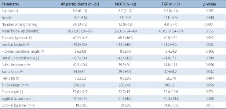
be-Table 1. Baseline carecteristic findings
Parameter All partipiciants (n=27) MCGR (n=12) TGR (n=15) p-value
Age (years) 8.6 (6–11) 8.7 (7–11) 8.5 (6–11) 0.282
Gender 18 F–9 M 7 F–5 M 11 F–4 M 0.448
Number of lengthening 8.0 (3–15) 12 (8–15) 4.8 (3–7) <0.001
Mean follow up (months) 38.7±8.8 (24–57) 36±6.6 (24–45) 40.8±10 (24–57) 0.186
Thoracic kyphosis (º) 49.2±14.3 48.5±16.3 49.8±13.1 0.922
Lumbar lordosis (º) –49.1±10.8 −43.9±10.4 −53.2±9.6 0.033
Proximal junctional angle (º) 8.6±4.8 8.9±4.9 8.4±4.9 0.806
Distal junctional angle (º) –13.7±10.4 −12.4±13.5 −14.8±7.3 0.788
Pelvic incidence (º) 42.5±10.4 39.5±9.1 44.8±11.1 0.096 Sacral slope (º) 34.1±8.1 29.9±5.9 37.4±8.2 0.032 Pelvic tilt (º) 8.5±8.2 9.6±8.8 7.6±7.9 0.494 T1–S1 range (mm) 296±38 280±48 309±21 0.056 Cobb angle (º) 52.6±12.5 52.7±15 52.6±10.6 0.574 Sagittal balance (mm) –15.7±27.9 –21.6±15.4 –10.3±34.8 0.558 Coronal balance (mm) 19±10.6 24±8.8 14.9±10.5 0.015
Values are presented as mean±standard deviation (range) or number (range). MCGR : magnetically control growing rod, TGR : traditional growing rod, F : female, M : male
tween groups was evaluated using chi-squre analysis. Mann Whitney U test was used for the comparisons between the two independent groups when the numerical variables were not normally distributed. Changes in findings between the preoperative, postoperative and the last follow up examintions were tested using repeated measures using analysis of variance with a Greenhouse-Geisser correction. Spearman’s correlation coefficient was used for evaluation of relationships between parameters. Statistical significance was accepted as p<0.05.
RESULTS
Age and gender distributions were similar in both groups. The baseline data and preoperative values of the patients are given in Table 1. The preoperative, early postoperative and fi-nal control radiological data of the patients who were operated with MCGR technique are given in Table 2 (Figs. 1 and 2), and those operated with TGR technique are given in Table 3 (Figs.
Table 2. MCGR radiological results in preoperative, postoperative and last follow-up period
Parameter Preoperative Postoperative Last follow-up p-value
Thoracic kyphosis (º) 48.5±16.3 27.8±13.0 23.1±6.7 <0.001
Lumbar lordosis (º) −43.9±10.4 −32.7±13.7 −39.0±10.8 0.071
Proximal junctional angle (º) 8.9±4.9 11.7±7.8 16.5±7.4 0.001
Distal junctional angle (º) −12.4±13.5 −8.5±13.5 −10.3±16.5 0.678
Pelvic incidence (º) 39.5±9.1 38.9±13.1 42.4±8.3 0.319 Sacral slope (º) 29.9±5.9 33.9±7.2 35.9±8.7 0.04 Pelvic tilt (º) 9.6±8.8 5.0±14.4 6.5±9.0 0.208 T1–S1 range (mm) 280.2±48.8 309.8±46.7 352.1±51.0 <0.001 Cobb angle (º) 52.7±15.0 27.8±13.0 32.2±15.4 <0.001 Sagittal balance (mm) −21.6±15.4 −13.7±36.8 −4.7±31.2 0.027 Coronal balance (mm) 24.0±8.8 10.3±8.0 13.0±8.5 0.001
Values are presented as mean±standard deviation. MCGR : manyetik control growing rod
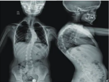
Fig. 1. Preoperative AP/lateral X-ray of patienttreatedwith MCGR technique. AP : anteroposterior, MCGR : magnetically controlled growing
rod. Fig. 2. Last follow-up AP/lateral X-ray of patienttreatedwith MCGR
technique. AP : anteroposterior, MCGR : magnetically controlled growing rod.
3 and 4). The comparison of the radiological findings of the two groups on the postoperative and final control visits is giv-en in Table 4.
No correlation was observed between TK, PJA, and DJA in the postoperative period and last postoperative visit in both patient groups. Correlation was not detected between postop-erative LL and DJA in both patients groups. In patients who underwent MCGR, the number of lengthening was positively correlated with postoperative PJA and negatively correlated with postoperative DJA (r=0.674, p=0.016; r=-0.766, p=0.004, respectively). There was a negative correlation detected be-tween PJA and DJA in the postoperative period in patients who underwent MCGR (r=-0.687, p=0.014).
In patients who underwent TGR, the number of lengthen-ing was detected positively correlated with postoperative and last postoperative PJA (r=0.683, p=0.010; r=0.687, p=0.005, respectively), and negatively correlated with postoperative and last postoperative DJA (r=-0.818, p<0.001; r=-0.542, p=0.037, respectively). In patients who underwent TGR operation, a negative correlation was detected between PJA and DJA in the postoperative period and last postoperative period (r=-0.528, p=0.043; r=0.520, p=0.047).
Table 3. TGR radiological results in preoperative, postoperative and last follow-up period
Parameter Preoperative Postoperative Last follow-up p-value
Thoracic kyphosis (º) 49.8±13.1 38.4±12.0 39.2±11.7 0.004
Lumbar lordosis (º) −53.2±9.6 −43.2±12.1 −47.0±11.9 0.009
Proximal junctional angle (º) 8.4±4.9 8.6±5.4 10.8±6.4 0.166
Distal junctional angle (º) −14.8±7.3 −14.1±11.5 −15.8±8.8 0.591
Pelvic incidence (º) 44.8±11.1 43.0±11.7 45.0±10.7 0.455 Sacral slope (º) 37.4±8.2 34.1±6.3 35.2±7.5 0.183 Pelvic tilt (º) 7.6±7.9 9.1±7.3 9.6±7.7 0.467 T1–S1 range (mm) 309.4±21.5 333.0±27.3 362.2±27.7 <0.001 Cobb angle (º) 52.6±10.6 30.4±10.2 26.5±12.5 <0.001 Sagittal denge (mm) −10.3±34.8 0.1±33.5 −3.0±40.6 0.628 Coronal denge (mm) 14.9±10.5 9.2±7.0 8.2±6.7 0.067
Values are presented as mean±standard deviation. TGR : traditional growing rod
Fig. 3. Preoperative AP/lateral X-ray of patienttreatedwith TGR technique. AP : anteroposterior, TGR : traditional growing rod.
Fig. 4. Last follow-up AP/lateral X-ray of patienttreatedwith TGR technique. AP : anteroposterior, TGR : traditional growing rod.
Superficial wound infection in one patient and screw pull out complication in three patients were encountered in the MCGR group. Superficial wound infection in three patients, deep tissue infection in one patient, 6-times rod breakages in four patients and 7-times screw pull out complications in six patients were occurred in TGR group.
DISCUSSION
In the TGR application, the requirement of periodic length-ening in the operation room every 6 to 9 months, and the re-quirement of a total of 6 to 18 scheduled surgical procedures from the beginning of treatment are the disadvantages of this
technique. In addition, there is an increased risk of complica-tions such as surgical wound complicacomplica-tions, the termination of the growth by stiff spine development (autofusion), deterio-rated chest development, and thoracic insufficiency syn-drome3,8). In MCGR application, it is thought that the increase
in the frequency of lengthening may reduce the development of the autofusion by meeting the spine growth more closely12).
There is an opinion that, with the more frequent lengthening in MCGR, which is possible as outpatient, the rods would more closely match up with spinal growth and possibly stress may be reduced throughout the implants1). In our study, in the
TGR technique, rod breakage in four patients (27%), screw pullout complication in six patients (40%) were encountered, while screw pullout complication in only three patients (25%) were encountered in patients who underwent surgery with MCGR technique.
Akbarnia et al.4) and Shah et al.23) have observed a decrease
in TK after the first implantation, however, they have ob-served an increase in the last follow-up. Similarly, LL has ini-tially decreased, and increased at the latest follow-up. In their study comparing TGR and MCGR, Akbarnia et al.5) reported
a decrease in postoperative value of TK compared to the pre-operative value in both groups, and a tendency to increase in the last follow up. In our study, in line with the literature, we have observed a decrease in the early postop period in TK and LL values, however, in the last follow up, there was an increase in the patient group treated with TGR, while the decrease was continuing in the patients treated with MCGR. Since the ky-phosis could not be given to the magnetic rod actuator part and this part coincided with the patient’s kyphosis region, we think that hypokyphosis might have been developed in the MCGR group.
In order to prevent proximal junctional kyphosis in patients whom they applied TGR, Akbarnia and Emans3) suggested
that rods should be countered in the shape of kyphosis and interspinal ligaments should be kept as strong as possible, up-per level should be limited at T2 level and the upup-per level should rarely be reached, and mentioned that in the presence of thoracic hyperkyphosis, the excessive correction of the rods may lead to implant failure after surgery. in their study in which they have applied MCGR and evaluated the proximal junctional kyphosis, Inaparthy et al.15) have emphasized that
male, syndromic, hyperkyphotic and younger patients should be closely followed-up in terms of proximal junctional
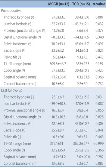
kypho-Table 4. Comparasion of MGRS and TGR radiological results in postoperative and last follow-up period
MCGR (n=12) TGR (n=15) p-value
Postoperative
Thoracic kyphosis (º) 27.8±13.0 38.4±12.0 0.001
Lumbar lordosis (º) −32.7±13.7 −43.2±12.1 0.032
Proximal junctional angle (º) 11.7±7.8 8.6±5.4 0.378
Distal junctional angle (º) −8.5±13.5 −14.1±11.5 0.340
Pelvic incidence (º) 38.9±13.1 43.0±11.7 0.497 Sacral slope (º) 33.9±7.2 34.1±6.3 0.825 Pelvic tilt (º) 5.0±14.4 9.1±7.3 0.478 T1−S1 range (mm) 309.8±46.7 333±27.3 0.139 Cobb angle (º) 27.8±13.0 30.4±10.2 0.420 Sagittal balance (mm) −13.7±36.8 0.1±33.5 0.366 Coronal balance (mm) 10.3±8.0 9.2±7.0 0.732 Last follow-up Thoracic kyphosis (º) 23.1±6.7 39.2±15.5 0.03 Lumbar lordosis (º) −39.0±10.8 −47.0±11.9 0.087
Proximal junctional angle (º) 16.5±7.4 10.8±6.4 0.056
Distal junctional angle (º) −10.3±16.5 −15.8±8.8 0.825
Pelvic incidence (º) 42.4±8.3 45.0±10.7 0.303 Sacral slope (º) 35.9±8.7 35.2±7.5 0.941 Pelvic tilt (º) 6.5±9.0 9.6±7.7 0.463 T1−S1 range (mm) 352.1±51 362.2±27.7 <0.999 Cobb angle (º) 32.2±15.4 26.5±12.5 0.366 Sagittal balance (mm) −4.7±31.2 −3.0±40.6 0.282 Coronal balance (mm) 13.0±8.5 8.2±6.7 0.083
Values are presented as mean±standard deviation. MCGR : manyetik control growing rod, TGR : traditional growing rod
sis (PJK) development. In our study, we found that the in-crease in PJA was higher in the MCGR group in the last follow up, although it did not reach statistical significance. It is evi-dent that the PJA values increased as the TK values of the pa-tients decreased. Although there was an increase in PJA values in the early postoperative period and last postoperative period in patients who underwent TGR operation, there was no cor-relation between TK and PJA. Since inadequate kyphosis was given to the actuator part in the MCGR group, patients have less kyphosis angle than the TGR group in postoperative and future postoperative follow-up. We think that this situation may be effective in the increase of PJA in MCGR group in the follow-ups. Besides, we think that further lengthening in the MCGR group compared to the TGR group may be the reason for distractive forces to increase more in the adjacent segments and higher increase rate in PJA. It should be kept in mind that repeated distraction below the existing upper thoracic kypho-sis may worsen the upper deformity. Due to the small number of patients, we could not subgroup patients according to pre-operative kyphosis values and this is one of the limitations of our study. Another limitation of our study was that the PJK values were higher in the MCGR patient group at the last fol-low-up compared to the TGR patient group, but we could not find a statistically significant value due to the insufficient number of patients and follow-up time.
Lowe et al.20) have reported that preoperative thoracolumbar
kyphosis was a risk factor for postoperative DJK development. Shah et al.23) have indicated that DJA showed the same pattern
as LL and correlated with the change in LL and that DJA closely reflected the change in LL. In our study, we observed that both of our groups had the same correlative pattern be-tween LL and DJA, however, this change was not statistically significant.
Some studies have shown that coronal and sagittal plane deformity can develop after initial surgery in both single and dual GR techniques25). In a study, which included 67 patients
and examined the effect of serial lengthening on sagittal bal-ance (SB) in TGR, it was reported that the balbal-ance tended to come to neutral in the early postoperative period and it was found to be statistically significant, however, this significance was reported to be lost in future follow-up period19). We
ob-serve in our study that the SB and coronal balance change tended to come to neutral in the MCGR group but may not provide information about the superiority of methods to each
other due to small number of patients in the groups and short follow-up periods.
Pelvic incidence (PI) is defined independently of position and has been shown to play an important role in maintaining sagittal alignment9,18). The presence of abnormal PI is a risk
factor for sagittal imbalance and failure following adult scoli-osis surgery11,17). In their recent study, Atici et al.7) stated that
pelvic parameters (PI, pelvic tilt, and sacral slope) remained unchanged in patients with EOS treated with GR. Shah et al.23)
have found a 5-degree increase in PI compared to preoperative value, however, they have reported that this was not cally significant. In our study, although we observed a statisti-cally significant increase in the sacral slope in our follow-up in the MCGR group, we did not find any significant changes in other spinopelvic parameters and spinopelvic parameters in the TGR patient group. As a result, we have not determined statistically significant difference between the MCGR group and the TGR group in terms of spinal and spinopelvic param-eters in the short-term follow-up.
Sankar et al.22) have shown that there was an inconsistency
between the expected and the obtained net T1–S1 length in the case of repeated lengthening. They attributed this to the hardening of the spine due to autofusion. The effects of this phenomenon on the sagittal profile are still unclear. We pro-vided statistically significant lengthening of T1–S1 distance in both groups in the follow-up period. When we compared the surgical technique, we could not find a statistically significant difference between the groups. Both methods are effective and successful in providing increase in length. In both methods, it should be considered that the planned and actual lengthening will not be the same.
Limitations of our study were; limited number of patients, being retrospective in the nature, short-term follow-up peri-ods and performance of the two procedures by two different surgical teams and clinics.
Studies on TGR procedures have shown that wound and implant complication rates are as high as 58%8,16). In their
multicentric study, Choi et al.12) have shown that MCGR
re-sults in a lower rate of infections compared to TGR (11% in TGR versus 4% in MCGR). Rod breakage and screw pull out are frequently observed complications with TGR use and it was seen that the rod type or corset usage did not prevent these problems8). It is thought that this is a problem regarding
implant, and is caused by spanning unfused spinal seg-ments12). On the other hand, the MCGR is considered not
eliminating implant-related common complications such as rod breakage or screw pull out12). The absence of lengthening
after some lengthening procedures is specific to MCGR, how-ever, does not always indicate a problem with the device12).
Also in our study, implant-related complications were higher in the TGR group. However, planning and follow-up of the treatment (TGR or MCGR) according to the normal pediatric spinal sagittal parameters may help in preventing the compli-cations.
CONCLUSION
When evaluating MCGR and TGR techniques in their own, we can obtain significant changes in Cobb angle, and T1–S1 values when we look at preoperative, early postoperative and last follow up values. In the evaluation of sagittal pelvic pa-rameters, we observed the lack of significant difference be-tween the two groups. MCGR can cause hypokyphosis and PJK in a minimum 2-year follow-up period. In order to pre-vent proximal junctional kyphosis, magnetic rods with differ-ent kyphotic degrees might be manufactured by the company and compatible magnetic rods with pelvic parameter and lumbar lordosis can be placed. The implant-related complica-tions were less in the MCGR group. However, larger case groups and longer follow-up periods, may aid in the under-standing the superiority of one method to other in terms of complications.
ConFliCts oF interest
No potential conflict of interest relevant to this article was reported.
inForMed Consent
Informed consent was obtained from all individual partici-pants included in this study.
aUthor ContriBUtions
Conceptualization : SE Data curation : ONÖ Formal analysis : YA Funding acquisition : ÇÖ Methodology : BPProject administration : SE Visualization : BP, ONÖ Writing - original draft : SE, BP Writing - review & editing : A, ÇÖ
References
1. Agarwal A, Agarwal AK, Jayaswal A, Goel V : Smaller ınterval distrac-tions may reduce chances of growth rod breakage without ımpeding desired spinal growth: a finite element study. Spine Deform 2 : 430-436, 2014
2. Akbarnia BA, Cheung K, Noordeen H, Elsebaie H, Yazici M, Dannawi Z, et al. : Next generation of growth-sparing techniques: preliminary clini-cal results of a magneticlini-cally controlled growing rod in 14 patients with early-onset scoliosis. Spine (PhilaPa 1976) 38 : 665-670, 2013 3. Akbarnia BA, Emans JB : Complications of growth-sparing surgery in
early onset scoliosis. Spine (PhilaPa 1976) 35 : 2193-2204, 2010 4. Akbarnia BA, Marks DS, Boachie-Adjei O, Thompson AG, Asher MA :
Dual growing rod technique for the treatment of progressive early-onset scoliosis: a multicenter study. Spine (PhilaPa 1976) 30(17 Suppl) : S46-S57, 2005
5. Akbarnia BA, Pawelek JB, Cheung KM, Demirkiran G, Elsebaie H, Emans JB, et al. : Traditional Growing rods versus magnetically controlled growing rods for the surgical treatment of early-onset scoliosis: a case-matched 2-year study. Spine Deform 2 : 493-497, 2014
6. Alanay A, Dede O, Yazici M : Convex instrumented hemiepiphysiodesis with concave distraction: a preliminary report. Clin Orthop Relat Res
470 : 1144-1150, 2012
7. Atici Y, Akman YE, Erdogan S, Sari S, Yavuz U, Carkci E, et al. : The ef-fect of growing rod lengthening technique on the sagittal spinal and the spinopelvic parameters. Eur Spine J 24 : 1148-1157, 2014
8. Bess S, Akbarnia BA, Thompson GH, Sponseller PD, Shah SA, El Sebaie H, et al. : Complications of growing-rod treatment for early-onset scoliosis: analysis of one hundred and forty patients. J Bone Joint Surg Am 92 : 2533-2543, 2010
9. Boulay C, Tardieu C, Hecquet J, Benaim C, Mouilleseaux B, Marty C, et al. : Sagittal alignment of spine and pelvis regulated by pelvic incidence: standard values and prediction of lordosis. Eur Spine J 15 : 415-422, 2006
10. Campbell RM Jr, Smith MD, Hell-Vocke AK : Expansion thoracoplasty: the surgical technique of opening-wedge thoracostomy. Surgical
tech-nique. J Bone Joint Surg Am 86-A Suppl 1 : 51-64, 2004
11. Cho W, Mason JR, Smith JS, Shimer AL, Wilson AS, Shaffrey CI, et al. : Failure of lumbopelvic fixation after long construct fusions in patients with adult spinal deformity: clinical and radiographic risk factors: clinical article. J Neurosurg Spine19 : 445-453, 2013
12. Choi E, Yazsay B, Mundis G, Hosseini P, Pawelek J, Alanay A, et al. : Im-plant complications after magnetically controlled growing rods for early onset scoliosis: a multicenter retrospective review. J Pediatr Orthop
37 : e588-e592, 2017
13. D’Astous JL, SandersJO : Casting and traction treatment methods for scoliosis. Orthop Clin North Am 38 : 477-484, v, 2007
14. Eltorai AEM, Fuentes C : Magnetic growth modulation in orthopaedic and spine surgery. J Orthop 15 : 59-66, 2018
15. Inaparthy P, Queruz JC, Bhagawati D, Thakar C, Subramanian T, Nnadi C : Incidence of proximal junctional kyphosis with magnetic expansion control rods in early onset scoliosis. Eur Spine J 25 : 3308-3315, 2016 16. Kabirian N, Akbarnia BA, Pawelek JB, Alam M, Mundis GM Jr, Acacio R,
et al. : Deep surgical site ınfection following 2344 growing-rod proce-dures for early-onset scoliosis: risk factors and clinical consequences. J
Bone Joint Surg Am 96 : e128, 2014
17. Le Huec JC, Faundez A, Dominguez D, Hoffmeyer P, Aunoble S : Evi-dence showing the relationship between sagittal balance and clinical outcomes in surgical treatment of degenerative spinal diseases: a litera-ture review. Int Orthop 39 : 87-95, 2015
18. Legaye J, Duval-Beaupère G, Hecquet J, Marty C : Pelvic incidence: a fundamental pelvic parameter for three-dimensional regulation of spinal sagittal curves. Eur Spine J 7 : 99-103, 1998
19. Li WJ, Sun ZJ, Guo SG, Qiu GX, Zhang JG, Shen JX, et al. : The effect of growing Rod treatment on coronal balance during serial lengthening surgeries in early onset scoliosis. BMC Musculoskelet Disord 17 : 158, 2016
20. Lowe TG, Lenke L, Betz R, Newton P, Clements D, Haher T, et al. : Distal junctional kyphosis of adolescent idiopathic thoracic curves following anterior or posterior instrumented fusion: incidence, risk factors, and prevention. Spine (PhilaPa 1976) 31 : 299-302, 2006
21. Moe JH, Kharrat K, Winter RB, Cummine JL : Harrington instrumentation without fusion plus external orthotic support for the treatment of difficult curvature problems in young children. Clin Orthop Relat Res 185 : 35-45, 1984
22. Sankar WN, Skaggs DL, Yazici M, Johnston CE 2nd, Shah SA, Javidan P, et al. : Lengthening of dual growing rods and the law of diminishing returns. Spine (PhilaPa 1976) 36 : 806-809, 2011
23. Shah SA, Karatas AF, Dhawale AA, Dede O, Mundis GM Jr, Holmes L Jr, et al. : The effect of serial growing rod lengthening on the sagittal profile and pelvic parameters in early-onset scoliosis. Spine (Phila Pa
1976) 39 : E1311-E1317, 2014
24. Takaso M, Moriya H, Kitahara H, Mimani S, Takahashi K, Isobe K, et al. : New remote-controlled growing-rod spinal instrumentation possibly ap-plicable for scoliosis in young children. J Orthop Sci 3 : 336-340, 1998 25. Thompson GH, Akbarnia BA, Kostial P, Poe-Kochert C, Armstrong DG, Roh J, et al. : Comparison of single and dual growing rod techniques fol-lowed through definitive surgery: a preliminary study. Spine (PhilaPa