RESEARCH ARTICLE
Histological examination of the skin and AgNOR parameters of matrix pili cells in the
chinchilla
Yasemin Oznurlu*, Ilhami Celik, Emrah Sur, Tugba Ozaydin Özet
Öznurlu Y, Çelik I, Sur E, Özaydın T. Çinçilyada deri ve kıl matriks pili hücrelerinde AgNOR parametrelerinin histolo-jik incelemesi. Eurasian J Vet Sci, 2011, 27, 1, 39-43 Amaç: Çinçilyalarda derinin histolojisi ve kıl matriks pili hücrelerinde AgNOR sayısının ışık mikroskobik seviyede incelenmesi amaçlandı.
Gereç ve Yöntem: Onbeş adet ergin ve sağlıklı çinçilyadan alınan deri örnekleri tamponlu formol salin solüsyonunda tespit edildi ve rutin histolojik yöntemlerle parafinde blok-landı. Bloklardan alınan kesitler Crossmon’un üçlü boyama-sı, Gordon ve Sweet’in retiküler iplik boyamaboyama-sı, Verhoef’in elastik iplik boyaması ve AgNOR boyama yöntemleriyle bo-yandı. Derinin toplam kalınlığı ile epidermis ve dermis ka-lınlıkları, birim alandaki (1 mm2) ortalama kıl folikül sayı-ları ve germinal matriks pili hücrelerinde AgNOR sayısı ve alanı belirlendi.
Bulgular: Germinal matriks hücrelerinin çekirdeklerinde-ki AgNOR’ların kahverengi-siyah lekeler halinde 1 ile 4 adet arasında değişen sayılarda olduğu tespit edildi. Çinçilyalar-da birleşik kıl foliküllerinin bir ya Çinçilyalar-da iki primer folikül çev-resinde çok sayıda sekonder folikülden oluştuğu gözlendi. Sekonder foliküllerin primer foliküllere oranı (S/P) 13/1 olarak tespit edildi.
Öneri: Daha geniş populasyonlarda ve mevsimsel değişik-likler göz önüne alınarak yapılacak çalışmalarda, kıl folikül-lerinin sayı ve dağılımı ile germinal matriks hücrelerinde-ki AgNOR parametrelerinin belirlenmesi seleksiyonda kürk kalitesi için önemli bir kriter olabilir.
Abstract
Oznurlu Y, Celik I, Sur E, Ozaydin T. Histological examina-tion of the skin and AgNOR parameters of matrix pili cells in the chinchilla. Eurasian J Vet Sci, 2011, 27, 1, 39-43 Aim: In this study, histology of the skin and number and area of the argyrophilic nucleolar organiser regions (AgNORs) of the germinal matrix epithelial cells of the chinchilla were investigated by light microscopic techniques.
Materials and Methods: Skin samples were taken from 15 adult healthy chinchilla, fixed in formol saline and im-mersed in parafine. Tissue sections were stained with Crossman’s trichrome, Gordon and Sweet’s reticuler fiber, Verhoef’s elastic fibre and AgNOR staining methods. The to-tal thickness of epidermis and dermis, hair follicle numbers per square milimetres, and number and area of AgNORs of the germinal matrix epithelial cells were measured.
Results: AgNORs were seen as 1-4 black dots which distrib-uted in the nuclei of the germinal matrix cells of the hair follicles. The chinchilla compound hair follicle consisted of one or two primary follicles surrounded by multiple smaller secondary follicles. The ratio of secondary to primary hair follicles (S/P) was 13/1.
Conclusion: In further studies on a larger population sea-sonal differences, determination of number and distribu-tion of hair follicules and AgNOR parameters in the germi-nal matrix cell should be used as significant criteria for fur quality in selection.
Department of Histology, Veterinary Faculty, Selcuk University, Campus, 42075, Konya, Turkey
Received: 27.06.2010, Accepted: 10.08.2010 *[email protected]
Anahtar kelimeler: Çinçilya, deri, kıl follikülleri, AgNOR Keywords: Chinchilla, skin, hair follicle, AgNOR
Journal of Veterinary Sciences
Introduction
The long-tailed chinchilla (Chinchilla lanigera), also called the Chilean, Coastal, or Lesser chinchilla, is one of two species of rodents from the genus Chinchilla. The fur from chinchillas is popular in the fur trade due to its extremely soft feel, because they have about 60 hairs sprouting from each hair follicle (Spotorno et al 2004).
As in all animal species, the chinchilla skin also con-sists of two main layers an outer epithelial layer, epi-dermis and a deeper connective tissue layer, epi-dermis (Ozfiliz et al 1997, Dellmann and Eurell 1998, Moore et al 1998). The dermis consisted of primarily of dense irregular connective tissue with a felt work type I collagen, some elastic and reticular fibres em-bedded in an amorphous ground substance. The der-mis is divided into a superficial papillary layer that blends into a deep reticular layer without a clear line of demarcation. Although, the basic architecture of the skin is similar in all mammals, differences exist, however, in the thickness of the epidermis and der-mis between species and within the same species in various regions of the body (Atlee et al 1997, Moore et al 1998).
Hair follicles are classified as primary and secondary follicles. The primary follicles have a large diameter and rooted deep in the dermis and usually associated with sebaceous and sweat glands. A hair fibre that emerges from a primary follicle is called a primary hair fibre. The secondary follicles are smaller in di-ameter; their roots are superficially located and may have a sebaceous gland but lack a sweat gland and arrector pili muscle. Their hair fibres are secondary hair fibres. Hair follicles are located either individu-ally or in groups and form compound follicles, as in the animals that live in wet and cold climates (Koul et al 1987, Atlee et al 1997, Dellmann and Eurell 1998, Moore et al 1998, Astı and Kurtdede 2002).
Nucleolus organizing regions (NORS) are the loops of DNA containing ribosomal RNA genes and pro-teins in these regions are easily visualized as silver-stained black dots (AgNORS) in the cell nucleus with colloidal silver since they are argyrophilic (Cabrini et al 1992, Orrea et al 2001).Variations in the number and normal distribution pattern of AgNORs might in-dicate qualitative and quantitative changes in protein synthesis activity of the cell (Watchler et al 1986). An increase in the number of AgNORs in interphase nu-clei indicates cellular hyperactivity, and might give a valuable knowledge on the proliferation rate, differ-entiation process and secretory activity of a given cell. These changes may also be associated with the pro-cesses involved in malignant transformation (Crocker and Nar 1987, Orrea et al 2001, Sur et al 2003, Guler et al 2005).
Some structural and morphometrical features of the skin have been known to be closely related to the
leather quality and fur yield of the animal. The aim of this study was to determine some histological, mor-phometric features and AgNOR parameters of germi-nal matrix epithelial cells in the skin of chinchilla.
Material and Methods
Animals and skin samples
Skin samples were taken from 15 adult male and ap-parently healthy chinchillas. The chinchillas were kept under optimal feeding and management condi-tions of Research Farm of Selcuk University, Veteri-nary Faculty. Because of the high seasonal variation in hair fibre growth of chinchilla, punch skin biopsies were performed in the same month (November). The 3 mm diameter skin samples were taken from the lumbar dorsum of each animal after anaesthesia by local injection with 2% lidocaine and use of disinfec-tant thereafter. The chinchillas received humane care according to the criteria outlined in the “Guide for the Care and Use of Laboratory Animals” prepared by the National Institutes of Health.
Histological procedures
The samples were fixed in 10% buffered formalin and divided into two pieces. Following the fixation, all samples were processed by means of the rou-tine histological methods and immersed in paraffin blocks. One piece of the each sample was examined paralel to the surface of skin, while the other pieces were examined vertical to the surface of skin. The 6 μm thick sections were taken and a couple of sections from each block were stained by means of Masson’s trichrome, and reticular and elastic fibre stains (Cull-ing et al 1985). For AgNOR stain(Cull-ing, the sections were incubated for 30 min at 37 0C in the dark for 10-12
minutes in a fresh solution of one part of 1% gelatin (Sigma, USA) in 1% formic acid (Merck, Germany) with two parts of 50% aqueous silver nitrate III (Mer-ck, Germany). Stained sections were dehydrated in graded ethyl alcohol, cleaned in xylene and covered with a synthetic mounting medium. The AgNORs were visualized as intranuclear black dots under light microscope (Korek et al 1991, Pich et al 1994).
Histological evaluation
All specimens were examined under the light micro-scope [Nikon Eclipse E-400 equipped with a digital camera head 5M) and camera control unit (DS-L1), Nikon, Japan]. The histological evaluation and measurements were performed on the digital images. The following parameters were determined: i) Thick-ness of each skin layer, ii) Number of the hair follicles in a unit tissue area (1 mm2), iii) Mean secondary
folli-cle/primary follicle ratio (S/P) of compound follicles, iv) Mean nucleus area of the germinal matrix cells, v) Mean AgNOR area and AgNOR number per nucleus of the germinal matrix cells, vi) Relative AgNOR area (percentage of AgNOR area in a given nucleus area) of
germinal matrix cells was calculated.
Results
Vertical sections showed that the skin of chinchil-las consisted of two layers an outer epidermis and a deeper connective tissue layer, the dermis (Figure 1). In all sections, it was determined that epidermis was very thin and formed with three layers such as stra-tum germinativum, strastra-tum granulosum and strastra-tum corneum. The stratum corneum was thin, whereas the stratum lucidum were not seen. The mean total epi-dermal thickness was 29.6 μm.
Figure 1. Vertical sections of skin of the chinchilla. 1:Epidermis, 2- 4- 5: Collagen fibres in dermis, 3: Hair follicles. Trichrome stain. Bar: 100 µm.
The dermis was possessed a superficial papillary lay-er that blended into a deep reticular laylay-er without a clear line of demarcation. There were a few elastic fi-bers and very thin bouquet of collagen fibres extend-ing parallel to just beneath the epidermis. The reticu-lar fibres mostly condensed in close proximity to the basal cells of the epidermis and formed a close felt work of fibrils that inserted into basal lamina beneath the epidermis and extended perpendicularly into the dermis as anchoring fibrils. The bulbs of the hair fol-licles were located in stratum papillare. Hypodermis was rich in adipocytes.
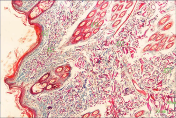
Figure 2. Cross sections of compound hair follicles of the chinchilla. PF: Primary Follicle SF : Secondary Follicles. Trichrome stain. Bar: 50 µm.
The hair follicles opened with their long axes to the skin’s surface at a narrower angle than the right an-gle. The primary follicles were distinguished with their large diameter (67.71 μm) and their roots were placed deep in the dermis, whereas secondary fol-licles were smaller (35.44 μm) and the roots located superficially. The chinchilla had compound hair fol-licles. Compound hair follicles constituted of one or two primary follicles and multiple smaller secondary follicles located around them (Figure 2). Both prima-ry and secondaprima-ry hair follicles were formed with me-dulla, cortex and cuticula. The number of primary and secondary hair follicles in mm2 was found to be 5.69
and 72.59, respectively.
AgNORs were seen as 1-4 black dots distributed in the nuclei of the germinal matrix cells of the hair follicles, although some of them were located at close proxim-ity to the nuclear envelope (Figure 3). AgNOR num-bers of the germinal matrix cells were 2.63. AgNOR and nucleus area of the germinal matrix cells were 1.79 µm2 and 24.31 µm2. Relative AgNOR area was
7.36 µm2.
Figure 3. Sections of hair bulbs of the chinchilla. AgNORs (arrows) are seen as black dots in the nuclei of germinal matrix cells. AgNOR staining. Bar: 15 µm.
Discussion
The histological observations have revealed that the Chinchilla display histological characteristics of the thin skin and similar to share common structural properties of skins of the other animal species (Soko-lov 1982). The skin of Chinchilla had a thin epidermis containing stratum germinativum, stratum granulo-sum and stratum corneum. The stratum lucidum lay-ers were absent. Similar findings were reported for rabbit skin by previous researchers (Sokolov 1982, Yağcı at al 2006). Yağcı et al (2006) have showed that the epidermal thickness was 13.9 μm for males and 11.66 μm in females of the white New Zealand rabbit, whereas it was 28 μm and 22 μm in Lepus tolai and Lepus timidus rabbits, respectively (Sokolov 1982). The dermis consists primarily of dense irregular con-nective tissue with a feltwork collagen, elastic and re-ticular fibers embedded in an amorphous ground sub-stance. The dermis can be divided into a superficial
papillary layer that blends into a deep reticular layer without a clear line of demarcation. The mean thick-ness of dermis in chinchilla was 1323.28 μm (stratum papillare [945.28 μm] and stratum reticulare [378.00 μm]). Yağcı et al (2006) have found that the mean dermis thickness was 1494.4 μm in the females, and 1754.8 μm in the males of the white New Zealand rab-bits and it was 454 μm in Lepus timidus rabrab-bits and 412 μm in Lepus tolai rabbits (Sokolov 1982). Gül et al (2005) have found that the mean dermis thickness was 842.6 μm in the back and 365.65 μm in the cheek of the wistar albino rats. Thomas (2005) have re-ported that significant effects of age, diet, or age-diet interaction were observed in respect of the thickness of epidermis, dermis, epidermal nuclear number, col-lagen percentage fraction, and area fraction of capil-laries in rat.
Many differences exist in the arrangement of the hair follicles among the animal species. They are located either individually or in groups. The individual hair follicles, which are also called simple follicles, have only one hair emerging to the surface. The com-pounds follicles are clusters of several hair follicles located in the dermis, and usually consist of one primary hair follicle and several secondary follicles (Atlee et al 1997, Dellmann and Eurell 1998, Moore et al 1998). The chinchilla had compound hair follicles. Compound hair follicles constituted of one or two pri-mary follicles and multiple smaller secondary follicles located around them. Gül et al (2005) have reported that the race, age, sex and different body regions had significant effects on the number of hair follicle and dermal papilla in the wistar albino rats. However, Yağcı et al (2006) reported that there was not any sex derived difference in both primary and secondary fol-licle numbers of the white New Zealand rabbit. In this study, the number of primary and secondary hair fol-licles in mm2 was found to be 5.69 and 72.59 respec-tively. The fiber quality of wool-producing species is often expressed in terms of the secondary-to primary- hair ratio (S/P ratio) (Atlee et al 1997). Oznurlu et al (2009) have reported that the ratio of secondary to primary hair follicles was higher in the Angora rab-bit (19/1) than the New Zealand rabrab-bit (5/1). In this study, the ratio of secondary to primary hair follicles (S/P) was 13/1.
Nucleolus organizer regions (NORs) are the specific DNA regions containing genes that code for the syn-thesis of rRNA. The amount of AgNOR proteins is used as a marker of proliferation, since it is low in G1 phase and high in the S-G2 phase. Therefore, a higher Ag-NOR value indicates that the major part of the cells is in the S-G2 phase and correlatively few are in the G1 phase, suggesting a rapid cell cycle (Field et al 1984, Pession et al 1991). Moreover, silver staining has been suggested to reveal transcriptionally active NORs (Or-rea et al 2001) and variations in the normal pattern of AgNORs might indicate qualitative and quantitative
changes in protein synthesis (Watchler et al 1986). Because that an increase in the number of AgNORs in interphase nuclei indicates cellular hyperactivity, the researchers (Crocker and Nar 1987, Cabrini et al 1992, Orrea et al 2001, Guler et al 2005). Also based on the findings of this technique administrated other tissues (muscle, mammary gland, ovary follicle, etc.), it is possible to correlate the growth performance, egg and milk production traits (Aydın 2004). Oznurlu et al (2009) reported that AgNOR number, absolute and relative AgNOR areas were higher in Angora rabbit which is famous for both growth and quality of hair fibre than that of the white New Zealand rabbit. The mean AgNOR number, absolute and relative AgNOR areas were reported as 2.5, 5.3 µm2 and 19.1 µm2
re-spectively in the Angora rabbit; however, these values were found as 1.7, 3.8 µm2 and 13.2 µm2 respectively
in the New Zealand rabbit. In this study, AgNOR num-bers of the germinal matrix cells was 2.63. AgNOR and nucleus area of the germinal matrix cells were 1.79 µm2 and 24.31 µm2 respectively. Relative AgNOR area
was 7.36 µm2.
The germinal matrix, which proliferates and produces the hair and the inner root sheath via the signal, has close homologies of the stratum basale of the epider-mis (Stenn and Paus 2001). A negative but high corre-lation between fibre diameter and follicle density was previously illustrated and selection efforts on wool-producing activities of skin would predominantly af-fect follicle density and fibre charecteristics (Moore et al 1998). Oznurlu et al (2009) reported a positive co-orelation between AgNOR number and fibre density in Angora rabbits which have high wool production. Poyraz et al (2005) reported that performing selec-tion using some morphological characteristics highly correlated with some pelt traits could contribute to improved fur quality for the production or breeding of chinchillas.
Conclusion
The present study confirms the presence of species specific characteristics and breed-dependent varia-tions of the thickness of sublayers and hair follicle number in a unit area of the chinchilla skin. Finally, our results suggested that determination of number and distribution of hair follicules and AgNOR param-eters in the germinal matrix cell can be used as signifi-cant criteria for fur quality in selection.
References
Astı R, Kurtdede N, 2002. Investigation on the skin structu-re of Hampshistructu-re Down x Akkaraman, Awassi and Kon-ya Merino crossbred (F1 and B1) sheep. Turk J Vet Anim Sci, 26, 701-708.
Atlee BA, Stannard AA, Fowler ME, Willemse T, Ihrke PJ, Olivry T, 1997. The histology of normal llama skin. Vet Dermatol, 8, 165-176.
Aydın F, 2004. Determination of the distribution of silver stained nucleolar organizing regions in various tissues of broilers and laying hens. PhD Thesis, SU Healthy
Sci-ence Institute, Konya, Turkey.
Cabrini R, Schwint A, Mendez A, Femopase P, Lanfranchi H, Itoiz M, 1992. Morphometric study of nucleolar or-ganizer regions in human oral normal mucosa, papillo-ma and squamous cell carcinopapillo-ma. J Oral Pathol Med, 21, 257-259.
Crocker J, Nar P, 1987. Nucleolar organizer regions in lymphomas. J Pathol, 151, 111-118.
Culling CFA, Allison RT, Barr WT, 1985. Connective Tissue, In: Cellular Pathology Technique, Eds; Culling CFA, Al-lison RT, Barr WT, 4th edition, Butterworths, London, UK, pp: 164-179.
Delmann HD, Eurell JA, 1998. Integument, In: Textbook of Veterinary Histology, Eds; Delmann HD, Eurell JA, Lip-pincot Williams and Wilkins, Baltimore, Maryland, USA, pp: 303-332.
Field DH, Fitzgerald PH, Sin FY, 1984. Nuucleolar silver sta-ining pattern related to cell cycle phase and cell gene-ration of PHA stimulated human lymphocytes. Cytobi-os, 41, 23-33.
Gül M, Eşrefoğlu M, Seyhan M, 2005. Histomorphometric characteristics of the skin in wistar albino rats. J Derma-tol, 15, 136-140.
Guler N, Uckan S, Celik I, Oznurlu Y, Uckan D, 2005. Expressi-on of fas and fas-ligand and analysis of argyrophilic nuc-leolar organizer regions in squamous cell carcinoma: re-lationships with tumour stage and grade, and apoptosis. Int J Oral Maxillofac Surg, 34, 900-906.
Korek G, Martin H, Wenzelides K, 1991. A modified method for the detection of nucleolar organizer regions (Ag-NORs). Acta Histochem, 90, 155-157.
Koul GL, Biswas JC, Somvanshi R, 1987. Follicle and fibre characteristics of Indian pashmina goats. Res Vet Sci, 43, 398-400.
Moore GPM, Jackson N, Isaacs K, Brown G, 1998. Pattern and morphogenesis in skin. J Theo Biol, 191, 87-94. Orrea SC, Tomasi VH, Schwint AE, Itoiz ME, 2001. Modified
silver staining of nucleolar organizer regions to impro-ve the accuracy of image analyses. Biotech Histochem, 76, 67-73.
Ozfiliz N, Ozer A, Yakışık M, Erdost H, 1997. A histological and morphometrical comparative study on the skin of Kıvırcık and Karacabey Merino sheep. Turk J Vet Anim Sci, 21, 125-133.
Oznurlu Y, Çelik I, Sur E, Telatar T, Ozparlak H, 2009. Com-parative skin histology of the White New Zealand and Angora rabbits: Histometrical and immunohistochemi-cal evaluations. JAVA, 8, 1694- 1701.
Pession A, Farabegoli F, Trere D, Novello F, Montanaro L, Sperti S, Rambelli F, Derenzini M, 1991. The Ag-NOR proteins and transcription and duplication of ribosomal genes in mammalian cell nucleoli. Chromosoma, 100, 242-250.
Pich A, Chiarle R, Chiusa L, Palestro MD, 1994. Argyrophi-lic nucleolar organizer region counts predict survival in thymoma. Cancer, 74, 1568-1574.
Poyraz O, Akinci Z, Onbasilar EE, 2005. Phenotypic correla-tions among some traits in chinchilla lanigera produced in Turkey. Turk J Vet Anim Sci, 29, 381-384.
Sokolov VE, 1982. Mammal skin. University of California Press, Berkeley, Los Angeles, London, UK, pp: 177-182. Spotorno AE, Zuleta CA, Valladares JP, Deane AL, Jimenez JE,
2004. Chinchilla Laniger. Mammalian Species, 758, 1-9. Stenn KS, Paus R, 2001. Controls of hair follicle cycling.
Physical Review, 81, 450-481.
Sur E, Celik I, Oznurlu Y, Aydin MF, Sen I, Ozparlak H, 2003. Enzyme histochemistry and AgNOR numbers in the pe-ripheral blood leukocytes of 6 month-old Kangal bred Anatolian shepherd dogs. Rev Med Vet, 154, 591-598. Thomas JR, 2005. Effects of age and diet on rat skin
histo-logy. Laryngoscope, 115, 405-411.
Watchler F, Hopman AHN, Wiegant J, Schwarzacher G, 1986. On the position of nucleolus organizer regions (NORs) in the interphase nuclei: studies with a new, nonauto-radiographic in situ hybridization method. Exp Cell Res, 167, 227-240.
Yağcı A, Zık B, Uğuz C, Altunbaş K, 2006. Histology and morphometry of White New Zealand rabbit skin. Ind Vet J, 83, 876-880.