REGULAR ARTICLES
The role of goats as reservoir hosts for bovine herpes virus 1
under field conditions
Sibel Gür1
&Nural Erol2&Orhan Yapıcı3&Mehmet Kale4&Mehmet Tolga Tan2&Turhan Turan5& Mehmet Atilla Çakmak6
&Cemil Tosun6&Simay Yılmaz7&Abuzer Acar8&Işınsu Özenli7&Coşkun Gür9 Received: 16 June 2018 / Accepted: 2 November 2018 / Published online: 22 November 2018
# Springer Nature B.V. 2018 Abstract
Bovine herpesvirus 1 (BoHV1) is the cause of economically significant viral infections in cattle. Respiratory symptoms associ-ated with the infection are known as Infectious Bovine Rhinotracheitis (IBR). Sheep and goats are less sensitive to the infection although their role in inter-species viral transmission under field conditions is subject to controversy. The objective of this study was to investigate seroprevalence of BoHV1 infections in cattle, sheep, and goats raised together for at least a year. Blood serum samples were taken from 226 cattle, 1.053 sheep, and 277 goats from 17 small- to medium-scale farms. BoHV1-specific antibody presence and titers were determined using virus neutralization test. In total, 73 of the 226 cattle (32.3%) were seropositive. The infection was detected in 13 of the 17 farms. Infection rates ranged from 5.8 to 88.8%. Only one of the 1053 sheep (0.09%) was seropositive. However, 58 of the 277 (20.9%) goats were seropositive. Goat samples taken from 8 of the 17 farms were seropositive with infection rates ranging from 17 to 38.9%. Statistical analysis showed a significant correlation in infection rates between cattle and goats but not sheep. These results suggest that goats may be more sensitive to the BHV1 infection than sheep and the role of goats as possible reservoirs for BoHV1 in the control and eradication of BHV1 in cattle should be considered in future studies.
Keywords BHV1 . Cattle . Goat . Sheep . Reservoir
Introduction
Bovid herpesvirus type 1 (BoHV1) is the cause of Infectious Bovine Rhinotracheitis/Infectious Pustuler Vulvovaginitis/ Infectious Pustuler Balanopostitis (IBR/IPV/IPB) complex disease (Roizman et al. 1981). Herpesviruses are large, enveloped, double-stranded DNA viruses (Wyler et al.
1989). The infection was identified asBcoital exantheme^ by Büchner and Trommsdorf in Germany in the nineteenth century, and the cause of the infection was discovered in 1928 by Reisinger and Reimann (Muylkens et al. 2007). BoHV1 infection is still seen in the vast majority of the world and is one of the most important infectious diseases leading to economic losses in cattle. Infection in cattle has been eradicat-ed in Denmark, Switzerland, Finland, Sweradicat-eden, Austria, and parts of Italy. Many European countries have implemented control programs.
According to the latest genome analysis of the BoHV1 viral polypeptides, the virus has five subtypes classified within three groups. Types 1 and 2a are responsible for respiratory disorders and 2b is the cause of IPV and IPB. Infection with * Sibel Gür
1 Department of Virology, Faculty of Veterinary Medicine, Afyon Kocatepe University, Afyonkarahisar, Turkey
2
Department of Virology, Faculty of Veterinary Medicine, Adnan Menderes University, Aydın, Turkey
3
Department of Virology, Faculty of Veterinary Medicine, Selcuk University, Konya, Turkey
4 Department of Virology, Faculty of Veterinary Medicine, Mehmet Akif Ersoy University, Burdur, Turkey
5
Department of Virology, Faculty of Veterinary Medicine, Cumhuriyet University, Sivas, Turkey
6
Ministry of Food, Agriculture and Livestock, Afyonkarahisar, Turkey
7 İşlek LLC, Afyonkarahisar, Turkey 8
Department of Internal Diseases, Faculty of Veterinary Medicine, Afyon Kocatepe University, Afyonkarahisar, Turkey
subtypes 3a and 3b results in encephalitis (Wentink et al.
1993). The virus can be transmitted via oral or genital routes. Soon after acute infection, proviral DNA genome integrates into nerve cells and remains in the trigeminal or sacral gangli-ons for life, a general characteristic of alphaherpesviruses (Guy and Potgieter1985). Respiratory and digestive system disorders co-exist in newborns and youngs. Subclinical or single system disorder is generally seen in acute phase in immune competent adults.
In addition to cattle and buffaloes which serve as the main hosts, seropositivity and usually moderate disorders have been identified in many ruminant species. IgM titer reaches the maximum level at the end of the second week and the highest IgG titer can be determined at 2–4 weeks post-infection (Guy and Potgieter1985). The half-life of antibodies (Abs) specific to BoHV1 is limited to approximately by months. Re-activation is particularly prevalent in the presence of immuno-suppressive factors. After a natural infection, the animals be-come permanently seropositive (Rusch et al.1981). Routine controls involve Ab detection since, excluding the acute phase and the re-activation periods, no free virions can be found in the organisms. BoHV1 has been isolated from healthy and diseased sheep and goats in both natural cases and experimen-tal studies (Trueblood et al.1978; Hage et al.1997; Six et al.
2001; Mahmoud and Ahmed 2009; Tuncer-Göktuna et al.
2016).
Since the first serologic detection of BoHV1 in Turkey in 1971 (Gürtürk et al.1974) and antigen detection in 1987 (Burgu and Akça1987), many studies conducted in the coun-try revealed high infection rates especially in intensive closed system cattle enterprises (Alkan et al.2005; Tan et al.2006; Gençay et al.2009; Yıldırım et al.2009; Aslan et al.2015; Özgünlük and Yıldırım2017). Studies on sheep and goats were mostly done in the last decade and seropositivity rates are less than 10% in majority of the studies (Yeşilbağ et al.
2003; Albayrak et al. 2007; Çabalar and Özgünlük2014; Çeribaşı et al.2016; Yılmaz and Coşkun2016).
Even though sheep and goat are not as sensitive to the BoHV1 infection as cattle and buffalo, inter-species transmis-sion is possible and there is evidence to suggest that small ruminants may serve as reservoir hosts. In this study, BoHV1 infection was serologically investigated in sheep, goats, and cattle housed in the same farm for at least a year to evaluate the role of sheep and goats as reservoir hosts for the infection under field conditions.
Materials and methods
Sampled animals
Samples were collected only from farms where at least two of the cattle, sheep, or goats were raised together in the same
farm for at least a year. Blood serum samples were taken from 17 small to medium size private farms. There were cattle, sheep, and goats in the first eight farms and only cattle and sheep in the remaining farms (Table1). The cattle were held in adjoin-ing barns but pasture and drinkadjoin-ing water troughs were shared. All of the sampled farms were located in the Afyonkarahisar province in Western Anatolia (38° 75′ 68″ N–30° 53′ 87″ W) except that one farm (i.e., farm no. 5) was located in Kutahya, an adjacent province. All animals were adults (≥ 1 year of age) and clinically healthy during the sampling.
The gender of the animals was ignored during sampling although the vast majority were female. There were no de-tailed medical records of the animals available but the vaccine and health status of the animals in recent years were obtained from farm owners or veterinarians. All animals in the studied herds were not vaccinated against the BoHV1. There were no significant health problems except that reproductive problems were present in some of the cattle. None of the animals were vaccinated for BoHV1. In total, 1.556 blood serum samples were collected from (with number of animals in parentheses) cattle (226), sheep (1053), and goats (277).
Cell culture
Madin-Darby Bovine Kidney (MDBK) cell culture (ATCC, CCL-22) was used for virus propagation, titration, and neu-tralization test. Eagle’s minimal essential medium (EMEM) containing 2–10% fetal calf serum (FCS) was used as the culture medium.
Test virus
The BColorado^ reference strain obtained from Ankara University, Faculty of Veterinary Medicine, Department of Virology, was used as a control virus in serological tests. Tissue culture infective dose 50% (TCID50= 10−5/0.1 mL) of the virus suspension was determined by the Spearman and Kaerber method. Titer value (100DCID50) of the virus was 102.5/0.1 mL.
Virus neutralization test
Due to its high sensitivity and specificity, the standard virus neutralization test (VNT) was used to detect antibodies against BoHV1. Fifty microliters each of the serum samples and virus suspension was added in two wells of tissue culture plates and incubated for 2 h at 37 °C with 5% CO2. Fifty microliters of cell suspension (300.000 cells/mL) was added and the sam-ples were incubated for 24–48 h. Plates were evaluated on the basis of micro-morphology of the cells using an inverted mi-croscope. Ab-positive serum samples were diluted serially (1/1, 1/2, 1/4...1/128) and re-tested to determine antibody titer values [i.e., Serum Neutralisation50(SN50)].
Results
Serological findings
BoHV1-specific Abs were detected in 13 of the 17 farms tested. Infection rates were between 5.8 and 88.8%. 32.3% (73/226) of the cattle were seropositive. All of the 1053 sheep samples were seronegative except that one of the animals had 1/2 titer. Seropositivity in goats was detected in 4 of the 8 farms tested with infection rates ranging from 17 to 38.9%. In total, 58 of the 277 (20.9%) goats tested were seropositive (Table1). Both the cattle and the goats were totally seroneg-ative in two of the farms.
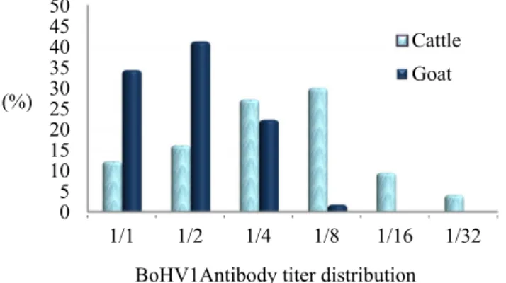
In re-tested samples to detect Ab titer values, BoHV1-specific Ab titers in cattle were between 1/1 and 1/32. The mean titer was 1/8. The titer in goats ranged from 1/1 to 1/8 and the mean titer was 1/2 (Fig.1).
Statistical analysis
Statistical analysis performed using Cohen’s kappa coefficient (Landis and Koch1977) showed a significant correlation (69.6%) in seropositivity rates between cattle and goats in seven enterprises. Data from nine cattles in farm no. 8 were not included in the analyses because the cattle were kept in isolation without any contact with sheep or goats.
Discussion
BoHV1 infection is one of the most economically significant infectious diseases in large ruminants. The same concern is not valid for small ruminants that are less sensitive to the infection. Wentink et al. (1993) stressed that cattle are the only significant source of viral spread. However, there are reports suggesting that small ruminants may play a role in inter-species viral transmission and spread of infection in eradica-tion and control studies of BoHV1 in cattle (Yeşilbağ and Bilge-Dağalp2006; Yeşilbağ and Güngör 2009). However, the role of small ruminants in spread of BoHV1 infection has not been established in the current literature.
The objective of this study was to investigate the BoHV1 infection in cattle, sheep, and goat that have been raised togeth-er for one or more years in the same farm undtogeth-er same housing conditions. All but one the farms sampled were located in the Afyonkarahisar province, one of the largest beef cattle breeding sites in Turkey. The province houses less small ruminants due to the climatic characteristics in the region. Housing dual or triple species in the same farm is rare and performed only in rural areas of Turkey. BoHV1 infection was detected in 32.3% (73/226) of cattle. Thirteen of the 17 farms where cattle were present were seropositive.
Clinical symptoms of BoHV1 are generally difficult to di-agnose due to latency, subclinical features, and nonspecific symptoms of the infection. Reported seropositivity rates in samples randomly taken from cattle in different regions of Turkey are 51.6% (285/552) in the Central Region (Gençay et al. 2009), 40.1% (288/718) in the South Eastern Anatolia (Özgünlük and Yıldırım 2017), 61.50% (163/265) in Northeastern Anatolia (Yıldırım et al.2009), 19.5% (61/313) in Western Anatolia (Tan et al.2006), and 41.3% (254/615) in various provinces throughout Turkey (Aslan et al.2015). Alkan et al. (2005) tested 13.011 cattle from 31 herds in various re-gions of Turkey. Antibodies specific to the virus were detected in 30 herds and 53.2% of the animals were seropositive.
In this study, only one sheep from farm 16 was seropositive although the sheep shared grasslands with cattle. Interestingly, Table 1 BoHV1-specific antibody data in sheep, goats, and cattle
Farm No. Cattle Sheep Goat
NA Ab(+) % NA Ab(+) % NA Ab(+) %
1 51 3 5.8 35 – – 6 – – 2 8 3 37.5 127 – – 41 7 17 3 4 2 50 58 – – 66 12 18.1 4 13 11 84.6 80 – – 56 16 28.5 5 7 – – 25 – – 10 – – 6 11 6 54.5 71 – – 59 23 38.9 7 4 – – 27 – – 24 – – 8 9 8 88.8 56 – – 15 – – 9 7 – – 43 – – 10 15 – – 166 – – 11 20 5 25 46 – – 12 13 1 7.6 64 – – 13 3 1 33.3 28 – – 14 10 4 40 75 – – 15 24 15 62.5 36 – – 16 22 11 50 85 1 1.1 17 5 3 60 31 – – Total 226 73 32.3 1.053 1 0.09 277 58 20.9 NA, number of animals tested
0 5 10 15 20 25 30 35 40 45 50 1/1 1/2 1/4 1/8 1/16 1/32 (%)
BoHV1Antibody titer distribution Cattle Goat
however, there was a significant correlation in BoHV1 infection rates in goats and cattle. In farm nos. 2, 3, 4, and 6, the rate of infection in the goats was approximately half of the rates detect-ed in cattle. Both goat and cattle in farms 5 and 7 were complete-ly seronegative. In farm no. 1, a very low infection rate was detected in cattle and all the goats were negative. A significant correlation between goats and cattle was obtained in data from the first seven farms. Data from nine cattle in farm no. 8 were excluded from the analyses because the animals were housed in an isolated area without any contact with the small ruminants.
As seen in Fig.1, titers of Abs against BoHV1 ranged from 1/1 to 1/32 (mean 1/8) in cattle. The titers ranged from 1/1 to 1/8 (mean 1/2) in goats. The peak value was lower in goats than in cattle as expected. Excluding farm no. 8, cattle and small rumi-nants shared same pasture and adjacent barn facilities. Possible route of transmission of the virus includes aerosol transmission and direct contact with acutely viremic animals. The three spe-cies used the same pasture for 8 to 9 months a year and shared drinking water. Common use of tools and equipment in the farms and farm workers may have also transmitted viral infec-tions. Half-life of the antibodies in alphaherpesviral infections is short. In most of the farms, the studied species were reared together for more than a year, and this period is sufficient for reactivation of the virus, at least in cattle.
BoHV1 was first isolated by Trueblood et al. (1978) from the trachea of a lamb with respiratory disorders. Later on, the virus was isolated from clinically healthy and ill sheep and goats (Rosadio et al.1984; Tolari et al.1990; Kálmán and Egyed2005; Yang et al.2008; Tuncer-Göktuna et al.2016). The virus specific Ab presence has been reported from differ-ent parts of the world (Goyal et al.1988; Maiga and Sarr1992; Suresh and Suribabu1993; Yılmaz and Coşkun2016).
Susceptibility of ruminant species to BoHV1 is variable. It seems that the goats are more sensitive than sheep in both experimental (Wafula et al. 1985) and natural infections (Yang et al. 2008; Mahmoud and Ahmed2009; Yeşilbağ
and Güngör2009). Total negativity in sheep (Brako et al.
1984) and 0.5% (2/377) seropositivity in ewes (Goyal et al.
1988) have been reported. In Egypt, a study involving 1.600 small ruminants reported that the seropositivity rates were higher in goats (27.6%) than in sheep (23.8%) (Mahmoud and Ahmed 2009). Seropositivity rates in Korea (13.7%, 110/ 804) (Yang et al.2008) have been reported. There are also vice versa reports. Kálmán and Egyed (2005) reported 47% and 29.5% in sheep and goats, respectively, in Hungary. As for the countries neighboring Turkey, BoHV1 seropos-itivity rates in Iraq and Syria have not been published but the infection is common in Iran. Shirvani et al. (2012) detected 72% (462/642) seropositivity in cattle. Bahari et al. (2013) investigated 492 cattle from 41 herds. Thirty-four of the herds and 58.7% of the animals tested were seropositive. Mahmoud and Ahmed (2009) reported 23.8% (257/1.096) and 27.6 (139/ 504) seropositivity in sheep and goats, respectively. First
emergence of clinical BoHV1 infection in Saudi Arabia was reported by Abu Elzein et al. (2008). Recently, Al-Hammadi (2016) reported 50% seropositivity in cattle samples taken from various regions in the county.
There are only few studies investigating transmission dy-namics between species. In an experimental study by Hage et al. (1997), sheep were infected and kept in the same barn with calves. All sheep showed nasal discharge and sero-conversion and some of them excreted the virus between 8th and 15th days post-infection. All calves showed mild symp-toms and seroconverted. Considering viral excretion time scale, it was concluded that, of the 5 calves, one was infected by sheep and the others were infected by the first infected calf. In the second phase of the study, sheep treated with dexameth-asone re-excreted the virus for 6 to 9 days starting 3–4 days after onset of the treatment. No evidence was found to prove sheep to sheep and calves to sheep transmission.
Another experimental study was carried out using goats. Nasal discharge, cough, dyspnea, adenitis, and diarrhea-like clin-ical disorders were developed in the animals inoculated with the virus and virus excretion was confirmed in 2 to 10 days post-inoculation. Seroconversion and virus re-activation as a result of dexamethasone treatment were noted (Six et al.2001). Viral re-activation after steroid administration was also reported by Tolari et al. (1990). However, some other authors have reported con-flicting findings (Pirak et al.1983; Wafula et al.1985).
Caprine herpesvirus (CpHV1) was isolated from newborns with enteritis (Saito et al.1974). Subsequent studies reported the presence of the virus in many parts of the world. The path-ogenesis of CpHV1 and BoHV1 infections is known to be very similar. Latency was successfully achieved as a result of the experimental infection, but virus re-activation was detected nei-ther in cattle (Six et al.2001) nor in sheep (Papanastasopoulou et al.1991). Clinical manifestations and virulence are restricted to the goats (Engels et al.1992). The difficulty with the sero-logic diagnosis of alphaherpesviruses is the cross-reactivity be-tween CpHV1 and BoHV1 due to antigenic similarity, in both standard ELISA and serum neutralization tests (Martin et al.
1990; Lyaku et al. 1992). It is not possible to serologically discriminate the two viruses. However, CpHV1 infection has not yet been reported in Turkey. There is no clinical evidence for alphaherpesviral infection in goats but reproductive and other system problems have been reported in cattle. These ob-servations collectively suggest that the identified antibodies are most likely raised against BoHV1.
Majority of the BoHV1 studies that have been conducted in small ruminants in Turkey are based on serological screening. No clinical cases have been reported. The BoHV1 antigen was detected in lung tissue from pneumonic sheep and goats as 5.1% and 7.5%, respectively (Çeribaşı et al. 2016). Tuncer-Göktuna et al. (2016) detected BoHV1 antigen in both an aborted calf and a lamb fetus. Yeşilbağ et al. (2003) reported that 5.52% of the 615 goats from five provinces in the Marmara
and West Anatolia Regions were seropositive. Yeşilbağ and Bilge-Dağalp (2006) reported that 2.44% of 1.024 sheep from six of the 14 provinces throughout Turkey were seropositive. Albayrak et al. (2007) reported seropositivity in 20 of the 1.146 (1.74%) sheep samples taken from eight provinces in Northern Turkey. In more recent studies investigating infection rates in sheep, 1.4% (8/564) of the animals from six farms (Çabalar and Özgünlük2014) and 2.27% (5/220) of the animals from eight farms (Yılmaz and Coşkun2016) were seropositive. In another study conducted in both species, seropositivity rates were 9.8% in sheep and 38.2% in goats (Yeşilbağ and Güngör 2009).
Almost all of the studies conducted in Turkey or various regions of the world investigated BoHV1 seropositivity in animals raised in herds that were physically separate. According to the literature, sheep and goats are partially sen-sitive to the infection. It is now generally accepted that sheep are less likely than goats to infect cattle. The role of goats in transmitting the infection is not clear although majority of serological surveys show slightly higher infection rates in goats than sheep. Our results suggest that sheep have no role as a reservoir host for BoHV1 under field conditions. Seropositivity rates in cattle and goat were 25.5% and 20.9%, respectively in data taken from the first seven farms. An apparent correlation was confirmed statistically.
In summary, our data show that BoHV1 can be transmitted between cattle and goats. Although the main source of the virus is cattle, possible role of the goats as reservoir host for the virus under field conditions should be considered. It is advisable to consider goats when planning BoHV1 control and eradication studies in cattle. However, sheep does not seem to play any role in BoHV1 transmission.
Acknowledgments The authors appreciate the help from Veterinarians Gökçe Elveren, ErsenŞahanoğlu, and Halil İbrahim Önder in sample collection.
Funding information This study received financial support from the Afyon Kocatepe University, Scientific Research Commission (Approval No. 09.VF.05/2017).
Compliance with ethical standards
The study complied with the Ethical Standard of Animal Ethics Committee of the Afyon Kocatepe University, Afyon, Turkey.
Conflict of interest The authors declare that they have no conflict of interest.
References
Abu Elzein, E. M. E., Housawi, F. M. T., Al-Afaleq, A. I., Al-Musa, J., 2008. Emergence of clinical infectious Bovine Rhinotracheitis in Eastern Saudi Arabia. Revue d’élevage et de médecine vétérinaire des pays tropicaux, 61, 11–13.
Albayrak, H., Yazıcı, Z., Okur-Gümüşova, S., 2007. Seroprevalence to bovine herpesvirus type 1 in sheep in Turkey. Veterinarski Arhiv, 77(3), 257–263.
Al-Hammadi, M. A., 2016. Serological Surveillance of Infectious Bovine Rhinotracheitis virus, Bovine viral diarrhea virus and Bovine Parainfluenza-3 virus in Saudi Arabia. Alexandria Journal of Veterinary Sciences, 51, 48–53.
Alkan, F., Burgu,İ., Bilge Dağalp, S., Yıldırım, Y., Gençay, A., Güngör, B., Ataseven, V. S., Akça, Y., 2005. The seroprevalence of BHV-1 infection on selected dairy cattle herds in Turkey.
Aslan, M. E., Azkur, A. K., Gazyağcı, S., 2015. Epidemiology and ge-netic characterization of BVDV, BHV-1, BHV-4, BHV-5 and Brucella spp. infections in cattle in Turkey. Journal of Veterinary Medical Science, 77, 1371–1377.
Bahari, A., Gharekhani, J., Zandieh M., Sadeghi-Nasab, A., Akbarein, H., Karimi-Makhsous, A., Yavari, M., 2013. Serological study of bo-vine herpes virus type 1 in dairy herds of Hamedan province, Iran. Veterinary Research Forum, 4, 111–114.
Brako, E. E., Fulton, R. W., Nicholson, S. S., Amborski, G. F., 1984. Prevalence of bovine herpesvirus-1, bovine viral diarrhea, parainfluenza-3, goat respiratory syncytial, bovine leukemia, and bluetongue viral antibodies in sheep. American Journal of Veterinary Research, 45(4): 813–816.
Burgu,İ., Akça, Y., 1987. First isolation of IBR virus in Turkey. Tropical Animal Health and Production, 19, 56.
Çabalar, M., Özgünlük,İ., 2014. Van İlindeki Koyun Sürülerinde Bovine Herpesvirus-1 Antikorlarının Prevalansı. Harran Universitesi Veteriner Fakultesi Dergisi, 3(2), 89–92.
Çeribaşı, A. O., Çeribaşı, S., Özkaraca, M., 2016. Immunohistochemical detection of bovine herpesvirus type 1 and bovine adenovirus type 3 antigens in frozen and paraffinized lung sections of pneumonic sheep and goats. Veterinarski Arhiv, 86(1), 9–21.
Engels, M., Palatini, M., Metzler, A.E., Probst, U., Kihm, U., Ackermann, M., 1992. Interactions of bovine and caprine herpesviruses with the natural and foreign hosts. Veterinary Microbiology, 33, 69–78. Gençay, A., Bilge Dağalp, S., Şahna, K., Pınar, D., Başaran, Z., 2009.
Kayseri Bölgesindeki Sığırlarda Bovine Herpesvirus Tip 1 (BHV-1) Enfeksiyonunun Seroprevalansı. Fırat Üniversitesi Sağlık Bilimleri Veteriner Dergisi, 23, 47–52.
Goyal, S. M., Khan, M. A., McPherson, S. W., Robinson, R. A., Boylan, W. J., 1988. Prevalence of antibodies to seven viruses in a flock of ewes in Minnesota. American Journal of Veterinary Research, 49(4), 464–467.
Gürtürk, S., Finci, E., Burgu,İ., 1974. Yurdumuz sığırlarında enfeksiyöz bovine rhinotracheitis (IBR) üzerine araştırmalar. Ankara Universitesi Veteriner Fakültesi Dergisi, 21, 34–46.
Guy, J. S., Potgieter, L. N. D., 1985. Bovine herpesvirus-1 infection of cattle. Kinetics of antibody formation after intranasal exposure and abortion induced by the virus. American Journal of Veterinary Research, 46, 893–898.
Hage, J. J., Vellema, P., Schukken, Y. H., Barkema, H. W., Rijeswik, F. A. M., van Oirschot, J. T., Wentink, G. H., 1997. Sheep do not have a major role in bovine herpesvirus 1 transmission. Veterinary Microbiology, 57, 41–54.
Kálmán, D., Egyed, L., 2005. PCR detection of Bovine Herpesviruses from nonbovine ruminants in Hungary. Journal of Wildlife Diseases, 41, 482–488.
Landis, J. R., Koch, G. G., 1977. The measurement of observer agree-ment for categorical data. Biometrics, 33(1), 159–174.
Lyaku, J. R., Nettleton, P. F., Mardsen, H., 1992. A comparision of sero-logical relationship among five ruminant alphaherpesviruses by ELISA. Archives of Virology, 124, 333–341.
Mahmoud, M. A., Ahmed, S. A., 2009. Prevalence of Bovine Herpesvirus-1 in Sheep and Goats in Egypt. Global Veterinaria, 3, 472–479.
Maiga, S., Sarr, J., 1992. Epidemiological survey of the main respiratory viruses of small ruminants in Mali. Revue D’Elevage Et De Medecine Veterinaire Des Pays Tropicaux, 45, 15–17.
Martin, W. B., Catrucci, G., Frigeri, F., Ferrari, M., 1990. A serological comparision of some animal herpesviruses. Comparative Immunology and Microbiology of Infectious Diseases, 13, 75–84. Muylkens, B., Thiry, J., Kirten, P., Schynts, F., Thiry, E., 2007. Bovine
Herpesvirus 1 infection and infectious bovine rhinotracheitis. Veterinary Research, 38, 181–209.
Özgünlük,İ., Yıldırım, Y., 2017. Güneydoğu Anadolu Bölgesindeki Sığırlarda Bovine Herpes Virus 1 (BHV 1) ve Bovine Viral Diarrhea Virus (BVDV) Enfeksiyonlarının Serolojik Olarak Araştırılması. Harran Üniversitesi Veteriner Fakültesi Dergisi, 6, 152–157.
Papanastasopoulou, M., Koptopoulos, G., Lekkas, S., Papadopoulos, O., Ludwig, H., 1991. An experimental study on the pathogenicity of the caprine herpesvirus type 1. Comparative Immunology and Microbiology of Infectious Diseases, 14, 47–53.
Pirak, M., Thiry, E., Brochier, B., Pastoret, P. P, 1983. Infection expérimentale de la chèvre bpar le virus de la rhinotrachéite infectieuse bovine (bovine herpes virus 1) et tentative de réactivation virale. Annales de Médecine Vétérinaire, 159, 1103– 1106.
Roizman, B., Carmichael, L. E., Deinhart, F., De-The, G., Nahmias, A. J., Plowright, W., Rapp, F., Takahashi, M., Wolf, K., 1981. Herpesviridae. Definition, provisional nomenculature and taxono-my. Intervirology, 16, 201–217.
Rosadio, R. H., Evermann, M. S. J. F., Mueller, G. M., 1984. Spectrum of naturally occuring disease associated with herpesvirus infection of goats and sheep. Agricultural Practice, 5, 20–27.
Rusch, P., Engels, M., Berchtold, M., Wyler, R., 1981. Untersuchungen über den Titerverlauf virusneutralisierender Antikörper nach akuter IBR. Schweizer Archiv Fur Tierheilkunde, 123, 419–247. Saito, J. K., Gribble, D. H., Berrios, P. E., Knight, H. D., McKercher, D.
G., 1974. A new herpesvirus isolate from goats: Preliminary report. American Journal of Veterinary Research, 35, 847–848.
Shirvani, E., Lotfi, M., Kamalzadeh, M., Noaman, V., Bahriari, M., Morovati, H., Hatami, A., 2012. Seroepidemiological study of bo-vine respiratory viruses (BRSV, BoHV-1, PI-3V, BVDV, and BAV-3) in dairy cattle in central region of Iran (Esfahan province). Tropical Animal Health and Production, 44, 191–195.
Six, A., Banks, M., Engels, M., Ros Bascuňana, C., Ackermann, M., 2001. Latency and reactivation of bovine herpesvirus 1 (BHV-1) in goats and of caprine herpesvirus 1 (CapHV-1) in calves. Arch. Virol., 146, 1325–1335.
Suresh, P., Suribabu, T., 1993. Serological survey of bovine herpesvirus 1 (BHV1) in sheep in Andrha Pradesh. Indian Journal of Animal Science, 63, 146–147.
Tan, M. T., Yıldırım, Y., Erol, N., Güngör, B., 2006. The Seroprevalence of Bovine Herpes Virus type 1 (BHV-1) and Bovine Leukemia Virus (BLV) in Selected Dairy Cattle Herds in Aydın Province, Turkey. Turkish Journal of Veterinary and Animal Science, 30, 353–357. Tolari, F., White, H., Nixon, P., 1990. Isolation and reactivation of bovid
herpesvirus 1 in goats. Microbiologica, 13, 67–71.
Trueblood, M. S., Swift, B. L., McHolland-Reymond, L., 1978. A bovine herpesvirus isolated from sheep. Canadian Journal of Comparative Medicine, 42, 97–99.
Tuncer-Göktuna, P., Alpay, G., Öner E. B., Yeşilbağ, K., 2016. The role of herpesviruses (BoHV-1 and BoHV-4) and pestiviruses (BVDV and BDV) in ruminant abortion cases in western Turkey. Tropical Animal Health Production, 48, 1021–1027.
Wafula, J. S., Mushi, E. Z., Wamwayi, H., 1985. Reaction of goats to infection with infectious bovine rhinotracheitis virus. Research in Veterinary Science, 39, 84–86.
Wentink, G. H., Van Oirschot, J. T., Verhoeff, J., 1993. Risk of infection with bovine herpesvirus 1: A review. Veterinary Quarterly, 15, 30–33. Wyler, R., Engles, M., Schwyzer, M., 1989. Infectious bovine rhinotracheitis/ vulvovaginitis (BHV-1). In: G. Wittmann (eds), Herpesvirus diseases of cattle, horses and pigs. Developments in Veterinary Virology. Boston: Kluwer Academic Publishers, 1–172. Yang, D. K., Hwang, I. J., Kim, B. H., Kweon, C. H., Lee, K. W., Kang, M. I., Lee, C. S., Cho, K. O., 2008. Serosurveillance of Viral Diseases in Korean Native Goats (Capra hircus). Journal of Veterinary Medical Science, 70(9), 977–979.
Yeşilbağ K, Bilge-Dağalp S, 2006. Koyunlarda bovine herpesvirus -1 enfeksiyonunun seroprevalansı. Ankara Universitesi Veteriner Fakültesi Dergisi, 53, 141–143.
Yeşilbağ, K., Güngör, B., 2009. Antibody prevalence against respiratory viruses in sheep and goats in North-Western Turkey. Tropical animal health and production, 41, 421–425.
Yeşilbağ K, Bilge Dağalp S, Okur-Gümüşova S, Güngör B, 2003. Studies on herpesvirus infections of goats in Turkey: prevalence of antibod-ies to bovine herpesvirus 1. Revue des Médecine Véterinaire, 154, 12, 772–774.
Yıldırım, Y., Yılmaz, V., Faraji Majarashin, A. R., 2009. Kuzeydoğu Anadolu Bölgesi Sınır İllerinde Bulunan Sığırlarda Viral Solunum Sistemi Enfeksiyonlarının Seroprevalansı. Kafkas Üniversitesi Veteriner Fakültesi Dergisi, 15, 601–606.
Yılmaz V, Coşkun N, 2016. Investigation of Bovine Herpes Virus Type 1 Infection in Sheep in the Kars Province of Turkey. Harran Universitesi Veteriner Fakultesi Dergisi, 5(1), 40–43.