Address for correspondence:
Dr. Filiz Kızılırmak, Istanbul Medipol University, Medipol Mega Hastaneler Kompleksi, Bağcilar, Istanbul, Turkey, e-mail: [email protected]
Received: 03.03.2015 Accepted: 25.02.2016 Available as AoP: 23.05.2016
Myocardial injury biomarkers after radiofrequency
catheter and cryoballoon ablation for atrial
fibrillation and their impact on recurrence
Filiz Kizilirmak, Tayyar Gokdeniz, Haci Murat Gunes, Gultekin Gunhan Demir, Beytullah Cakal,
Gamze Babur Guler, Ekrem Guler, Fatih Erkam Olgun, Fethi Kilicaslan
Istanbul Medipol University, Istanbul, Turkey
A b s t r a c t
Background: Myocardial injury induced by catheter ablation (CA) for atrial fibrillation (AF) leads to elevated biomarker levels.
Aim: This prospective study examined levels of myocardial injury biomarkers (creatinine kinase [CK], myocardial bound for CK [CK-MB], and troponin I [TnI]) and their impact on AF recurrence following two different ablation strategies, namely: cryobal-loon ablation (CBA) and radiofrequency ablation (RFA). We also aimed to evaluate the relationship between AF recurrence after CA and other clinical, echocardiographic and procedural parameters.
Methods: We enrolled 98 patients with AF, 21% of whom had persistent AF and 79% had paroxysmal AF. 58% of patients underwent CBA, and 42% underwent RFA. CK, CK-MB, and TnI levels were measured before and 6 h after the procedure. Patients had follow-up visits three, six, and nine months after the index procedure. Biomarker levels were compared between the patients with and without AF recurrence.
Results: Post-ablation CK (postCK), post-ablation CK-MB (postCKMB), and post-ablation TnI (postTnI) levels were significantly high in the CBA and RFA groups (p < 0.001 for all). TnI elevation (DTnI) was correlated with age (p = 0.033) and median temperature reached during ablation (p < 0.005) in the CBA group, while it was correlated with application time in the RFA group (p < 0.001). Multivariate analysis in the CBA group revealed age and left atrium diameter as positive independent predictors (p = 0.029 and p = 0.046), and DTnI as a negative independent predictor for AF recurrence (p = 0.001). Elevated cardiac biomarkers were not associated with AF recurrence in the RFA group (p > 0.05).
Conclusions: The levels of all cardiac biomarkers were elevated after CBA and RFA. Elevated TnI levels after CBA were independent negative predictors of AF recurrence. Measurement of TnI levels after CBA may be useful for the prediction of better clinical outcome.
Key words: catheter ablation, atrial fibrillation, myocardial injury biomarkers
Kardiol Pol 2017; 75, 2: 126–134
INTRODUCTION
Catheter ablation (CA) is an established therapy for patients with drug-refractory atrial fibrillation (AF) [1]. Pulmonary vein (PV) isolation is the cornerstone of ablation strategies for AF. PV isolation may be established via point-by-point radiofrequency ablation (RFA) or balloon-based cryoballoon ablation (CBA).
Various markers of myocardial injury are released into systemic circulation due to myocardial necrosis induced by CA. Markers including creatinine kinase (CK), myocardial bound for
CK (CK-MB), and cardiac troponin I (TnI) have been previously used as surrogates for effective ablation lesion formation [2, 3]. Conflicting results were obtained in studies investigating myo-cardial injury markers following CBA and RFA [4–6].
In this study, we aimed to analyse myocardial cell injury markers measured after RFA and CBA procedures. We also aimed to evaluate the relationship of these markers and other clinical, echocardiographic, procedural parameters with AF recurrence after CA.
METHODS Study population
The study comprised a total of 98 patients with AF refrac-tory or intolerant to antiarrhythmic therapy. Twenty-one (21%) patients had persistent AF and 77 (79%) patients had paroxysmal AF. A 256-slice computed tomography (CT) and three-dimensional reconstruction of the left atrium-PV was performed in order to visualise the PV anatomy and variation before the procedure. Complex PV anatomy was defined as the presence of two more PV ostia on each side (right or left). RFA was scheduled for patients with complex PV anatomy, and CBA was scheduled for the remaining patients.
Patients with severe anaemia, renal failure (creati-nine > 2 g/dL), moderate or severe valvular disease, active infection, malignancy, acute coronary syndrome, and those un-dergoing electrical cardioversion were excluded from the study.
Echocardiographic measurements
All echocardiographic measurements were performed accord-ing to the American Society of Echocardiography Guidelines [7].
Ablation procedure
The cryoballoon procedure was performed similarly to the CBA technique described elsewhere [8, 9]. Right femoral vein, left femoral vein, and left femoral artery punctures were performed with Seldinger technique in patients who had CBA. A 6 French (F) decapolar catheter was placed in the coronary sinus (CS) via the left femoral vein. A diagnostic catheter was advanced to the aortic root via the left femoral artery in order to mark the aorta during transseptal puncture. A 7 F long-sheath was advanced to the superior vena cava over a 0.38-inch guidewire from the right femoral vein. Transseptal puncture was performed with a Brockenbrough needle (St. Jude Medical) under fluoroscopic guidance. Transoesophageal echocardiography was used for selected patients with difficult puncture. A steerable 12 F sheath (FlexCath, Medtronic) was advanced to the left atrium.
We used a 28-mm cryoballoon (Arctic FrontTM Medtronic Cryocath and Aortic Front Advance) for the ablation pro- cedure. The balloon was introduced into the PV ostium over the Achieve guidewire (Medtronic Ablation Frontiers, LLC, Carlsbad, CA), which is utilised for mapping PV potentials before, during, and after cryo applications. Contrast me-dium was injected to the distal site of the balloon in order to visualise occlusion through the Arctic Front catheter. Cryoap-plication was delivered for 4 min per apCryoap-plication, and two applications were done for each PV. If PV potentials were still present, one extra cryoballoon application was attempted as needed. Before targeting the right PVs, the decapolar CS catheter was positioned in the superior cava for continuous phrenic nerve stimulation during cryoapplication. After the procedure, exit and entrance block of all PVs was confirmed by pacing manoeuvres.
The RFA procedure was performed similarly to the RFA technique described elsewhere [10, 11]. Two right femoral vein, one left femoral vein, and one left femoral artery punc-tures were performed with Seldinger technique in patients who had RFA. A 6 F decapolar catheter was placed in the CS via the left femoral vein. A diagnostic catheter was advanced to aortic root via left femoral vein in order to mark the aorta during transseptal puncture. The transseptal puncture was per-formed with a Brockenbrough needle (St. Jude Medical) under fluoroscopic guidance. Transoesophageal echocardiography was used for selected patients with difficult puncture. Two catheters were introduced via a transseptal puncture into the left atrium; a circumferential PV mapping catheter (Lasso TM, Biosense and Webster, Inc., CA, USA) and a 4-mm cooled-tip ablation catheter (ThermoCool Navi-Star, Biosense-Webster, Inc., CA, USA or Medtronic SPRINKLR). An electro-anatomic mapping system was used to guide the ablation (NavX; St. Jude Medical, St. Paul, MN, USA and CARTO; Biosense Webster, Diamond Bar, CA, USA). The circular mapping catheter was positioned close to the PV ostium, and point-by-point RFA was performed to encircle the right and left PVs. Radiofrequency energy was applied in a power-controlled mode with a power limit of 35 W (30 W at the posterior wall) and a maximal temperature of 45°C. At each point, a radiofrequency current was applied until a voltage of < 0.1 mV was achieved, with a maximum of 30 s per point. The endpoint of PV isolation was confirmed using entrance and exit blocks. Reconfirmation of PV isolation was performed 20 min after ablation for each PV.
For both procedures, as soon as transseptal puncture was achieved, bolus heparin (100 U/kg) was administered. Throughout the procedure, the activated clotting time was monitored every 30 min, and additional heparin boluses were given to maintain activation clotting time between 275 and 300 s.
Blood sampling and Biomarker measurements Blood samples (pre-ablation CK [preCK], pre-ablation CK-MB [preCKMB], pre-ablation troponin I [preTnI]) were obtained on admission one day before the index procedure. Post-proce-dural blood samples (post-ablation CK [postCK], post-ablation CK-MB [postCKMB], and post-ablation TnI [postTnI]) were collected 6 h after the index procedure. Blood samples were centrifuged within 30 min. CK was determined with Cobas 6000 device (Roche) by photometric method, and CK-MB and TnI were determined with an AQT90 device (Radiometer) by immunoassay method. Cut-off values for CK, CK-MB, and TnI were 192 U/L, 7.2 µg/L, and 0.023 µg/L, respectively.
Follow-up
Patients were scheduled for follow-up visits at three, six, and nine months after CA. AF recurrence was defined as the pres-ence of any AF episode lasting more than 30 s on 12-lead electrocardiogram (ECG) or 24-h ambulatory ECG
monitor-ing at each visit. Patients were treated with propafenone or amiodarone for six weeks following ablation. All patients were orally anticoagulated for three months following ablation, and those with a CHA2DS2VASc score ≥ two received continuous oral-anticoagulant therapy. Procedural success was defined as the absence of any atrial arrhythmia lasting longer than 30 s at six weeks after discontinuing after antiarrhythmic drug therapy.
Statistical analysis
SPSS 17.0 statistical software (SPSS Inc., Chicago, IL, USA) was used for statistical analysis. Continuous variables were
expressed as mean ± standard deviation or median and interquartile range as appropriate. Categorical variables were expressed as percentages. The Kolmogorov-Smirnov test was used to test normality of distribution of continuous vari-ables. Group means for continuous variables were compared with the use of Student’s t-test or the Mann-Whitney U test, as appropriate. Pearson or Spearman correlation analysis was used for assessing correlation between DTnI (postTnI -preTnI) and continuous variables depending on Gaussian distributions. The c2 test examined the correlation between categorical variables
and continuous variables. To account for the non-Gaussian Table 1. Comparison of clinical and laboratory characteristics of cryoballoon ablation (CBA) and radiofrequency ablation (RFA) groups
CBA (n = 57) RFA (n = 41) P Age [years] 53 ± 12 56 ± 10 0.199 Gender (male) 28 (49.1%) 22 (53.7%) 0.812 Hypertension 25 (43.9%) 25 (61%) 0.142 DM 17.5% 5 (12.2%) 0.575 CAD 10.5% 8 (19.5%) 0.249 LAD [cm] 3.7 ± 0.4 4 ± 0.5 0.022 LVEF [%] 64 ± 2 63 ± 6.7 0.098 Persistent AF 5 (8.8%) 17 (41.5%) < 0.001 Recurrence 8 (14%) 18 (43.9%) 0.001 Ablation procedure
Procedure time [min] 73 [91–61] 136 [152–77] < 0.001
Fluoroscopic time [min] 20 ± 6.4 37 ± 7.3 < 0.001
Application time [min] 42 ± 6.9 44 ± 6.2 0.113
Application number 8 ± 0.8 35.7 ± 7.9 < 0.001 Temperature [ºC] –42.5 ± 1.7 42.6 ± 1.3 Laboratory PreCK [U/L] 112.65 ± 67.45 115.58 ± 92.09 0.856 PreCKMB [µg/L] 13.23 ± 13.39 9.61 ±9.07 0.138 PreTnI [µg/L] 0.1 ± 0.29 0,09 ± 0.27 0.847 Medications Beta-blocker 39 (68.4%) 30 (73.2%) 0.777 ACEI 6 (10.5%) 6 (14.6%) 0.550 ARB 10 (17.5%) 9 (22%) 0.613 CCB 6 (10.5%) 9 (22%) 0.158 Amiodarone 8 (14%) 8 (19.5%) 0.582 Propafenone 18 (31.6%) 12 (29.3%) 0.982 OAD 7 (12.3%) 4 (9.8%) 0.757 Warfarin 5 (8.8%) 10 (24.4%) 0.047 Dabigatran 12 (21.1%) 12 (29.3%) 0.487 Rivaroxaban 0 (0%) 2 (4.0%) 0.173 Balloon type
Arctic FrontTM Medtronic Cryocath 22 (38.6%)
Aortic Front Advance 35 (61.4%)
ACEI — angiotensin converting enzyme inhibitors; AF — atrial fibrillation; ARB — angiotensin receptor blockers; CAD — coronary artery disease; CCB — calcium channel blockers; DM — diabetes mellitus; LAD — left atrium diameter; LVEF — left ventricular ejection fraction; OAD — oral anti- diabetic; PreCK — pre-ablation creatinine kinase; PreCKMB — pre-ablation myocardial bound for creatinine kinase; PreTnI — pre-ablation troponin I
distribution of DTnI, a log10 {x+1} transformation was made. Variables with a p value ≤ 0.05 and those that were previously shown to be associated with AF recurrence including age, left atrium diameter (LAD), persistent AF, hypertension (HT), diabetes mellitus (DM), and fluoroscopic time were selected for logistic regression analysis. Logistic regression analysis was performed to find independent associates of recurrence. A p value ≤ 0.05 was considered statistically significant.
RESULTS
Table 1 shows a comparison of clinical and laboratory char-acteristics of CBA and RFA groups. The CBA and RFA groups were similar in terms of age, gender, HT, DM, coronary artery disease, and ejection fraction. LAD and persistent AF frequency were significantly higher in the RFA group than in the CBA group. Similarly, the AF recurrence rate was signifi-cantly higher in the RFA group compared to the CBA group.
Procedural time, fluoroscopic time, and application number were significantly lower in the CBA group than in the RFA group. The levels of post-CK, post-CKMB, and post-TnI were higher in the CBA group compared to the RFA group. Plasma levels of CK, CK-MB, and TnI measured after ablation in the CBA and RFA groups were higher than pre-procedural levels, respectively (Table 2). Figure 1 shows post-procedural changes in TnI levels after CBA and RFA.
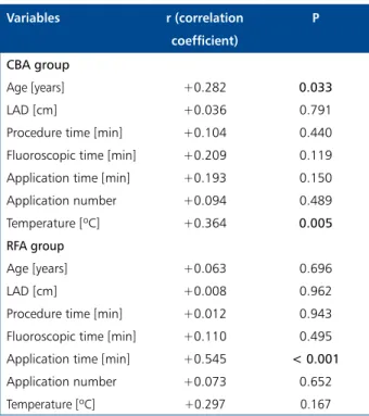
Table 3 shows correlation analysis of DTnI with clinical and laboratory parameters in the CBA and RFA groups, show-ing that only age and procedural temperature were signifi-cantly correlated with DTnI in the CBA group. Figure 2A, B shows the relationship between DTnI and temperature. In the RFA group, correlation analysis of DTnI with clinical and procedural variables revealed only application time in correla-tion with DTnI (p < 0.001). Figure 2B shows the relacorrela-tionship between DTnI and application time.
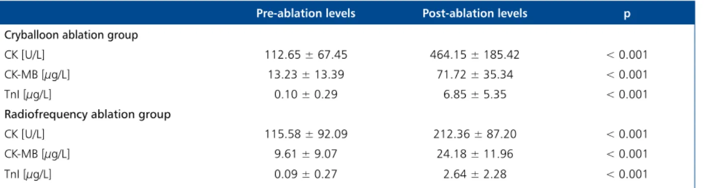
Table 2. Post-procedural changing of cardiac biomarkers
Pre-ablation levels Post-ablation levels p Cryballoon ablation group
CK [U/L] 112.65 ± 67.45 464.15 ± 185.42 < 0.001
CK-MB [µg/L] 13.23 ± 13.39 71.72 ± 35.34 < 0.001
TnI [µg/L] 0.10 ± 0.29 6.85 ± 5.35 < 0.001
Radiofrequency ablation group
CK [U/L] 115.58 ± 92.09 212.36 ± 87.20 < 0.001
CK-MB [µg/L] 9.61 ± 9.07 24.18 ± 11.96 < 0.001
TnI [µg/L] 0.09 ± 0.27 2.64 ± 2.28 < 0.001
CK — creatinine kinase; CK-MB — myocardial bound for creatinine kinase; TnI — troponin I
Figure 1.A. Post-procedural changes in troponin I (TnI) levels after cryoballoon ablation; B. Post-procedural in TnI levels after radiofrequency ablation
Table 3. Association between troponin I elevation (DTnI) and clinical, procedural variables in the cryoballoon ablation (CBA) and radiofrequency ablation (RFA) groups
Variables r (correlation coefficient) P CBA group Age [years] +0.282 0.033 LAD [cm] +0.036 0.791
Procedure time [min] +0.104 0.440
Fluoroscopic time [min] +0.209 0.119 Application time [min] +0.193 0.150
Application number +0.094 0.489
Temperature [ºC] +0.364 0.005
RFA group
Age [years] +0.063 0.696
LAD [cm] +0.008 0.962
Procedure time [min] +0.012 0.943
Fluoroscopic time [min] +0.110 0.495 Application time [min] +0.545 < 0.001
Application number +0.073 0.652
Temperature [ºC] +0.297 0.167
LAD — left atrium diameter
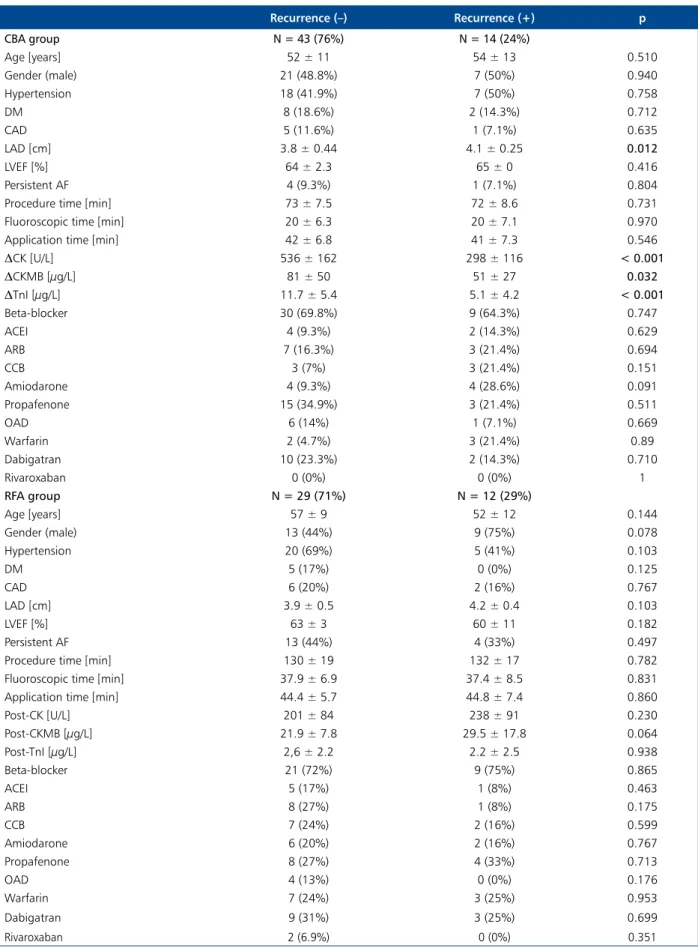
Table 4 shows clinical and laboratory characteristics and a comparison of patients with and without recurrence in the CBA and RFA groups. The AF recurrence rate in CBA group was 14 (24%). When patients with and without recurrence in CBA group were compared, LAD, DCK, DCKMB, and DTnI parameters were significantly different while other parameters
Figure 2. A. The relationship between troponin I elevation (DTnI) and temperature in the cryoballoon ablation group; B. The relationship between DTnI and application time in the radiofrequency ablation (RFA) group
5 2 25 6 4 10 TnI [ g/L] D TnI [ g/L]
Temperature [°C] Application time [min]
–38 –40 –42 –44 20 40 60
15 20
A B
were not. The AF recurrence rate in the RFA group was found to be 43%. When patients with and without AF recurrence in the RFA group were compared, there was no difference with regard to all parameters, including elevated cardiac biomarkers.
Multivariate analysis performed among variables includ-ing age, LAD, fluoroscopic time, persistent AF, HT, DM, and DTnI in the CBA group detected LAD and age as positive independent predictors, and DTnI as a negative independent predictor for AF recurrence (Table 5).
Figure 3 shows pre-ablation and post-ablation values of DTnI in the CBA group with and without recurrence.
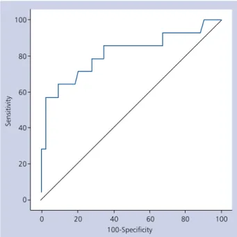
The optimal cut-off value of DTnI for predicting CBA efficacy was > 10.8, yielding a specificity of 90.7% and a sen-sitivity of 64.3% (AUC: 0.816, 95% CI 0.691–0.906) (Fig. 4).
DISCUSSION
Our study demonstrated elevated levels of CK, CK-MB, and TnI in both groups following CA. Among procedural parameters, median temperature reached during CBA was correlated with DTnI. The increase in CK, CK-MB, and TnI levels following CA was associated with lower AF recurrence rate in CBA group. This relationship was also supported in the multivariate analysis.
In the RFA group, application time, which was one of the procedural parameters, correlated with DTnI. The increase in CK, CK-MB, and TnI levels after RFA was not associated with AF recurrence. Catheter ablation causes the release of myocardial injury markers into systemic circulation [12, 13]. There are many studies investigating the alterations in plasma levels of these markers in the literature so far. One of these studies reported at least a 20-fold increase in troponin T (TnT) levels in all patients after RFA, but TnT levels after CA were not
Table 4. Clinical and laboratory characteristics and comparison of patients with and without recurrence in the cryoballoon ablation (CBA) and radiofrequency ablation (RFA) groups
Recurrence (–) Recurrence (+) p CBA group N = 43 (76%) N = 14 (24%) Age [years] 52 ± 11 54 ± 13 0.510 Gender (male) 21 (48.8%) 7 (50%) 0.940 Hypertension 18 (41.9%) 7 (50%) 0.758 DM 8 (18.6%) 2 (14.3%) 0.712 CAD 5 (11.6%) 1 (7.1%) 0.635 LAD [cm] 3.8 ± 0.44 4.1 ± 0.25 0.012 LVEF [%] 64 ± 2.3 65 ± 0 0.416 Persistent AF 4 (9.3%) 1 (7.1%) 0.804
Procedure time [min] 73 ± 7.5 72 ± 8.6 0.731
Fluoroscopic time [min] 20 ± 6.3 20 ± 7.1 0.970
Application time [min] 42 ± 6.8 41 ± 7.3 0.546
DCK [U/L] 536 ± 162 298 ± 116 < 0.001 DCKMB [µg/L] 81 ± 50 51 ± 27 0.032 DTnI [µg/L] 11.7 ± 5.4 5.1 ± 4.2 < 0.001 Beta-blocker 30 (69.8%) 9 (64.3%) 0.747 ACEI 4 (9.3%) 2 (14.3%) 0.629 ARB 7 (16.3%) 3 (21.4%) 0.694 CCB 3 (7%) 3 (21.4%) 0.151 Amiodarone 4 (9.3%) 4 (28.6%) 0.091 Propafenone 15 (34.9%) 3 (21.4%) 0.511 OAD 6 (14%) 1 (7.1%) 0.669 Warfarin 2 (4.7%) 3 (21.4%) 0.89 Dabigatran 10 (23.3%) 2 (14.3%) 0.710 Rivaroxaban 0 (0%) 0 (0%) 1 RFA group N = 29 (71%) N = 12 (29%) Age [years] 57 ± 9 52 ± 12 0.144 Gender (male) 13 (44%) 9 (75%) 0.078 Hypertension 20 (69%) 5 (41%) 0.103 DM 5 (17%) 0 (0%) 0.125 CAD 6 (20%) 2 (16%) 0.767 LAD [cm] 3.9 ± 0.5 4.2 ± 0.4 0.103 LVEF [%] 63 ± 3 60 ± 11 0.182 Persistent AF 13 (44%) 4 (33%) 0.497
Procedure time [min] 130 ± 19 132 ± 17 0.782
Fluoroscopic time [min] 37.9 ± 6.9 37.4 ± 8.5 0.831
Application time [min] 44.4 ± 5.7 44.8 ± 7.4 0.860
Post-CK [U/L] 201 ± 84 238 ± 91 0.230 Post-CKMB [µg/L] 21.9 ± 7.8 29.5 ± 17.8 0.064 Post-TnI [µg/L] 2,6 ± 2.2 2.2 ± 2.5 0.938 Beta-blocker 21 (72%) 9 (75%) 0.865 ACEI 5 (17%) 1 (8%) 0.463 ARB 8 (27%) 1 (8%) 0.175 CCB 7 (24%) 2 (16%) 0.599 Amiodarone 6 (20%) 2 (16%) 0.767 Propafenone 8 (27%) 4 (33%) 0.713 OAD 4 (13%) 0 (0%) 0.176 Warfarin 7 (24%) 3 (25%) 0.953 Dabigatran 9 (31%) 3 (25%) 0.699 Rivaroxaban 2 (6.9%) 0 (0%) 0.351
associated with the number of radiofrequency lesions, radio-frequency time, and procedural time [12]. Wójcik et al. [13] detected correlation between median temperature reached during CBA and post-procedural maximum levels of CK and CK-MB (p < 0.05). In addition, they detected correlation between CK-MB and total cryoapplication time (p < 0.03). The present study demonstrated that increased levels of TnI after CA were correlated with median temperature in the CBA group and application time in the RFA group. Interruption or reduction of blood flow between cryoballoon and myocardial tissue due to improved contact force by cryoballoon might have led to lower temperatures. Improved contact of cryobal-Table 5. Multivariate analysis of independent predictors of atrial fibrillation (AF) recurrence in cryoballoon ablation group
P Odds ratio 95% CI Age 0.029 1.125 1.012–1.251 LAD 0.046 15.665 1.046–234.575 Fluoroscopic time 0.116 1.185 0.954–1.463 Persistent AF 0.188 0.091 0.03–3.230 Hypertension 0.571 1.953 0.192–19.827 DM 0.969 1.059 0.057–19.747 DTnI 0.001 1.668 1.233–2.255
CI — confidence interval; DM — diabetes mellitus; LAD — left atrium diameter; DTnI — troponin I elevation
loon and lower temperatures are thought to result in increased myocardial injury and TnI levels. Increased application time in the RFA group indicates greater lesion size and thus greater myocardial injury.
Several parameters are associated with AF recurrence following CA. Previous studies did not demonstrate a clear association between levels of myocardial injury biomarkers measured after ablation and AF recurrence. The study by Lim et al. [14] has shown that post-ablation TnT levels were associ-ated with early AF recurrence occurring within the first three days after ablation whereas there was no association with AF recurrence in three and six months. Casella et al. [6] found no association between post-CA levels of CK-MB and TnI with AF recurrence during the follow-up visits at one, three, six, and 12 months. In our study, we demonstrated that elevated levels of biomarkers were associated with lower recurrence rate in the CBA group.
However, elevated cardiac biomarkers were not associ-ated with AF recurrence in the RFA group. The different results in the CBA and RFA groups can be explained by differences in techniques and patient characteristics. Good contact of the cryoballoon catheter with myocardial tissue during CBA increases lesion size, and thus increased levels of myocardial injury biomarkers may suggest more effective ablation. Fur-thermore, more complex PV anatomy of patients in the RFA group than in the CBA group may have had a significant effect on recurrence rates. The degree of complexity in patients with complex PV anatomy may be associated with recurrence, and elevated levels of cardiac biomarkers may indicate effective ablation in patients without complex PV anatomy.
Figure 4. Receiver operating characteristic curve analysis for
troponin I elevation (DTnI) in the detection of post-ablation recurrence
Figure 3. Pre-ablation and post-ablation values of cardiac troponin I (TnI) in the cryoballoon ablation group with (A) and without (B) recurrence
A
Several studies have compared AF recurrence rates fol-lowing CBA and RFA. In a study, no significant difference was shown between the two ablation groups in terms of recur-rence rate, whereas the CBA group had shorter fluoroscopic and procedural times [15]. Another study reported similar procedural time and AF recurrence rate in both groups; however, ablation and fluoroscopic times were longer in the CBA group [16]. In our study, patients were not randomised at the start of the trial, but those without suitable anatomy for CBA were allocated into the RFA group after PV tomography and three-dimensional reconstruction. Moreover, LAD and persistent AF frequency were significantly higher in the RFA group than in the CBA group. The comparison between the CBA and RFA groups is limited because of baseline differences in our study. Further studies are required to compare the long-term outcomes between the two ablation techniques.
Limitations of the study
Patients were not randomised into CBA or RFA groups at the beginning of the study, and randomisation of the patients into these two groups based on complex anatomy determined by PV tomography imaging and three-dimensional reconstruc-tion is the main limitareconstruc-tion of our study. Besides patients with complex PV anatomy in RFA group were not categorised ac-cording to the degree of complexity. This may have affected the post-CA AF recurrence rates. Post-procedural single blood sample was obtained 6 h after the procedure in each patient, and no serial measurements were performed. Serial meas-urements could have determined peak levels of biomarkers more accurately. Finally, the relatively small sample size may be another limitation.
CONCLUSIONS
Levels of all cardiac biomarkers were elevated after CBA and RFA. Elevated TnI levels after CBA were independent negative predictors for AF recurrence. Measurement of TnI levels after CBA may be useful for the prediction of better clinical outcome.
Conflict of interest: none declared References
1. January CT, Wann LS, Alpert JS, et al. 2014 AHA/ACC/HRS Guideline for the Management of Patients With Atrial Fibrillation: A Report of the American College of Cardiology/American Heart As-sociation Task Force on Practice Guidelines and the Heart Rhythm Society. J Am Coll Cardiol. 2014; 64(21): 2305–237. doi: 10.1016/j. jacc.2014.03.022, indexed in Pubmed: 24685669
2. Madrid AH, del Rey JM, Rubí J, et al. Biochemical markers and cardiac troponin I release after radiofrequency catheter ablation: approach to size of necrosis. Am. Heart J. 1998; 136(6): 948–955, doi: 10.1016/s0002-8703(98)70148-6, indexed in Pubmed: 9842005.
3. Pudil R, Parízek P, Tichý M, et al. Use of the biochip microarray system in detection of myocardial injury caused by radiofrequency catheter ablation. Clin Chem Lab Med. 2008; 46(12): 1726–1728, doi: 10.1515/CCLM.2008.341, indexed in Pubmed: 19055449. 4. Kühne M, Suter Y, Altmann D, et al. Cryoballoon versus
radiofre-quency catheter ablation of paroxysmal atrial fibrillation: biomark-ers of myocardial injury, recurrence rates, and pulmonary vein reconnection patterns. Heart Rhythm. 2010; 7(12): 1770–1776, doi: 10.1016/j.hrthm.2010.08.028, indexed in Pubmed: 20817019. 5. Herrera Siklódy C, Arentz T, Minners J, et al. Cellular damage,
platelet activation, and inflammatory response after pulmonary vein isolation: a randomized study comparing radiofrequency ablation with cryoablation. Heart Rhythm. 2012; 9(2): 189–196, doi: 10.1016/j.hrthm.2011.09.017, indexed in Pubmed: 21920484. 6. Casella M, Dello Russo A, Russo E, et al. Biomarkers of myocardial injury with different energy sources for atrial fibrillation catheter ab-lation. Cardiol J. 2014; 21(5): 516–523, doi: 10.5603/CJ.a2013.0153, indexed in Pubmed: 24293166.
7. Lang RM, Bierig M, Devereux RB, et al. Chamber Quantification Writing Group, American Society of Echocardiography’s Guide-lines and Standards Committee, European Association of Echocar-diography. Recommendations for chamber quantification: a report from the American Society of Echocardiography’s Guidelines and Standards Committee and the Chamber Quantification Writing Group, developed in conjunction with the European Association of Echocardiography, a branch of the European Society of Cardiology. J Am Soc Echocardiogr. 2005; 18(12): 1440–1463, doi:10.1016/j. echo.2005.10.005, indexed in Pubmed: 16376782.
8. Neumann T, Vogt J, Schumacher B, et al. Circumferential pul-monary vein isolation with the cryoballoon technique results from a prospective 3-center study. J. Am. Coll. Cardiol. 2008; 52(4): 273–278, doi: 10.1016/j.jacc.2008.04.021, indexed in Pubmed: 18634982.
9. Van Belle Y, Janse P, Rivero-Ayerza MJ, et al. Pulmonary vein isola-tion using an occluding cryoballoon for circumferential ablaisola-tion: feasibility, complications, and short-term outcome. Eur Heart J. 2007; 28(18): 2231–2237, doi: 10.1093/eurheartj/ehm227, indexed in Pubmed: 17569680.
10. Deisenhofer I, Schmitt C. Ablation strategies in paroxysmal atrial fibrillation. In: Aliot E, Haissaguerre M, Jackman WM eds. Catheter ablation of atrial fibrillation. Blackwell, 2008: 136–162. 11. Ernst S, Ouyang F, Antz M, et al. Catheter ablation of atrial
fibril-lation. In: Aliot E, Haissaguerre M, Jackman WM eds. Techniques targeting the pulmonary veins. Blackwell, 2008: 117–123. 12. Haegeli LM, Kotschet E, Byrne J, et al. Cardiac injury after
percutaneous catheter ablation for atrial fibrillation. Europace. 2008; 10(3): 273–275, doi:10.1093/europace/eum273, indexed in Pubmed: 18174208.
13. Wójcik M, Janin S, Neumann T, et al. Which standard biomarkers are useful for the evaluation of myocardial injury after pulmo-nary vein isolation with cryoballoon? Kardiol Pol. 2011; 69(11): 1151–1155, indexed in Pubmed: 22090225.
14. Lim HS, Schultz C, Dang J, et al. Time course of inflammation, myo-cardial injury, and prothrombotic response after radiofrequency catheter ablation for atrial fibrillation. Circ Arrhythm Electrophy-siol. 2014; 7(1): 83–89, doi: 10.1161/CIRCEP.113.000876, indexed in Pubmed: 24446024.
15. Xu J, Huang Y, Cai H, et al. Is cryoballoon ablation preferable to radiofrequency ablation for treatment of atrial fibrillation by pulmonary vein isolation? A meta-analysis. PLoS ONE. 2014; 9(2): e90323, doi: 10.1371/journal.pone.0090323, indexed in Pubmed: 24587324.
16. Schmidt M, Dorwarth U, Andresen D, et al. Cryoballoon versus RF ablation in paroxysmal atrial fibrillation: results from the German Ablation Registry. J Cardiovasc Electrophysiol. 2014; 25(1): 1–7, doi: 10.1111/jce.12267, indexed in Pubmed: 24134539.
Cite this article as: Kızılırmak F, Gokdeniz T, Gunes HM, et al. Myocardial injury biomarkers after radiofrequency catheter and cryoballoon ablation for atrial fibrillation and their impact on recurrence. Kardiol Pol. 2017;75(2): 126–134, doi: 10.5603/KP.a2016.0089.
przezcewnikowej ablacji prądem o wysokiej
częstotliwości i krioablacji balonowej
z powodu migotania przedsionków
oraz ich wpływ na nawrót migotania
Filiz Kizilirmak, Tayyar Gokdeniz, Haci Murat Gunes, Gultekin Gunhan Demir, Beytullah Cakal,
Gamze Babur Guler, Ekrem Guler, Fatih Erkam Olgun, Fethi Kilicaslan
Istanbul Medipol University, Istanbul, Turkey
S t r e s z c z e n i e
Wstęp: Uszkodzenie miokardium w wyniku ablacji przezcewnikowej (CA) z powodu migotania przedsionków (AF) wiąże się z podwyższonymi stężeniami biomarkerów.
Cel: W tym prospektywnym badaniu zmierzono stężenia biomarkerów uszkodzenia miokardium (kinaza kreatynowa [CK]), izoenzym sercowy kinazy kreatynowej [CK-MB], troponina I [TnI]) oraz oceniono ich wpływ na nawrót AF po zabiegu ablacji wykonanym jedną z dwóch metod: krioablacji balonowej (CBA) i ablacji prądem o wysokiej częstotliwości (RFA). Autorzy zamierzali również ocenić zależność między nawrotem AF po CA a innymi parametrami klinicznymi, echokardiograficznymi i związanymi z metodą zabiegową.
Metody: Do badania włączono 98 chorych z AF, spośród których u 21% rozpoznano przetrwałe AF, a u 79% — napadowe AF. U 58% chorych wykonano CBA, a u 42% osób — RFA. Stężenia CK, CK-MB i TnI zmierzono przed zabiegiem i 6 godzin po zabiegu. Wizyty kontrolne odbyły się 3, 6 i 9 miesięcy po ablacji. Porównano stężenia biomarkerów u pacjentów z na-wrotem AF i bez nawrotu.
Wyniki: Zmierzone po ablacji stężenia CK (postCK), CK-MB (postCKMB) i TnI (postTnI) były istotnie wyższe w grupach CBA i RFA (p < 0,001 dla wszystkich porównań). Zwiększenie stężenia TnI (DTnI) w grupie CBA korelowało z wiekiem (p = 0,033) i medianą temperatury osiągniętej w czasie ablacji (p < 0,005), natomiast w grupie RFA korelowało z czasem aplikacji (p < 0,001). Analiza wieloczynnikowa danych pacjentów z grupy CBA wykazała, że wiek i średnica lewego przed-sionka były niezależnymi czynnikami predykcyjnymi dodatnimi (p = 0,029 i p = 0,046), a DTnI — niezależnym czynnikiem predykcyjnym ujemnym nawrotu AF (p = 0,001). Podwyższone stężenia biomarkerów sercowych nie wiązały się z nawrotem AF w grupie RFA (p > 0,05).
Wnioski: Po CBA i RFA stężenia wszystkich biomarkerów sercowych były podwyższone. Zwiększone stężenia TnI po CBA stanowiły niezależny czynnik prognostyczny ujemny nawrotu AF. Pomiary stężeń TnI po CBA mogą być użyteczne w progno-zowaniu lepszego efektu klinicznego.
Słowa kluczowe: ablacja przezcewnikowa, migotanie przedsionków, biomarkery uszkodzenia mięśnia sercowego
Kardiol Pol 2017; 75, 2: 126–134
Adres do korespondencji: