Dilemma in pediatric mandible fractures:
resorbable or metallic plates?
Gaye Taylan Filinte, M.D.,1 İsmail Mithat Akan, M.D.,2 Gülçin Nujen Ayçiçek Çardak, M.D.,1 Özay Özkaya Mutlu, M.D.,3 Tayfun Aköz, M.D.4
1Department of Plastic, Reconstructive and Aesthetic Surgery, Dr. Lutfi Kirdar Kartal Training and Research Hospital, Istanbul 2Department of Plastic, Reconstructive and Aesthetic Surgery, Medipol University Faculty of Medicine, Istanbul
3Department of Plastic, Reconstructive and Aesthetic Surgery, Okmeydani Training and Research Hospital, Istanbul 4Department of Plastic, Reconstructive and Aesthetic Surgery, Maltepe University Faculty of Medicine, Istanbul
ABSTRACT
BACKGROUND: The aim of this study was to compare the efficiency of resorbable and metallic plates in open reduction and inter-nal fixation of mandible fractures in children.
METHODS: Thirty-one patients (mean age, 8.05 years; range 20 months-14 years) were operated on various fractures of the man-dible (26 [60.4%] symphysis- parasymphysis, 12 [27.9%] condylar-subcondylar fractures, 5 [11.6%] angulus and ramus fractures). Twelve patients were treated with resorbable plates and 19 patients with metallic plates. Mean follow-up time was 41 months (11–74 months) in the metallic hardware group and was 22 months (8–35 months) in the resorbable plate group. Both groups were investigated for primary bone healing, complications, number of operations, and mandibular growth. The results were discussed below.
RESULTS: Both groups demonstrated primary bone healing. Minor complications were similar in both groups. The metallic group involved secondary operations for plate removal. Mandibular growth was satisfactory in both groups.
CONCLUSION: Resorbable plates cost more than the metallic ones; however, when the secondary operations are included in the total cost, resorbable plates were favourable. As mandibular growth and complication parameters are similar in both groups, resorb-able plates are favored due to avoidance of potential odontogenic injury, elimination of long-term foreign body retention and provi-sion of adequate stability for rapid bone healing. However, learning curve and concerns for decreased stability against heavy forces of mastication accompanied with the resorbable plates when compared to the metallic ones should be kept in mind.
Key words: Mandibular fractures; metallic plates; resorbable plates.
the hardware are all the factors taken into consideration. Majority of pediatric mandibular fractures can be managed with closed techniques using short periods of maxilloman-dibular fixation or training elastics alone.[5] Performing open
reduction and internal fixation of mandibular fractures in pe-diatric patients may not always be necessary. Lingual splints have been used successfully for pediatric mandibular fractures.
[6–8] It is an effective and safe procedure in selected cases.[9]
High osteogenic potential of pediatric mandible allows non-surgical management to be successful in younger patients with conservative approaches.[10] Maxillofacial surgeons generally
justify the use of plate- and screw-type internal fixation to be reserved for difficult fractures.[5] Specific subsets of
man-dibular fractures, including displaced fractures of the body or angle, fractures of the condylar neck with significant barriers to movement, complex fractures, and fractures in non-toothbear-ing areas necessiate open reduction and internal fixation.[11]
Address for correspondence: Gaye Taylan Filinte, M.D. Gözenç Sokak, Babadan Apartmanı., No: 2, D: 1, Erenköy, 34738 İstanbul, Turkey
Tel: +90 216 - 441 39 00/1908 E-mail: [email protected]
Qucik Response Code Ulus Travma Acil Cerrahi Derg 2015;21(6):509–513
doi: 10.5505/tjtes.2015.23922 Copyright 2015
TJTES
INTRODUCTION
Management of pediatric mandible fracture is substantially different from adult injuries. Evaluation and approach of pe-diatric mandibular fractures require several issues to be con-sidered. Presence of tooth buds and potential injury to future growth are among the issues complicating management.[1–4]
The use of resorbable plates is an increasingly attractive op-tion in the treatment of pediatric mandibular fractures.[11] It is
both well-tolerated and effective. It enables realignment and stable positioning of rapidly healing fracture segments while obviating any future issues secondary to long-term metal re-tention (Fig. 1).[12]
Major concerns for using resorbable materials in the maxillo-facial region are the strength of the material and its ability to withstand masticatory forces, and the extent of inflammation as the materials begin to degrade.[13]
We used both systems of metallic and resorbable hardware for fixation of pediatric mandible fractures. Limited number of cases and follow-up demonstrated no difference between the stability and healing capacity of the two systems. Resorb-able materials have the advantage of avoidance of secondary removal operations. Limited number of long-term studies and high cost when compared to the metallic hardware are among the drawbacks of biodegradable systems. However, ongoing studies demonstrating the advantages of the resorb-able plates indicate that they are going to be preferred more in the future.
MATERIALS AND METHODS
The study consisted of thirty-one pediatric mandible fracture cases arriving to our clinic between 2000 and 2011. Resorb-able plates (2.0 mm PLLA/PGA Lactosorb system, Jacksonville, Florida, USA) were used in twelve patients (ages, 20 months-11 years; mean, 6.9 years) and metallic plates in nineteen (ages, 4–14 years; mean, 9.2 years). Follow up of the metallic plate group was 41 months (11–74 months) and of the resorbable
plate group was 22 months (8–35 months). Both groups were compared according to infection rates, primary bone healing, mandibular growth and need for secondary surgery.
RESULTS
Thirty-one patients with 43 fractures of the mandible were enrolled in the study. Patient age ranged from 20 months to 14 years with a mean of 8.05 years.. Fractures included 26 (60.4%) symphysis-parasymphysis fractures, 12 (27.9%) con-dylar-subcondylar fractures, and 5 (11.6%) angulus and ramus fractures. Metallic plates and screws were used in nineteen (62.7%) patients with 27 fractures and resorbable plates and screws in twelve (37.2%) patients with 16 fractures. Inter-maxiller fixation were used in nine patients with metallic plates and in six patients with resorbable plates.
Fracture Union
No mobility in any fracture site was noted in either groups at the follow-up period. Follow-up was 41 months (11–74 months) in the metallic plate group and 22 months (8–35 months) in the resorbable plate group. There was no facial asymmetry in both groups in the follow-up period (Figs 2, 3).
Infection
Two of the 19 patients with metallic hardware demonstrated clinical signs of infection. One of the two responded well to oral antibiotherapy. However, the other one developed a sub-mental fistula and recieved drainage and incision and finally plate removal at the postoperative 13th week. No infection
was noted at the resorbable plate group. One patient with resorbable plate demonstrated granuloma formation at the subcutaneous tissue in the 4th postoperative month, which
was excised with local anesthesia.
Malunion
Minor occlusal deformity was noted in one patient at the sec-ond week control which was corrected by an additional one-week use of light guiding elastics. The parasymphysis fracture of the patient was reconstructed with metallic hardware.
Revisional Surgery
There was no need for revisional surgery for fracture healing
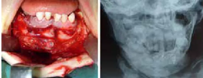
Figure 1. Upper left; Intraoperative view of the resorbable plate for
fixation of parasymphysis fracture of the mandible in a 3-year-old patient. Upper right; A 1-year follow-up lateral craniography. Lower; A 1-year follow-up panoramic radiograph. Note the well-healed fracture sites.
Figure 2. Left; Two resorbable plates for a parasymphysis and
symphysis fractures of the mandible in a 4-year-old patient. Right; An 18-month follow-up radiograph showing fracture alignment.
in both groups. Fistula that developed after infection in one patient was excised and sutured. Granuloma fomation was excised in the resorbable plate group.
Pain
One patient with symphysis fracture and metallic plate dem-onstrated discomfort due to mild pain with plate feeling un-der the skin.
DISCUSSION
Diagnosis and management of mandible fractures in the pe-diatric patient population can pose multiple challenges to the oral and maxillofacial surgeon.[14] There are some principles
that must be addressed when dealing with pediatric mandi-ble fractures. Pediatric mandimandi-ble is a dynamic structure that undergoes significant changes during development. In order to avoid undesirable outcomes, management of mandibular trauma requires knowledge of these changes over time.[15]
Presence of multiple tooth buds throughout the substance of the mandible as well as the potential injury to future growth complicate the management of these fractures.[1] One of the
main principles is to use the least amount of foreign material.
[16] The use of rigid fixation in children is controversial and
may cause growth retardation along cranial suture lines. Con-servative approaches with non-surgical management may be successful in pediatric population due to the high osteogenic potential in this population.[10] Children demonstrate a good
healing capacity.. These younger patients have the potential for restitutional remodeling, as opposed to sclerotic, func-tional remodeling seen in adults.[1] The majority of pediatric
mandibular fractures can be managed with closed techniques using short periods of maxillomandibular fixation or training elastics alone. Generally, the use of plate- and screw-type in-ternal fixation is reserved for difficult fractures.[5] Lingual splint
has been reported for the reduction, stabilization and fixation of a mandibular body fracture with a successful result.[9]
Pediatric mandible fractures, which are seen less frequently compared to those of adults, require a specific and differ-ent treatmdiffer-ent. Although less invasive methods are preferred mostly, internal fixation with open reduction should be con-sidered when required.[17] Rigid fixation of mandibular
frac-tures results in a faster bone healing, both by compression and lack of mobility between fracture segments.[18–23] Smartt et al.
have demonstrated that open reduction and internal fixation, when used judiciously, are indispensable in the treatment of specific subsets of mandibular fractures, including displaced fractures of the body or angle, fractures of the condylar neck with significant barriers to movement, complex fractures, and fractures in non-toothbearing areas. Open reduction should be performed cautiously, with minimal manipulation of over-lying soft tissues. When performed properly, it is a safe and versatile treatment modality.[11]
Metal plate-screw systems enable adequate fixation in bone healing process. Yet, their effects like limiting bone growth es-pecially in pediatric age group have prompted investigators to look for alternative fixation materials in the reconstruction of trauma and craniofacial anomalies.[24,25]
The ideal fixation system for stabilisation of an osteotomy or bone fracture would provide adequate strength initially to permit bone healing during function, and then, decrease in strength so that there isincreasing physiological force trans-ference to the bone. Biodegradable polymers can provide that while metals cannot.[13]
The most attractive characteristic of resorbable plates is that they obviate any potential impediments to long-term metal retention. They enable realignment and stable positioning of rapidly healing fracture segments. They are also quite well tolerated in this population.[14] Yerit et al. have
demonstrat-ed advantages of resorbabale materials in pdemonstrat-ediatric patients especially by faster mobilization and the avoidance of sec-ondary removal operations. Primary healing of the fractured mandible was observed in all of the thirteen patients, and malocclusion and growth restrictions did not occur.[26]
Tita-nium plates need to be removed; whereas, resorbable plates do not. Resorbable plates are radiolucent and allow full vi-sualization of the fractures on postoperative radiographs.[23]
They provide proper strength, and then, harmlessly degrade over time, until the load can be safely transferred to the healed bone. As there is no need for a removal operation, these biodegradable devices reduce the total treatment and rehabilitation time of the patient. Besides, they reduce costs related to this type of trauma.[23]
Figure 3. Upper; A 4-month follow-up of a
symphysis-parasym-physis fracture with screw sites visible on panoramic radiograph. Lower; The screw sites are healed with less visible holes after a total of 9-month follow-up.
The use of resorbable plates and screws for fixation of pe-diatric facial fractures is both well tolerated and effective. It enables realignment and stable positioning of rapidly healing fracture segments while obviating any future issues to long-term metal retention.[12]
Mandibular growth is not affected as demonstrated by the present and several other studies.[23] Complication rates are
comparable with nonresorbable plate fixation.
As we are experienced more with the use of resorbable hardware, we observed that the fixation strength of the re-sorbable hardware is not as powerful as that of the metallic hardware. Our first cases were reconstructed with 2-mm re-sorbable plates. It may be difficult to place the plates of this thickness in children younger than 5 years of age. It is also quite difficult to place the plates beneath the nerve in cases of parasymphysis fractures. Usually, there is not enough place for two plates. We hesitated using 1.5 mm plates, but there are examples of its usage in the literature.[12]
Muscle activity in mandibular ramus is considerable,[13] and
it is better to advice the patients to be cautious while chew-ing, especially in the first 4 weeks. Children’s adaptability to masticatory function increases with the development.[27]
Due to traumatic stress and pain, children usually adapt to soft diet better than adults. We did not offer a different diet to patients with the resorbable hardware. Both groups followed the same principles postoperatively. However, if concerns about resorbale plate stability occurs, some pre-cautions can be taken; additional intermaxiller fixation to open reduction, prolongation of soft diet regimen, and/or more visits postoperatively to earlier detection of the plate instability.
It is not faulty to say that we use resorbable plates to make unfavorable fractures favorable and keep the fracture edges in proper position. This is a kind of conservative approach, that is, we are in between a rigid fixation with metallic plates and maxillomandibular fixation alone.
The pediatric patient’s ability to heal and recovery of function are much beter compared to adults. Despite these advan-tages, certain unique characteristics should be appreciated.[12]
With a meticulous approach to these patients, final success is not so far away. The dilemma of pediatric mandible frac-tures is to choose the right therapy with the right hardware. Resorbable plates have been favoured in our clinic since we began to use them. We believe that with more studies per-formed in the future, resorbable plates will be preferred more than metallic plates and will be the first line in treatment of rigid fixation of mandibular fractures in the pediatric popula-tion.
Conflict of interest: None declared.
REFERENCES
1. Cole P, Kaufman Y, Izaddoost S, Hatef DA, Hollier L. Principles of pediatric mandibular fracture management. Plast Reconstr Surg 2009;123:1022–4.
2. Ferreira PC, Amarante JM, Silva PN, Rodrigues JM, Choupina MP, Silva AC, et al. Retrospective study of 1251 maxillofacial fractures in children and adolescents. Plast Reconstr Surg 2005;115:1500–8.
3. Davison SP, Clifton MS, Davison MN, Hedrick M, Sotereanos G. Pedi-atric mandibular fractures: a free hand technique. Arch Facial Plast Surg 2001;3:185–190.
4. Demianczuk AN1, Verchere C, Phillips JH. The effect on facial growth of pediatric mandibular fractures. J Craniofac Surg 1999;10:323–8. 5. Kushner GM, Tiwana PS. Fractures of the growing mandible. Atlas Oral
Maxillofac Surg Clin North Am 2009;17:81–91.
6. Hofer O. Newer knowledge in oral surgery. Fracture of the jaws. Zbl Zahn-Mund-Kieferheilk 1939;13:9–11.
7. Kruger E, Schilli W. Mandibular fractures. In: Oral and maxillofacial traumatology, vol. 1. Chicago: Quintessence Publishing 1982:211–393. 8. Hardin JC Jr. Proceedings: Triple fractures of the mandible with
flar-ing rami. Their treatment with lflar-ingual splints. Arch Otolaryngol 1973;98:387–8.
9. Binahmed A, Sansalone C, Garbedian J, Sándor GK. The lingual splint: an often forgotten method for fixating pediatric mandibular fractures. J Can Dent Assoc 2007;73:521–4.
10. Kocabay C, Ataç MS, Oner B, Güngör N. The conservative treatment of pediatric mandibular fracture with prefabricated surgical splint: a case report. Dent Traumatol 2007;23:247–50.
11. Smartt JM Jr, Low DW, Bartlett SP. The pediatric mandible: II. Manage-ment of traumatic injury or fracture. Plast Reconstr Surg 2005;116:28–41. 12. Eppley BL. Use of resorbable plates and screws in pediatric facial
frac-tures. J Oral Maxillofac Surg 2005;63:385–91.
13. Turvey TA, Proffit WP, Phillips C. Biodegradable fixation for craniomax-illofacial surgery: a 10-year experience involving 761 operations and 745 patients. Int J Oral Maxillofac Surg 2011;40:244–9.
14. Poore MC, Penna KJ. The use of resorbable hardware for fixation of pe-diatric mandible fracture. Case report. N Y State Dent J 2008;74:58–61. 15. Smartt JM Jr, Low DW, Bartlett SP. The pediatric mandible: I. A primer
on growth and development. Plast Reconstr Surg 2005;116:14–23. 16. Alagöz MS, Uysal AC, Sensoz O. An alternative method in
mandibu-lar fracture treatment: bone graft use instead of a plate. J Craniofac Surg 2008;19:411–20.
17. Eskitascioglu T, Ozyazgan I, Coruh A, Gunay GK, Yuksel E. Retrospec-tive analysis of two hundred thirty-five pediatric mandibular fracture cases. Ann Plast Surg 2009;63:522–30.
18. Cabrini Gabrielli MA, Real Gabrielli MF, Marcantonio E, Hochuli-Viei-ra E. Fixation of mandibular fHochuli-Viei-ractures with 2.0-mm miniplates: review of 191 cases. J Oral Maxillofac Surg 2003;61:430–6.
19. Ellis E 3rd, Walker L. Treatment of mandibular angle fractures using two noncompression miniplates. J Oral Maxillofac Surg 1994;52:1032–7. 20. Ellis E 3rd, Walker LR. Treatment of mandibular angle fractures using
one noncompression miniplate. J Oral Maxillofac Surg 1996;54:864–72. 21. Lamphier J, Ziccardi V, Ruvo A, Janel M. Complications of man-dibular fractures in an urban teaching center. J Oral Maxillofac Surg 2003;61:745–50.
22. Cawood JI. Small plate osteosynthesis of mandibular fractures. Br J Oral Maxillofac Surg 1985;23:77–91.
plates for the fixation of mandibular fractures: a prospective study. J Oral Maxillofac Surg 2007;65:89–96.
24. Cavuşoğlu T, Yavuzer R, Başterzi Y, Tuncer S, Latifoğlu O. Resorbable plate-screw systems: clinical applications. Ulus Travma Acil Cerrahi Derg 2005;11:43–8.
25. Fearon JA, Munro IR, Bruce DA. Observations on the use of rigid fixa-tion for craniofacial deformities in infants and young children. Plast
Re-constr Surg 1995;95:634–8.
26. Yerit KC, Hainich S, Enislidis G, Turhani D, Klug C, Wittwer G, et al. Biodegradable fixation of mandibular fractures in children: stability and early results. Oral Surg Oral Med Oral Pathol Oral Radiol Endod 2005;100:17–24.
27. Matsubara T, Ono Y, Takagi Y. A study on developmental changes of masticatory function in children. J Med Dent Sci 2006;53:141–8.
OLGU SUNUMU
Pediatrik mandibula kırıklarındaki ikilem: Eriyen plaklar mı yoksa metal plaklar mı?
Dr. Gaye Taylan Filinte,1 Dr. İsmail Mithat Akan,2 Dr. Gülçin Nujen Ayçiçek Çardak,1Dr. Özay Özkaya Mutlu,3 Dr. Tayfun Aköz4
1Dr. Lütfi Kırdar Kartal Eğitim ve Araştırma Hastanesi, Plastik Rekonstrüktif ve Estetik Cerrahi Kliniği, İstanbul 2Medipol Üniversitesi Tıp Fakültesi, Plastik Rekonstrüktif ve Estetik Cerrahi Anabilim Dalı, İstanbul 3Okmeydanı Eğitim ve Araştırma Hastanesi, Plastik Rekonstrüktif ve Estetik Cerrahi Kliniği, İstanbul 4Maltepe Üniversitesi Tıp Fakültesi, Plastik Rekonstrüktif ve Estetik Cerrahi Anabilim Dalı, İstanbul
AMAÇ: Bu çalışmanın amacı çocuklardaki açık redüksiyon ve internal fiksasyon ile tedavi edilen mandibula kırıklarında, emilebilen ve metal plakların etkinliğini karşılaştırmaktır.
GEREÇ VE YÖNTEM: Yaşları 20 ay-14 yıl (ortalama 8.05 yıl) arasında değişen 31 hasta mandibulanın farklı yerlerindeki kırıklar nedeniyle ameliyat edildi (26 [%60.4] simfisiz-parasimfisiz, 12 [%27.9] kondil-subkondil, 5 [%11.6] angulus ve ramus). On iki hasta eriyen plaklarla, 19 hasta ise metal plaklarla (titanyum) tedavi edildi. Ortalama takip süresi metal donanım kullanılan grupta 41 ay (11–74 ay), eriyen plak kullanılan grupta ise 22 ay’dı (8–35 ay). Her iki grup primer kemik iyileşmesi, komplikasyonlar, ameliyat sayısı ve mandibuladaki büyüme açısından incelendi. Bulgular aşağıda tartışıldı.
BULGULAR: Her iki grupta primer kemik iyileşmesi saptandı. Her iki gruptaki minör komplikasyonlar benzerdi. Metal donanım kullanılan grupta pak çıkartılması için ikinci operasyonlar gerçekleştirildi. Her iki gruptaki mandibula gelişimi tatmin ediciydi.
TARTIŞMA: Eriyen plaklar metal donanımlı plaklardan daha pahalı oldukları görüldü. İkinci operasyonların maliyeti göz önünde bulundurulduğunda ise eriyen plaklar daha avantajlıydı. Mandibula büyümesi ve komplikasyon parametreleri heriki grupta benzer olduğundan, eriyen plaklar; olası diş hasarının önlenmesi, uzun süreli yabancı cisim varlığının olmaması ve hızlı kemik iyileşmesi için gerekli olan yeterli sabitlik sağlaması gibi hususlara bağlı olarak tercih edilmektedir. Buna rağmen, emilebilir plakların kullanılması hususunda bir öğrenme periyodunun gerekliliği ve metal plaklarla karşılaştırıldığında, çiğneme kaslarının karşıt gücüne karşı düşük sabitlik sağladıkları gibi endişeler akılda tutulmalıdır.
Anahtar sözcükler: Eriyen plaklar; mandibula kırıkları; metal plaklar.
Ulus Travma Acil Cerrahi Derg 2015;21(6):509–513 doi: 10.5505/tjtes.2015.23922