Full Terms & Conditions of access and use can be found at
http://www.tandfonline.com/action/journalInformation?journalCode=ijmf20
The Journal of Maternal-Fetal & Neonatal Medicine
ISSN: 1476-7058 (Print) 1476-4954 (Online) Journal homepage: http://www.tandfonline.com/loi/ijmf20
A retrospective analysis of amniocenteses
performed for advanced maternal age and various
other indications in Turkish women
Nuri Danisman, Serkan Kahyaoglu, Sevki Celen, Inci Kahyaoglu, Zuhal
Candemir, Ahmet Yesilyurt & Esra Sukran Cakar
To cite this article: Nuri Danisman, Serkan Kahyaoglu, Sevki Celen, Inci Kahyaoglu, Zuhal Candemir, Ahmet Yesilyurt & Esra Sukran Cakar (2013) A retrospective analysis of amniocenteses performed for advanced maternal age and various other indications in Turkish women, The Journal of Maternal-Fetal & Neonatal Medicine, 26:3, 242-245, DOI: 10.3109/14767058.2012.733756
To link to this article: https://doi.org/10.3109/14767058.2012.733756
Accepted author version posted online: 01 Oct 2012.
Published online: 18 Oct 2012. Submit your article to this journal
Article views: 154
© 2013 Informa UK, Ltd.
ISSN 1476-7058 print/ISSN 1476-4954 online DOI: 10.3109/14767058.2012.733756
Objective: Prenatal cytogenetic diagnostic methods for the
diagnosis of fetal chromosomal anomalies have been used reli-ably over the last 40 years. Advanced maternal age has become a basic indication for amniocentesis. Methods: We examined the results of the chromosome analyses of 3485 women that had amniocentesis for any reason during their antenatal care in our perinatology clinic in 2007–2009. Amniocentesis was performed for advanced maternal age in 1456 women (41.8%) and for other reasons in the remaining 2029 women (58.2%). Chromosomal anomalies were examined numerically and structurally. Results: When the amniocentesis results of the patients were reviewed as numerically normal or abnormal; 40 (2.7%) of 1456 amniocen-tesis procedures performed for advanced maternal age, 5 (0.9%) of 531 procedures performed for an increased double-test risk and 14 (1.3%) of 1095 procedures performed for an increased triple test risk were found to have chromosomal aneuploidy.
Conclusions: Maternal age is still the most prevalent indication
for genetic amniocentesis other than positive prenatal screening tests. Among women with advanced maternal age, prenatal ultrasonography for soft markers of chromosomal aneuploidy accompanied with maternal serum biochemical screening tests should be evaluated during the decision making process of genetic amniocentesis.
Keywords: Amniocentesis, maternal age, prenatal diagnosis,
screening, aneuploidy
Introduction
Prenatal cytogenetic diagnostic methods for the diagnosis of fetal chromosomal anomalies have been used reliably over the last 40 years. Second trimester amniocentesis (AC) is the most used prenatal invasive diagnostic procedure. It was first developed as a diagnostic method in the late 1960s for the culture of the amniotic fluid sample. Clinical indications were changed over the years due to fetal loss rate associated with amniocentesis [1]. Because the relationship between advanced maternal age and fetal aneuploidy is well known, advanced maternal age has become one of the basic indications for amniocentesis [2]. Therefore, it is important to predict the risks of age-related fetal chromosomal anomalies. Today, advanced maternal age is not used as the only indication for amniocentesis, but it is increasing in importance as a component of prenatal screening test to detect fetal aneuploidies
[3]. Specific maternal biochemical serum markers were first used in 1980s for the detection of chromosomal anomalies. Second trimester maternal serum screening is the most performed test for the screening of Trisomy 21 and detects almost 75–80% of cases [4]. Recently, first trimester nuchal translucency (NT) and maternal age have been evaluated together, and with a 5% false positive rate, 75–80% of Trisomy 21 cases and other major chromosomal anomalies can be detected. The combination of first trimester biochemical markers and NT has a detection rate of 87–92%. Detection rate of fetal chromosomal anomalies is increased to 95% with integrated or sequential tests that are composed of first and second trimester serum biochemical markers, NT and maternal age. In most developed countries, second trimester prenatal ultrasonography, which is routinely performed as part of antenatal care, enables us to detect fetal structural anomalies and soft markers of Trisomy 21 [5]. In recent years, individual risk evaluation for fetal chromosomal anomalies has become an important part of prenatal genetic consultation [6]. As the chromosomal anomaly risk increases with the advanced maternal age, it is important to determine age-related chromosomal anomaly risks. In addition to being an indication for cytogenetic amniocentesis, maternal age is also used as an independent risk factor with biochemical serum markers [7]. In recent years, the number of women who delay pregnancy until after the age of 35 is increasing for many reasons. Because biological and environmental differences also influence fetal chromosomal anomaly risks, the risks from advanced maternal age in Turkish women are increasing in importance. A portion of pregnant women with advanced maternal age decide to undergo amniocentesis according to the results from their first and second trimester biochemical screening tests [8]. In our study, we retrospectively examined the results of chromosome analyses from 3485 women that had amniocentesis for any indication during their antenatal care in our perinatology clinic in 2007–2009. We investigated the fetal chromosomal anomaly detection rates based on specific indications.
Methods
We conducted this study between 2007 and 2009 using records from 3485 patients who were seen at Zekai Tahir Burak Women’s Health and Research Hospital Perinatology Clinic for their ante-natal care between 16–20 weeks’ gestation. Women who had
A retrospective analysis of amniocenteses performed for advanced
maternal age and various other indications in Turkish women
Nuri Danisman
1, Serkan Kahyaoglu
1, Sevki Celen
1, Inci Kahyaoglu
2, Zuhal Candemir
3,
Ahmet Yesilyurt
3& Esra Sukran Cakar
31Department of High Risk Pregnancy, Zekai Tahir Burak Women’s Health and Research Hospital, Ankara, Turkey, 2Department of Obstetrics and Gynecology, University of Ufuk, Ankara, Turkey, and 3Department of Genetics,
Zekai Tahir Burak Women’s Health and Research Hospital, Ankara, Turkey
Correspondence: Dr. Serkan Kahyaoglu, Department of High Risk Pregnancy, Zekai Tahir Burak Women’s Health and Research Hospital, Ankara, Turkey.
The Journal of Maternal-Fetal and Neonatal Medicine
2013
26
3
242
245
© 2013 Informa UK, Ltd.
10.3109/14767058.2012.733756
1476-7058
1476-4954
05052012 15September2012 21September2012A retrospective analysis of amniocenteses
N. Danisman et al.
A retrospective analysis of amniocenteses 243
© 2013 Informa UK, Ltd.
an amniocentesis performed for any indication were included. All pregnant women over the age of 35 and between 16 and 20 weeks’ gestation were offered amniocentesis. Gestational age was calculated using the first day of the patient’s last menstrual cycle. However, the ultrasonographic gestational age was used if the difference between the gestational ages calculated from the last menstrual period and first trimester crown-rump length was >3 days. Chromosomal anomalies were examined numerically and structurally. Numerical anomalies were classified as autosomal and sex chromosome anomalies, whereas structural anomalies were classified as translocation, deletion, and inversion and as balanced and unbalanced. Mosaic cases were described according to their basic abnormal cell series. The following were the indi-cations for amniocentesis in our study: advanced maternal age, increased NT, the presence of fetal anomaly on ultrasonographic examination, increased Down syndrome risk on double or triple test, increased Trisomy 18 risk on triple test, a bilateral choroid plexus risk and a history of a child with Down syndrome or another anomaly. Statistical analyses were performed using IBM SPSS 19.0 software (IBM SPSS 19 Inc., Chicago, IL, USA). The comparison of data was evaluated by using a chi-squared test. Fisher’s exact test was used to compare categorical variables.
p values <0.05 were considered statistically significant.
Results
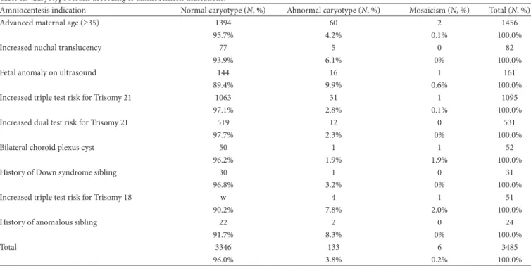
The distribution of indication for amniocentesis among 3485 patients included in the study is shown in Table І. Among all patients, amniocentesis was performed for the indication of advanced maternal age in 1456 women (41.8%) and for other reasons in the remaining 2029 women (58.2%). When the results of all amniocenteses were examined, normal karyotypes were observed in 3346 patients (96%), abnormal karyotypes were observed in (3.8%) and mosaicism was observed in six patients (0.2%) (Table ІІ) (p < 0.001).
When the results of the amniocenteses performed for advanced maternal age were compared with the results for all other indica-tions, 40 (2.7%) of 1456 ACs performed for advanced maternal age and 36 (1.8%) of 2029 ACs performed for all other reasons had abnormal karyotypes. The difference between two results was statistically significant (p = 0.05). When the results of the advanced maternal age group were compared with the women who had a calculated risk of 1/250 or higher based on the double and triple tests; chromosomal anomalies including chromosomal structural abnormalities were observed in 63 (4.2%) of 1456 ACs performed for advanced maternal age, 12 (2.3%) of 531 ACs performed based on an increased double-test risk and 32 (2.9%) of 1095 ACs performed for an increased triple test risk. The results were not
statistically significant (p = 0.15) (Figure 1). When the AC results of patients were grouped as numerically normal or abnormal, 40 (2.7%) of 1456 ACs performed for advanced maternal age, 5 (0.9%) of 531 ACs performed for an increased double-test risk and 14 (1.3%) of 1095 ACs performed for an increased triple test risk were observed to have abnormal karyotype number. The results were statistically significant (p = 0.006).
Discussion
In this study, we evaluated the results of 3485 amniocenteses performed in our hospital between 2007 and 2009 based on their indications. Prenatal cytogenetic diagnosis from amniotic fluid samples is a reliable test used for fetal karyotyping. Advanced maternal age is still the leading indication of amniocentesis [9–11]. Detection rate of chromosomal anomalies including chromosomal structural abnormalities for amniocenteses performed among patients with advanced maternal age was 4.2% in our study. The rate for other indications was 3.7%, and the difference between these rates was not statistically significant. On the other hand, when chromosomal aneuploidy detection rates for ACs performed for advanced maternal age and all other indications were compared, they were determined to be 2.7% and 1.8%, respectively, demonstrating a statistically significant differ-ence. When the AC results from advanced maternal age group was compared with those women with increased risks of double and/or triple tests, the difference in the incidence of chromo-somal abnormalities between these groups was not statistically significant. Chromosomal aneuploidy incidence in the advanced maternal age group were found to be significantly higher than women with increased risks of double and/or triple tests. According to these results, maternal age is the most important risk factor for chromosome number abnormalities, and in most of the antenatal clinics, it is still the most prevalent indication for AC other than positive prenatal screening tests. In our study, the ratio of ACs for advanced maternal age compared to other indications was 88%. There was a patient group with advanced maternal age and with abnormal double or triple test results that were offered AC but did not accept due to maternal and/or paternal reasons or because CVS was performed and the number of these cases could not be determined because of the retrospective design of this study. Although the use of first trimester screening tests has increased, the rate of CVS for prenatal diagnosis has not increased at the same rate. Clinical indications for AC in our study are similar to those in previous studies. In previous amniocentesis studies, the incidence of chromosomal anomalies varies between 1.0 % and 6.7% [6]. In our study, it was 4.0%, which is consis-tent with the current literature [10]. In the previous studies, fetal anomalies diagnosed by ultrasonography were found to have the highest positive predictive value for detection of chromosomal anomalies by performing prenatal amniocentesis [5]. In our study, fetal anomalies in prenatal ultrasonography were found to be the AC indication that had the gretatest percentage for detec-tion of chromosomal anomalies (10.5%) (Table II). In a study performed by Kagan et al., chromosomal anomalies were found in 15% of fetuses that had the congenital anomalies diagnosed by prenatal ultrasonography [7]. In contrast to our study; Kagan et al. evaluated the AC results of fetuses with fetal malformations with respect to qf-PCR and karyotyping results. Therefore, they found higher fetal chromosomal anomaly detection rates of ACs than those observed in our study. Because AC has a minimal risk of abortion even in experienced hands, prenatal screening tests with higher positive predictive values are needed [12]. Chromosomal
Table І. Numbers and percentages of various indications for amniocentesis (N, 3485).
Amniocentesis indication Number (N) Percent (%) Advanced maternal age (≥35) 1456 41.8 Increased nuchal translucency 83 2.4 Fetal anomaly on ultrasound 162 4.6 Increased triple test risk for Trisomy 21 1095 31.4 Increased dual test risk for Trisomy 21 531 15.2 Bilateral choroid plexus cyst 52 1.5 History of Down syndrome sibling 31 0.9 Increased triple test risk for Trisomy 18 51 1.5 History of anomalous sibling 24 0.7
244 N. Danisman et al.
anomaly detection rates for AC were higher in pregnant women younger than 35 years compared to women with advanced maternal age, and most chromosomal anomalies were detected in women younger than 35 years old [13,14]. In our study, increased NT and bilateral choroid plexus cysts were indications for AC, and detection rates were found to be 6.1% and 3.8%, respectively. Fetal chromosomal anomalies were detected in 9.8% of ACs performed for increased risk for Trisomy 18 as detected by a triple screening test. Fetal chromosomal anomalies were detected in 3.2% of ACs performed for a history of a previous fetus with aneuploidy. Fetal chromosome anomalies were detected in 8.3% of ACs performed in patients with a history of fetal anomalies, which is similar to the chromosomal anomaly detection rate of ACs performed for
increased Trisomy 18 risk based on the triple test results. The first trimester biochemical screening has become a routine part of clinical practice, and fewer clinicians suggest diagnostic tests only based on maternal age [15,16]. It is a difficult to determine which method for Down syndrome screening is most efficient. With the addition of ultrasonography to maternal serum biochemical markers and the use of new ultrasonographic markers, such as the nasal bone, detection rates will increase [17–22]. The inte-grated test offers the most effective and safe method of screening for women who seek prenatal care in the first trimester. The quadruple test is the best test for women who first seek prenatal care in the second trimester. Using the integrated test with an 85% detection rate, there would be six amniocentesis-related fetal losses per 100,000 women screened, compared with 35 using the combined test or 45 with the quadruple test [23]. Dommergues et al. evaluated the AC results of the 359 pregnant patients with advanced maternal age aged 38–47 and they found that all 7 cases of Down’s syndrome had increased NT measurements and/or increased chromosomal anomaly risk based on maternal serum screening results. They recommended proceeding with AC on a selective rather than routine basis in women over 38, based upon the results of noninvasive screening tests [24]. When cost-effectivity of prenatal diagnostic tests for fetal aneuploidy were considered; the combination of advanced maternal age, maternal serum screen and genetic sonogram have been found to result in the fewest procedure-related losses and lowest cost per Down syndrome case detected [25]. In the future, by increasing utiliza-tion of detecutiliza-tion methods like fetal chromosome identificautiliza-tion from cell free fetal DNA in the maternal plasma, invasive proce-dures like mid-AC will probably be used uncommonly for this purpose [26–28]. The noninvasive diagnostic tests based on cell free fetal DNA extracted from maternal plasma have promising detection rates so unnecessary invasive tests and associated fetal losses could be avoided in almost all women who have a normal fetus.
At the expense of investigating the incidence of chromosomal anomalies among pregnant women with advanced maternal age
Figure 1. Distribution of caryotype results among amniocentesis procedures performed for advanced maternal age, increased triple test risk, and increased dual test risk for aneuploidy.
Table ІІ. Caryotype results according to amniocentesis indications.
Amniocentesis indication Normal caryotype (N, %) Abnormal caryotype (N, %) Mosaicism (N, %) Total (N, %)
Advanced maternal age (≥35) 1394 60 2 1456
95.7% 4.2% 0.1% 100.0%
Increased nuchal translucency 77 5 0 82
93.9% 6.1% 0% 100.0%
Fetal anomaly on ultrasound 144 16 1 161
89.4% 9.9% 0.6% 100.0%
Increased triple test risk for Trisomy 21 1063 31 1 1095
97.1% 2.8% 0.1% 100.0%
Increased dual test risk for Trisomy 21 519 12 0 531
97.7% 2.3% 0% 100.0%
Bilateral choroid plexus cyst 50 1 1 52
96.2% 1.9% 1.9% 100.0%
History of Down syndrome sibling 30 1 0 31
96.8% 3.2% 0% 100.0%
Increased triple test risk for Trisomy 18 w 4 1 51
90.2% 7.8% 2.0% 100.0%
History of anomalous sibling 22 2 0 24
91.7% 8.3% 0% 100.0%
Total 3346 133 6 3485
96.0% 3.8% 0.2% 100.0%
A retrospective analysis of amniocenteses 245
© 2013 Informa UK, Ltd.
universally, we evaluated the AC results of women with advanced maternal age as a sole indication for genetic amniocentesis without considering the risk lowering or increasing effects of ultrasonographic soft markers for chromosomal aneuploidy that constitutes the major limitation of this study. The AC results of various fetal anomalies on prenatal ultrasonography that indi-cated an AC procedure to be performed have not been evaluated individually which is a limitation of this study. Besides, AC results of the patients with advanced maternal age having an increased aneuploidy risk based on serum biochemical tests and/or fetal malformations and/or soft markers of chromosomal anomalies have not been evaluated in this study that represents another limi-tation of our study worth to investigate in future studies. Third limitation of this study is the exclusion of the patients who have preferred to proceed with a CVS procedure after a first trimester screening for chromosomal aneuploidy that was intentionally conducted to compare the results of the “amniocentesis” proce-dures itself retrospectively.
In conclusion; despite the relatively high incidence of fetal chromosomal anomalies including chromosomal structural abnormalities (4.2%) in this study, that was detected from AC procedures performed for a sole indication of advanced maternal age resembles to favor genetic AC for women with advanced maternal age universally; increasing evidence in the literature reveals that maternal age alone seems to be a weak indicator of chromosomal anomalies when encountered with NT measure-ments within normal limits; with low dual, triple, quadruple, combined or integrated test result risks; and without fetal malfor-mations and/or soft markers for chromosomal anomalies. The integrated maternal serum screening test risk measurement that is recalculated with respect to the likelihood ratios of presence or absence of prenatal ultrasonographic markers related to chro-mosomal anomalies should be the most valuable method for a decision of offering an AC procedure to pregnant women with advanced maternal age.
Declaration of Interest: The authors report no conflict of interest.
References
1. Pitukkijronnakorn S, Promsonthi P, Panburana P, Udomsubpayakul U, Chittacharoen A. Fetal loss associated with second trimester amniocentesis. Arch Gynecol Obstet 2011;284:793–797.
2. Mademont-Soler I, Morales C, Clusellas N, Soler A, Sánchez A; Group of Cytogenetics from Hospital Clínic de Barcelona. Prenatal cytogenetic diagnosis in Spain: analysis and evaluation of the results obtained from amniotic fluid samples during the last decade. Eur J Obstet Gynecol Reprod Biol 2011;157:156–160.
3. Park IY, Kwon JY, Kim YH, Kim M, Shin JC. Maternal age-specific rates of fetal chromosomal abnormalities at 16-20 weeks’ gestation in Korean pregnant women >or=35 years of age. Fetal Diagn Ther 2010;27:214–221. 4. Kazerouni NN, Currier B, Malm L, Riggle S, Hodgkinson C, Smith
S, Tempelis C, et al. Triple-marker prenatal screening program for chromosomal defects. Obstet Gynecol 2009;114:50–58.
5. Offerdal K, Blaas HG, Eik-Nes SH. Prenatal detection of trisomy 21 by second-trimester ultrasound examination and maternal age in a non-selected population of 49 314 births in Norway. Ultrasound Obstet Gynecol 2008;32:493–500.
6. Tseng JJ, Chou MM, Lo FC, Lai HY, Chen MH, Ho ES. Detection of chromosome aberrations in the second trimester using genetic
amniocentesis: experience during 1995-2004. Taiwan J Obstet Gynecol 2006;45:39–41.
7. Kagan KO, Chitty LS, Cicero S, Eleftheriades M, Nicolaides KH. Ultrasound findings before amniocentesis in selecting the method of analysing the sample. Prenat Diagn 2007;27:34–39.
8. Hecht CA, Hook EB. The imprecision in rates of Down syndrome by 1-year maternal age intervals: a critical analysis of rates used in biochemical screening. Prenat Diagn 1994;14:729–738.
9. Langlois S, Duncan A. Use of a DNA method, QF-PCR, in the prenatal diagnosis of fetal aneuploidies. J Obstet Gynaecol Can 2011;33:955–960. 10. ACOG Committee on Practice Bulletins. ACOG Practice Bulletin No.
77: screening for fetal chromosomal abnormalities. Obstet Gynecol 2007;109:217–227.
11. Dailey T, Dale B, Cohen J, Munné S. Association between nondisjunction and maternal age in meiosis-II human oocytes. Am J Hum Genet 1996;59:176–184.
12. Savva GM, Morris JK, Mutton DE, Alberman E. Maternal age-specific fetal loss rates in Down syndrome pregnancies. Prenat Diagn 2006;26:499–504.
13. Druzin ML, Chervenak F, McCullough LB, Blatman RN, Neidich JA. Should all pregnant patients be offered prenatal diagnosis regardless of age? Obstet Gynecol 1993;81:615–618.
14. Ghosh S, Feingold E, Dey SK. Etiology of Down syndrome: Evidence for consistent association among altered meiotic recombination, nondisjunction, and maternal age across populations. Am J Med Genet A 2009;149A:1415–1420.
15. Driscoll DA, Morgan MA, Schulkin J. Screening for Down syndrome: changing practice of obstetricians. Am J Obstet Gynecol 2009;200:459. e1–459.e9.
16. Maulik D. New directions in prenatal care. J Matern Fetal Neonatal Med 2003;13:361.
17. Cuckle HS. Growing complexity in the choice of Down’s syndrome screening policy. Ultrasound Obstet Gynecol 2002;19:323–326. 18. Breathnach FM, Malone FD. Screening for aneuploidy in first and
second trimesters: is there an optimal paradigm? Curr Opin Obstet Gynecol 2007;19:176–182.
19. Nicolaides KH, Spencer K, Avgidou K, Faiola S, Falcon O. Multicenter study of first-trimester screening for trisomy 21 in 75 821 pregnancies: results and estimation of the potential impact of individual risk-orientated two-stage first-trimester screening. Ultrasound Obstet Gynecol 2005;25:221–226.
20. Nyberg DA, Souter VL, El-Bastawissi A, Young S, Luthhardt F, Luthy DA. Isolated sonographic markers for detection of fetal Down syndrome in the second trimester of pregnancy. J Ultrasound Med 2001;20:1053–1063.
21. Benn PA, Fang M, Egan JF. Trends in the use of second trimester maternal serum screening from 1991 to 2003. Genet Med 2005;7:328–331. 22. Rozenberg P, Bussières L, Chevret S, Bernard JP, Malagrida L, Cuckle
H, Chabry C, et al. Screening for Down syndrome using first-trimester combined screening followed by second-trimester ultrasound examination in an unselected population. Am J Obstet Gynecol 2006;195:1379–1387.
23. Wald NJ, Rodeck C, Hackshaw AK, Rudnicka A. SURUSS in perspective. BJOG 2004;111:521–531.
24. Dommergues M, Audibert F, Benattar C, Champagne C, Gomel V, Frydman R. Is routine amniocentesis for advanced maternal age still indicated? Fetal Diagn Ther 2001;16:372–377.
25. Hartnett J, Borgida AF, Benn PA, Feldman DM, DeRoche ME, Egan JF. Cost analysis of Down syndrome screening in advanced maternal age. J Matern Fetal Neonatal Med 2003;13:80–84.
26. Lau TK, Jiang F, Chan MK, Zhang H, Salome Lo PS, Wang W. Non-invasive prenatal screening of fetal Down syndrome by maternal plasma DNA sequencing in twin pregnancies. J Matern Fetal Neonatal Med 2012 Oct 4. [Epub ahead of print].
27. Lau TK, Chan MK, Salome Lo PS, Connie Chan HY, Kim Chan WS, Koo TY, Joyce Ng HY, Pooh RK. Clinical utility of noninvasive fetal trisomy (NIFTY) test - early experience. J Matern Fetal Neonatal Med 2012. 28. Clerici G, Donti E, Zacutti A, Di Renzo GC. Prenatal diagnosis in Italy.