ANALYSIS OF X CHROMOSOME INACTIVATION IN PRIMARY
AND SECONDARY SJOGREN SYNDROME
A THESIS SUBMITTED TO THE DEPARTMENT OF MOLECULAR BIOLOGY AND GENETICS AND THE INSTITUTE OF ENGINEERING AND SCIENCE OF
BILKENT UNIVERSITY IN PARTIAL FULFILLMENT OF THE REQUIREMENTS FOR THE DEGREE OF MASTER OF SCIENCE
BY MELDA KANTAR AUGUST, 2008
I certify that I have read this thesis and that in my opinion it is fully adequate, in scope and in quality, as a thesis for the degree of Master of Science.
___________________
Prof. Tayfun Özçelik
I certify that I have read this thesis and that in my opinion it is fully adequate, in scope and in quality, as a thesis for the degree of Master of Science.
__________________
Assist. Prof. Ali O. Güre
I certify that I have read this thesis and that in my opinion it is fully adequate, in scope and in quality, as a thesis for the degree of Master of Science.
__________________
Assist. Prof. Hüseyin Boyacı
Approved for the Institute of Engineering and Science
_____________________________ Director of Institute of Engineering and Science Prof. Dr. Mehmet Baray
ABSTRACT
Lack of evidence for the role of skewed X chromosome inactivation in the female predisposition to Sjogren Syndrome
Melda Kantar
M.S. in Molecular Biology and Genetics Supervisor: Prof. Dr. Tayfun Özçelik
August 2008, Pages 91
Sjogren Syndrome is an autoimmune disease with one of the highest prevalences and unknown etiology. The majority of the patients (~90%) are female similar to several other autoimmune diseases. Based on this observation, a hypothesis was proposed stating that X chromosome inactivation (XCI) could be involved in female predisposition to autoimmunity. XCI is a physiological mechanism which takes place early in development resulting in the transcriptional silencing of one of the pair of X chromosomes at random in each cell. A significant deviation from a random distribution of two cell populations with paternal and maternal X chromosome inactive is called skewed XCI. Skewing in the dendritic cell population involved in tolerance induction in the thymus was proposed to cause escape of autoreactive lymphocytes and result in autoimmunity (Immunol Today, 19, 352-7, 1998). Skewed XCI was observed in scleroderma (Arth Rheum 52, 1564-70, 2005) and autoimmune thyroiditis (Eur J Hum Genet 14, 791-7, 2006). But this observation is not true for all autoimmune diseases. For example, the XCI profiles of primary biliary cirrhosis patients are similar to normal controls (Hepatol Res 37, Suppl 3, 384-8, 2007). The aim of this study is to determine the XCI profiles of patients diagnosed with primary Sjogren Syndrome, manifesting exocrinopathy or secondary Sjogren Syndrome displaying additional systemic features. DNA was isolated from the peripheric blood samples of 78 Sjogren syndrome patients and 160 controls. XCI profile was determined by the genotyping of a polymorphism in the androgen receptor (AR) gene. For this analysis, restriction enzyme HpaII was used which does not cut methylated regions. Analysis was done with Genescan Abi Prism 310 or 8% polyacrylamide gel electrophoresis and densitometric analysis. Extreme skewing (>90%) of XCI was observed in 3 (5.9%) patients and 3 controls (2.4%) samples (P = 0.3651). Our findings do not support a role for skewed XCI in Sjogren Syndrome.
ÖZET
Sjogren sendromu ile X inaktivasyonu bozukluğu arasında bir ilişki bulunmuyor
Melda Kantar
Moleküler Biyoloji ve Genetik Yüksek Lisans Tez Yöneticisi: Prof. Dr. Tayfun Özçelik
Ağustos 2007, Sayfa 91
Sjogren sendromu etiyolojisi bilinmeyen ve prevalansı en yüksek olan otoimmün hastalıklardan biridir. Pek çok otoimmün hastalıkta olduğu gibi hastaların büyük bir çoğunluğu (~%90) kadındır. Bu gözlemden yola çıkarak, bozuk X inaktivasyonunun otoimmun hastalıkları tetikleyebileceği hipotezi ileri sürülmüştür (Immunol Today, 19, 352-7, 1998). X inaktivasyonu kadınlarda erken gelişme döneminde hücrelerdeki X kromozomu çiftinden rastgele birinin susturulmasıyla sonuçlanan normal bir fizyolojik olaydır. Hücrelerin önemli bir kısmında aynı X kromozomunun susturulması bozuk X inaktivasyonu olarak adlandırılır. Timusda, tolerans oluşumunda rol alan dendritik hücrelerdeki X inaktivasyonu bozukluğunun otoreaktif lenfositlerin kaçmasına ve otoimmüniteye neden olabileceği hipotezi ileri sürülmüştür. Skleroderma (Arth Rheum 52, 1564-70, 2005) ve otoimmün tiroiditis (Eur J Hum Genet 14, 791-7, 2006) hastalıklarında bozuk X inaktivasyonu gözlenmiştir. Fakat bu gözlem tüm otoimmün hastalıklar için geçerli değildir. Örneğin primer biliyer siroz hastalarının X inaktivasyonu profilleri normal kontroller gibidir (Hepatol Res 37, Suppl 3, 384-8, 2007). Bu çalışmanın amacı sadece salgı bezi patolojisi olan primer Sjogren Sendromu ve aynı zamanda başka sistemik patolojiler içeren sekonder Sjogren Sendromu tanısı alan hastalarda X-inaktivasyonu profillerinin belirlenmesidir. 78 hasta ve 160 kontrolün periferik kanından DNA izole edilmiştir. X inaktivasyon profili androjen reseptör geninde bulunan bir polimorfizmin genotiplemesi ile belirlenmiştir. Bu inceleme için metilli bölgeleri tanımayan bir restriksiyon enzimi olan HpaII ile kesim yapılmış ve hedef bölge çoğaltılmıştır. İnceleme Genescan Abi Prism 310 ya da %8 poliakrilamid jel elektroforezi ve densitometrik ölçümle yapılmıştır. İleri derecede (>%90) X inaktivasyonu bozukluğu 3 (%5.9) hasta ve 3 kontrol (%2.4) örneğinde gözlenmiştir (P = 0.3651). Bulgularımız X inaktivasyonu bozukluğu ile Sjogren Sendromu arasında bir ilişkinin varlığını desteklememiştir.
TO MY FAMILY,
ACKNOWLEDGEMENTS
It is my pleasure to express my deepest gratitude to my advisor Prof. Dr. Tayfun Özçelik for his scientific guidance and support all through my thesis. I especially want to thank him for introducing me to research..
I would like to adress my special thanks to Zeynep Özbalkan for obtaining the blood samples and coordinating the clinical studies.
I would like to thank Scientific and Technological Research Council of Turkey for the funding.
I would like to express my thanks to Elif Uz and Çiğdem Aydın Mustafa for guiding me in every step of my research and always being there for my questions and for their endless support in every obstacle I was faced to. A special thank to Elif Uz for her help in determining the XCI status of Sjogren Syndrome patients. I would like to thank all my group members Emre Onat, Şafak Çağlayan, Süleyman Gülsuner for their incredible help and support. I would like to thank Mustafa Gökhan Ertosun for helping me with the format of my thesis.
I would like to thank all MBG members for making Bilkent Molecular Biology Department an incredible place to live and work.
I would also like to thank my friends Hülya Budunoğlu, Hande Erkut, Cansu Aksu, Banu Demir, Emre Onat, Fuat Yağcı, Elif Yaman, Hande Kocak, Pelin Telkoparan, Esen Oktay, Gizem Tinçer, Ceren Sucularlı and all the others I have not mentioned for listening to my every problem during my academic and personal life. I want to thank my boyfriend, Onur Karakuş for sharing my life.
Lastly, I want to express my sincere love and thanks to my family for being there whenever I needed help and support. I want to thank my mother, Nujan Kocagöz and my father, Elvend Kantar for helping me through every struggle and especially my dear sister, Selda Kantar without whom life would be unbearable.
TABLE OF CONTENTS
ABSTRACT III ÖZET IV DEDICATION PAGE V ACKNOWLEDGEMENTS VI TABLE OF CONTENTS VII LIST OF TABLES X LIST OF FIGURES XI ABBREVIATIONS XII 1. INTRODUCTION 1 1.1.Sjogren Syndrome 1 1.1.1. History 1 1.1.2. Symptoms 3 1.1.3. Criteria for diagnosis 3 1.1.4. Treatment 5 1.1.5. Epidemiology 6 1.1.6. Classification as primary and secondary 6Sjogren Syndrome
1.1.7. Pathophysiology and Etiology 7
1.1.7.1 Genetic predisposition 12 1.1.7.2 Associated genes 13 1.1.7.3 Associated antibodies 14 1.1.7.4 Viruses 16 1.1.8 .Research to relate microchimerism 17 and Sjogren Syndrome
1.2. X Chromosome Inactivation 18 1.2.1 X dosage compensation 18 1.2.2. Discovery of mammalian dosage compensation 18
1.2.3. Features of Inactive X 20 1.2.4. Clonal Nature of XCI 20 1.2.5. Timing of XCI 21 1.2.6. Master control region of XCI and its components 22 1.2.7. Steps of XCI 24 1.2.8. Genes escaping XCI 26 1.2.9. Primary and secondary causes of skewed XCI 27 1.3.Self tolerance and Autoimmunity 28
1.3.1 Immune system 28 1.3.1.1 Clonal theory of Jerne and Burnett 28 1.3.1.2 Self tolerance 28 1.3.2 Autoimmunity 29
1.3.2.1 Genes associated with autoimmunity 30 1.3.2.2 X-linked genes implicated in immunity 32 1.3.2.3 Molecular Mimicry 32 1.3.2.4 Female predominance in autoimmunity 33
1.3.2.4.1 Hormones 35 1.3.2.4.2 Microchimerism 36 1.3.2.4.3 X chromosome Monosomy 37 1.4 Skewed X inactivation and autoimmunity 38 1.4.1 Kast and Stewart hypothesis 38
1.5. Aim and Strategy 40
2. MATERIALS AND METHODS 41 2.1 Samples 41 2.1.1 Control Samples 41 2.1.2 Sjogren Syndrome patient Samples 41
2.2 Materials 42 2.2.1 Primers 42 2.2.2 Enzymes 42
2.2.3 Oligonucleotide 44 2.2.4 Chemicals Reagents, Kits 45 2.2.4 Standard solutions and buffers 46 2.3 Methods 47 2.3.1 Sample collection 47 2.3.2 DNA Isolation from venous blood 47 2.3.4 AR genoyping assay 48
2.3.4.1. Restriction Enzyme Digestion 50 2.3.4.2 Polymerase chain reaction (PCR) 51 2.3.4.3 Visualization 51
2.3.4.3.1 Agarose gel electrophoresis 51 2.3.4.3.2 Polyacrylamide gel 52
electrophoreis (PAGE)
2.3.4.4 Densitometric and Statistical Analysis 52
3. RESULTS 53
4. DISCUSSION 56
5. FUTURE PERSPECTIVES 61
6. REFERENCES 62
LIST OF TABLES
Table 1.1 Timeline of important advances in delineating Sjogren Syndrome 2 Table 1.2 Current criteria for Sjogren Syndrome diagnosis 4 Table 1.3 Prevalence of sicca features and associated Sjogren Syndrome 7
in systemic autoimmune diseases
Table 1.4 Polymorphisms of interleukins and other genes associated 14 with Sjogren Syndrome
Table 1.5 Autoantibodies described in Sjogren Syndrome patients 15 and their corresponding prevelances
Table 1.6 Ratio of female:male occurence of autoimmune disorders 34 Table 2.1 List of chemicals, reagents, kits used in this study 45 Table 2.2 Characteristics of Sjogren patients Table 3.1 Proportion of Sjogren Syndrome patients and controls with 54
LIST OF FIGURES
Figure 1.1 Minor salivary gland biopsy photomicrographs 8 Figure 1.2 Steps of the Sjogren Syndrome pathogenesis according 10
to the epithelithis model
Figure 1.3 Models in relation to XCI timing in the embryo 22 Figure 1.4 Elements of Xic 24 Figure 1.5 Different mechanisms of self tolerance 29 Figure 1.6 Incidence of autoimmune disorders by sex 35 Figure 1.7 Consequences of skewed XCI in thymic cells 39 Figure 2.1 AR genotyping amplicon 43
Figure 2.2 Sizes of the fragments of pUC Mix Marker 8 and 44 MassRuler DNA Ladder Mix and appearance on gel electrophoresis. Figure 2.3 Establishing random and skewed XCI with AR genotyping assay 49 Figure 3.1 XCI inactivation status in Sjogren Syndrome patients 54
ABBREVIATIONS
ANA Antinuclear antibodies Abs Antibodies
AID Autoimmune disease
AIDS Acquired immunodeficiency disease AITD Autoimmune tyroid disease
AIRE Autoimmune regulator
ALPS Autoimmune lymphoproliferative syndrome AQP Aquaporin
APC Antigen presenting cell AR Androgen receptor BAFF B cell survival factor bp Base pair
BCR B cell receptor Csk c-src tyrosine kinase CTLA-4 Cytotoxic T lymphocyte antigen 4 DEMS Dry eyes and mouth symptoms ddH2O Deionized water
dNTP Deoxynucleotide triphosphate DNA Deoxyribonucleic acid
EBV Epstein barr virus
EDTA Ethylenediaminetetraacetic acid EtBr Ethidium bromide
EtOH Ethanol
G6PD Glucose 6-phosphate dehydrogenase GVHD Graft versus host disease
HBV Hepatitis B virus
HLA Human leukocyte antigen HIV Human immunodeficiency virus HSV-1 Herpes simplex virus Type I
HTLV Human T lymphotrophic virus HIGM1 Hyper-IgM syndrome type I Ig Immunoglobulin
IgVλ Immunoglobulin variable λ IFN-γ Interferon γ
IL Interleukin
IPEX Immune dysregulation, polyendocrinopahty, enteropathy L1 LINE1
Mb Megabase
MCTD Muscle connective tissue disease MgCl2 Magnesium chloride
MHC Major histocompatibility complex MBL Mannose binding lectin
mM Millimolar ml Milliliter µl Microliter MS Multiple sclerosis kb Kilobase L1 Line1 elements NF-қB Nuclear factor kappa B
PAGE Polyacrylamide gel electrophoresis PI3K Phosphatidylinositol-3-kinase PBC Primary biliary cirrhosis PCR Polymerase chain reaction PcG Polycomb-group proteins PI3K Phosphoinositide 3 phosphatide PLP Proteolipid protein
PKC Phosphokinase C RA Rheumatoid arthritis RE Restriction enzyme RF Rheumatoid factor
SCID Severe combined immunodeficiency syndrome SCLE Subacute cutaneous lupus erthematosus
SDS Sodium dodecyl sulphate SLE Systemic lupus erythramatosus SNP Single nucleotide polymophism SH3 Src homology 3
SSc Systemic sclerosis T1D Type 1 diabetis
TAE Tric-acetic acid-EDTA TCR T cell receptor
TEMED N, N, N, N-tetramethyl-1-2, diaminoethane Th1 T helper 1
Th2 T helper 2
TLR Toll like receptor TNF Tumour necrosis factor Tris Tris aminomethan
WAS Wiskott-Aldrich syndrome Xa Active X
XCI X chromosome inactivation Xce X chromosome controlling element Xi Inactive X
Xic X-inactivation center Xist X-inactive specific element
Xite X-inactivation intergenic transcription element XEDA-ID X-linked recessive anhydortic ectodermal dysplasia with
immunodeficiency
XLA X-linked agammaglobulinemia XLP X-linked lymphoproliferative syndrome
CHAPTER I: INTRODUCTION
1.1. Sjogren Syndrome
Sjogren Syndrome is a chronic and systemic autoimmune disease characterized by progressive lymphocytic infiltration of polyglandular tissue and subsequent tissue destruction. This results in the functional impairment of salivary and lacrimal glands, and causes keratoconjunctivitis sicca, which refers to dryness of the eyes and xerostomia meaning dryness of the mouth. The ease and safety of a minor salivary gland biopsy enables study of the molecular biology of this autoimmune exocrinopathy. With microscopic examination, the lymphocytic replacement of the epithelium and lymphoepithelial lesion can be observed. It is an inflammatory rheumatic disease with main features in the eyes and mouth (Ramos-Casals, 2005; Kassan, 2004; Schwartz, 2007; Fox, 2000).
1.1.1 History
Sjogren Syndrome is a relatively newly identified disease. Its histology was described and a link with arthritis was reported in the late 19th and early 20th centuries as single case reports. In 1933, Henrick Sjogren reported the association of xeropthalmia meaning dry eyes and xerostomia with polyarthritis in 19 cases. In the early years, research was mainly on the opthalmology of Sjogren Syndrome. The clinical spectrum of Sjogren Syndrome in 1956 was defined in 62 subjects. The rheumatologic aspect of Sjogren Syndrome was heavily emphasized when the distinction between primary and secondary Sjogren Syndrome was first described in 1965. In 1970s, autoantibodies characteristic of Sjogren Syndrome were described. In 1990s, the diagnostic tests became a routine procedure and research on disease-modifying drugs started (Venables, 2004). Table 1.1 shows the timeline of advances in delineating Sjogren Syndrome.
Table 1.1 Timeline of important advances in delineating Sjogren Syndrome
(Venables et al.,2004)
Year Progress
1888 Case reports of Hadden and Rowlands
1892 Case report of Miculicz (histology described)
1926 Description of three patients by Gougerot
1928 Case report of Houwer (link with arthritis)
1933 Description of nineteen patients with keratoconjuctivitis sicca by Sjogren
1946 Translation of Sjogren thesis to English
1953 “Sjogren syndrome” established as a term in literature
1965 Primary and secondary Sjogren Syndrome distinguished
1970 Anti-Ro (SS-A) and anti-La (SS-B) in Sjogren Syndrome is determined
1980 Extraglandular symptoms described
1990 Routine application of diagnostic tests
1990 Trials of disease-modifying drugs
2002 Consensus diagnosis criteria
Sjogren Syndrome is a milder disease in comparison to rheumatoid arthritis (RA) or systemic lupus erythematosus (SLE). There was not a strict classification criteria for the syndrome until lately. Its low mortality and underdiagnosis prevented the disease from being a primary research area and caused Sjogren patients to be underdiognosed and undertreated (Venables, 2004).
1.1.2 Symptoms
The symptoms in relation to the eyes are more serious than the symptoms of the mouth. Underlying collagen can be revealed as a result of erosions in the conjunctiva leading to the appearance of filamentary keratitis. More dental care is a sign of xerostomia. Parotid swelling occurs in half of the patients. Other exocrine glands can be involved effecting skin, vagina, gastrointestinal tract, colon and rectum. 70% of the patients complain of fatigue.
Systemic features include renal involvement, neurological involvement, vascular involvement, cutenous involvement, pulmonary involvement, muscuskeletal involvement, gastroenterologic involvement, congenital heart block and lymphoma (Venables, 2004; Kassan, 2008; Ramos-Casals, 2005).
1.1.3 Criteria for diagnosis
Until recently, a consensus international diagnosis criteria for Sjogren Syndrome was unavailable. Because of its heterogeneous nature, Sjogren Syndrome is hard to diagnose. It has been hard to achieve a consensus because there are diseases that mimic Sjogren Syndrome such as age-related atrophy, chronic anxiety, chronic fatigue syndrome, fibromyalgia and dry eyes and mouth symptoms (DEMS). Previously, there were `Copenhagen`, `Californian` and `European` criteria. The revised American-European consensus criteria was established in 2002 (Vitali, 2002). The application of this criteria was essential both for clinical diagnosis and research.
The current criteria is as designated in Table 1.2. Four of the listed observations including either the fourth or sixth one is needed to diagnose a patient as Sjogren Syndrome. The observations include objective signs and symptoms of dryness. Evaluation of symptoms is based on a short questionaire. Ocular signs are evaluated based on Schirmer`s test or Rose Bengal staining. Oral signs are evaluated with scintiagraphy, sialography or a test for salivary flow rate. One observation that indicates the inflammatory nature of the disease is the
characteristic appearance of a minor salivary gland biopsy with focal inflammation. Focal lymphocytic infiltrates are expected in the minor salivary gland biopsy resulting in a focus score of more than 1 (A cluster of 50 or more lymphocytes is called a focus). Another observation that indicates inflammation is the presence of characteristic autoantibodies, rheumatoid factor or anti-nuclear antibodies in the serum. To prevent confusion with dry eyes and mouth symptoms (DEMS), these observations for determining the inflammatory nature of the disease are necessary (Venables, 2004).
Table 1.2 Current Criteria for Sjogren Syndrome diagnosis (Venables et al, 2004)
Ocular symptoms
1. Have you had daily, persistance, troublesome dry eyes for more than 3 months 2. Do you have a recurrent sensation of sand and gravel in the eyes
I
3. Do you use a tear substitutive more than 3 times a day Oral symptoms
1. Have you had a daily feeling of dry mouth for more than 3 months
2. Have you had recurrently or persistantly swollen salivary glands as an adult II
3. Do you frequently drink liquids to aid in swallowing dry foods III Positive Schirmer`s I test or Rose Bengal score
IV Abnormal lower lip biopsy (focus score >= 1)
V Positive result for unstimulated whole salivary flow (<= 1.5ml in 15 min) VI Antibodies to Ro (SS-A) or La (SS-B), or both
In diagnosis, it is important to show that the salivary dysfunction roots from an inflammation. Salivary dysfunction can also be caused by drugs, infection, head and neck radiation treatment, acquired immune deficiency syndrome (AIDS), preexisting lymphoma, sarcoidosis, graft versus host disease (GVHD), use of anticholinergic drugs and the autonomic nervous system. These are exclusions to diagnosis (Venables, 2004).
1.1.4 Treatment
Until recently, the treatment was only topical and symptomatic to improve moisture; decrease inflammation and prevent damage. These include performing general measures and using tear and saliva substitutes and mucolytic agents. Recently, research on disease-modifying drugs have been intensified with the recognition that Sjogren Syndrome is a disease of considerable morbidity. Currently a more aggressive approach to therapy is applied with topical and systemic treatment. The disease-modifying drugs include secretagogues and immunomodulatory drugs.
If Sjogren Syndrome is a consequence of the destruction of glands, secretagogues can not be a rational form of treatment. However there is currently increasing evidence that hypofunction of the glands is caused by inflammation and dryness is caused by this hypofunction. Sialogogues, pilocarpine and cevimeline hydrocholorine are both cholinergic agents with muscarinic agonist activity and are of proven benefit for Sjogren Syndrome (Vivino, 2001 ; Fife, 2002).
It seems logical to use immunomodulatory drugs because most of the morbidity is caused by widespread disruption of the immune system. There is supportive evidence for the use of hydroxycholoroquine sulphate. These can be effective to cure most disabling features such as fatique (Fox, 1996).
There is limited research on the use of biological agents for the treatment of Sjogren Syndrome. There are trials of a recombinant interferon-alpha, tumour necrosis factor antibody Infliximab and B cell antibodies such as anti-CD20 antibody Rituximab (Cummins, 2003; Serge, 2002; Bradley, 2003).
Anti-inflammatory agents and cytotoxic drugs are used to treat systemic complications (Venables, 2004).
1.1.5 Epidemiology
The prevalance of Sjogren Syndrome have been controversial based on the lack of a consensus diagnostic criteria (Thomas, 1998). An estimated 2 to 4 million people in the United States have this syndrome. The heterogeneity of its clinical manifestations is a barrier against its diagnosis in some cases. Approximately 1 million people have an established diagnosis of Sjogren Syndrome. No racial predisposition to the syndrome is known. Females are affected more than males in a 9:1 ratio. It is a disease of middle-aged or elderly women although it can occur at any age. It has a low pediatric prevelance. Approximately 60% of the Sjogren patients have secondary syndromes. Secondary Sjogren Syndrome results in death, especially in cases with myelopathy (Schwartz, 2007; Kassan, 2004).
1.1.6 Classification as primary and secondary Sjogren Syndrome
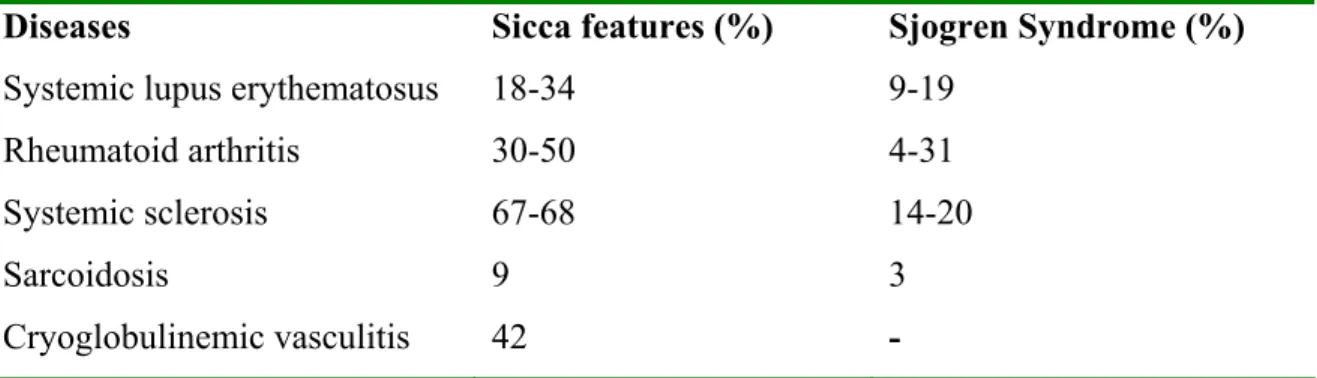
The presence of autoimmune inflammatory exocrinopathy and sicca symptoms alone is designated as primary Sjogren Syndrome. The occurence of keratoconjuctivitis sicca and xerostomia in association with another connective tissues diseases is designated secondary Sjogren Syndrome. These associated autoimmune diseases could be rheumatoid arthritis, systemic lupus erythematosus, scleroderma (SSc), muscle connective tissue disease (MCTD), subacute cutaneous lupus erythematosus (SCLE), polymyositis, sarcoidosis, systemic vasculitides and antiphospholipid syndrome (Ramos-Casals, 2007). The prevelance of Sjogren Syndrome in other autoimmune diseases is shown in Table 1.3.
Table 1.3 Prevalence of sicca features and associtated Sjogren Syndrome in
systemic autoimmune diseases (Ramos-Casals et al, 2007)
Diseases Sicca features (%) Sjogren Syndrome (%)
Systemic lupus erythematosus 18-34 9-19 Rheumatoid arthritis 30-50 4-31 Systemic sclerosis 67-68 14-20
Sarcoidosis 9 3
Cryoglobulinemic vasculitis 42 -
Sjogren Syndrome patients have a 44 times increased risk of developing B cell lymphoma (Kassan, 1978). Lymphoma is the cause of death in one of five Sjogren patients (Ioannidis, 2002; Bolstad, 2002). The prevalence of malignant lymphoma in Sjogren Syndrome patients is 4.3%, being mostly low-grade marginal zone B-cell lymphoma. Whether this lymphoma is primary or secondary to Sjogren Syndrome is unknown. (Yamamoto, 2002).
1.1.7. Pathophysiology and Etiology
The etiology of Sjogren Syndrome is unknown. It is a complex disease, in other words, both genetic predisposition and environmental factors have a role in the pathogenesis. The aetiopathogenesis is probably sequential leading to selective dysfunction of target organs caused by migration of lymphocytes (Ramos-Casals, 2005). In a minor salivary biopsy, the infiltrated lymphocytes can be observed as in Figure 1.1.
Sjogren Syndrome is generally accepted a T-cell mediated disease. The infiltrating cells in the glands are mostly T cells. T helper cells (CD4+) are more commonly seen than cytotoxic T cells (CD8+) in a ratio of 5:3. In labial salivary glands of Sjogren Syndrome patients, 2% of the infiltrating mononuclear cells were reported to be dendritic cells (Xanthou, 1999). Additionally, ductal and
co-stimulatory molecules (Monoussakis, 1999). The majority of the T cells in the
glands express CD45RO, which is a feature of activated or memory cells. T helper (Th1) cytokines such as interleukin 2 (IL-2), interleukin 10 (IL-10) are
produced in much higher amounts in comparison to normal controls. There is also an increase in T helper 2 (Th2) cytokines such as interleukin 4 (IL-4) and interleukin 5 (IL-5). There is additionally an increase in cytokines interleukin 1 (IL-1), interleukin 6 (IL-6), interleukin 10 (IL-10), tumor necrosis factor (TNF) and interferon gamma (IFN-γ) expressed by the tissue (Fox, 1994, Ohyama, 1996). A few T cells proliferate in the area. It is possible that memory T cells are semiactivated and the other T cells are probably suppressed in response to the strong immune response (Yamamoto, 2003).
Figure 1.1 Minor salivary gland biopsy photomicrographs A) 50X, Sjogren
Syndrome B) 50X, normal C) D) 200X, Sjogren Syndrome E) F) 500X, Sjogren Syndrome (Fox et al., 2000)
It seems likely that Sjogren Syndrome results from glandular destruction so infiltrating cytotoxic T cells are candidates for the pathogenesis. Both apoptosis with perforin/granzyme B and Fas/Fas ligand pathways were implicated with the etiology. It was shown that cytotoxic T cells expressing integrin localize around acinar epithelial cells that express E-cadherin (Fujihare, 1999). However the findings in relation to glandular apoptosis in Sjogren Syndrome patients in comparison to controls is controversial (Ohlson, 2001;Yammato 2003).
Several features of the disease can result from B cell stimulation and hyperglobulinemia. 20% of the infiltrating population consists of B cells. Antigen-driven, germinal center-type B cell response takes place within the salivary glands of Sjogren syndrome patients. Immunoglobulin G (IgG) isotype is extraordinarily seen more than immunoglobulin A (IgA) in the glands and in the serum. Immunoglobulin variable λ (IgVλ) light chain usage in primary Sjogren Syndrome was researched and it was found that there were differences in V-J recombination in Sjogren Syndrome patients from controls. It was concluded that there are defects in B-cell selection and maturation; immunoglobulin receptor editing and mutational targetting. There is B cell clonal expansion with antigen stimulation (Scott, 1998;Yammato, 2003).
Figure 1.2 Steps of the Sjogren Syndrome pathogenesis according to epithelithis
model (Ramos-Casals et al, 2005)
The current aetiopathogenic hypothesis of Sjogren Syndrome is autoimmune epithelitis. According to this theory, intrinsic influences such as genetic make-up and/or extrinsic influences such as environmental factors form the background for the pathogenesis. The initiation occurs as a result of an altered immune system incapable of discriminating between ‘foreign’, ‘self’ molecules and /or altered self antigens of the gland epithelium. This results in autoimmunity. The establishment process includes an abnormal immune response with abnormal T cell dysfunction and B cell hyperreactivity. This results in lymphocytic destruction forming histopathological lesions of acinar and ductal epithelial cells. It may also cause change in cytokine and chemokine secretion leading to perpetuation of the response. Then mechanisms of tissue damage such as apoptosis are activated leading to chronic inflammation with fibroses and loss of secretory function. Other mechanisms involved can be enhanced proteolytic mechanisms or altered epithelial repair. Apoptosis is thought to be related to formation of further abnormal autoantigens (Ramos-Casals, 2005). An overview of this model is given in Figure 1.2.
There are several potential mechanisms that are related to the etiology of Sjogren Syndrome: (1) Unsuccessful deletion of autoimmune T cells in the thymus. (2) Expression of increased levels of cell adhesive molecules in high endothelial venules leads to homing of autoimmune lymphocytes to glands. (3) Increase in expression of human leukocyte antigens (HLAs) in aiding increased presentation of antigens to lymphocytes. (4) In the gland, lymphocyte activation by their interaction with HLA-DR, cell adhesion molecules, and co-stimulatory factors. (5) Circulating autoantibodies against the ribonucleoproteins Ro and La (6) Inflamatory response is perpetuated by the secretion of pro-inflammatory cytokines by lymphocytes and epithelial cells. (7) Anti-apoptotic markers such as Bcl-2 and Bcl-x in the lymphocytes and apoptotic markers such as Fas and Bax in the epithelium are upregulated (Fox, 2000).
The role of apoptosis in autoimmune diseases including Sjogren Syndrome has been a major research area. Apoptosis has been proposed to have two aspects in the Sjogren Syndrome pathogenesis: (1) Increase in apoptosis in the ductal epithelial cells of the salivary glands was suggested to cause salivary decrease. (2) Accumulation of lymphocytes and displacement of functional acinar cells result from lymphocytes escaping apoptosis because of the defects of in the pathway.
Another major hypothesis on the etiology of Sjogren Syndrome is autonomic dysfunction. It was seen that half of the acinar cells remain histologically intact when patient biopsies were observed. So hypofunction rather than destruction of glandular epithelial cells is proposed as a theory for the secretory problem in Sjogren Syndrome. In one model, the gland can not receive enough neural signals from medulla. This can result from decreased neural innervation. Decreased neural axon-specific protein 9.5 and synaptophysin was observed with immunohistology (Konttinen, 1992). Release of acetylcholine is needed for glandular secretion. Cytokines can be toxic to the nerves and prevent the release of acetylcholine. It was shown in vitro and in transgenic mice that interleukin 1 and tumor necrosis factor α are toxic to nerves (Main, 1993). Another model suggests that acinar cells respond in a limited manner to neural
input, in other words there are problems in post signal transduction. Decrease of protein kinase C (PKC) isoforms involved in secretory response were reported for epithelial and myoepithelial cells (Törnwall 1997; Campbell, 1995). Antibodies against muscarinic M3 receptors were also found in Sjogren Syndrome patients (Borda, 1996; Venables, 2004; Fox, 2000).
Interactions of ductal and acinar cells with matrix may be important in the etiology of the disease. These interactions mediate response to cytokines, growth signals and hormones; they aid in homeostasis, regeneration and function of the cells. It was reported that the receptors on epithelial cells and some cytokines are expressed at higher amounts in these patients (Fox, 1994, Ohyama, 1996). It was also reported that cell-matrix interactions are important for secretory functions of to glandular cells in response muscarinic M3 agonists (Laurie, 1996).
The higher prevalence of Sjogren Syndrome in perimenopausal women suggest that hormonal factors can have a role in the disease etiology. The research on this field is controversial (Ramos-Casals, 2005).
1.1.7.1 Genetic predisposition
Based on animal models, familial aggregation and candidate gene association studies, a genetic predisposition to Sjogren Syndrome was suggested. Several families with more than two cases of Sjogren Syndrome have been identified and presence of multiple autoimmune diseases in families and in an individual have frequently been reported (Lichtenfeld, 1976; Koivukangas, 1973; Mason, 1971; Boiling, 1983, Sabio, 1983; Tanaka, 2001). Approximately 35% of Sjogren Syndrome patients have relatives with other autoimmune diseases. Limited number of case reports is present on twin concordance of primary Sjogren Syndrome. Reported twins have very similar phenotypes including clinical presentation, serological data, specificity of immune response to Ro/SSA and biopsy focus score (Scofield, 1997; Besena, 1991; Kogo, 1980; Bolstad, 2000; Bolstad, 2002).
1.1.7.2. Associated genes
Many genes were implicated in Sjogren Syndrome etiology and it is a polygenic disease. The best documented genetic risk factors of autoimmune diseases are polymorphic major histocompatibility complex (MHC) genes. For Sjogren Syndrome, MHC class II genes are of importance, especially HLA-DR and HLA-DQ alleles. HLA class II haplotypes, the DRB*03-DQB1*02 were frequently reported to be linked with the disease (Kang, 1993). These distinct polymorphisms of HLA alleles were associated with differences in autoantibody repertoire, especially anti-Ro (SS-A) and anti-La (SS-B) antibodies (Gottenberg, 2003; Davies, 2001). Link with MHC class I genes was also reported. The higher frequency haplotype found was HLA-A24 (Loiseau, 2001).
Lately, research has focused on finding association between Sjogren Syndrome and polymorphic genes that encode molecules involved in the immune system, especially cytokines. Cyokines mediate and regulate immune and inflammatory responses and they were suggested to have a role in autoimmune diseases including Sjogren Syndrome pathogenesis. Polymorphism of cytokines researched are interleukin 10, interleukin 4, interleukin 6, tumor necrosis factor α and tumor growth factor β (Ramos-Casals,2005; Hulkkonen, 2001; Gottenberg, 2004; Pertovaara, 2004; Youn, 2000).
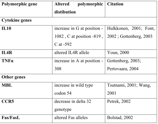
Additional candidate gene studies were also undertaken. Sjogren Syndrome was associated with the polymorphisms of mannose binding lectin (MBL), Fas/FasL and receptor mononuclear cell attracting chemokine receptor, CCR5. (Tsutsumi, 2001, Wang, 2001, Bolstad, 2000, Petrek, 2002; Ramos-Casals,2005). Table 1.4 lists the non-MHC genes reported to be associated with Sjogren Syndrome.
Table 1.4 Polymorphisms of interleukins and other genes associated with
Sjogren Syndrome
Polymorphic gene Altered polymorphic distibution
Citation
Cytokine genes
IL10 increase in G at position -1082 , C at position -819 , C at -592
Hulkkonen, 2001; Font, 2002 ; Gottenberg, 2003
IL4R altered IL4R allele Youn, 2000
TNFα increase in A at position -308
Gottenberg, 2003; Pertovaara, 2004
Other genes
MBL increase in wild type codon 54 Tsutnami, 2001; Wang, 2001 CCR5 decrease in delta 32 genotype Petrek, 2002
Fas/FasL altered Fas alleles Bolstad, 2002
There are recently investigated candidate antigens for a role in Sjogren Syndrome etiology. The most important of these is aquaporins, plasma membrane transporters of water. There is controversial results on the aberrant cytoplasmic localization of aquaporin 5 (AQP-5) channel proteins in this disease (Tsubota, 2001; Steinfeld, 2002; Beroukas, 2001). There is one report on the reduced expression of aquaporin 3 (AQP-3) in acinar cells in primary Sjogren Syndrome patients (Waterman, 2003 ; Ramos-Casals, 2005).
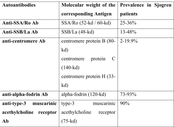
1.1.7.3 Associated antibodies
Table 1.5 Autoantibodies described in Sjogren Syndrome patients and their
corresponding prevalences (Nakamura et al, 2006)
Autoantibodies Molecular weight of the corresponding Antigen
Prevalence in Sjogren patients
Anti-SSA/Ro Ab SSA/Ro (52-kd / 60-kd) 25-36%
Anti-SSB/La Ab SSB/La (48-kd) 13-48%
anti-centromere Ab centromere protein B (80-kd) centromere protein C (140-kd) centromere protein H (33-kd) 2-19.9% anti-alpha-fodrin Ab alpha-fodrin (120-kd) 73-93% anti-type-3 muscarinic acethylcholine receptor Ab type-3 muscarinic acethylcholine receptor (75-kd) 90%
Sjogren Syndrome is an autoimmune disease, and a wide variety of auto-antibodes have been described in these patients (Table 1.5).
In Sjogren Syndrome, especially Ro/SS-A and to a lesser extent anti-La/SS-B and anti-centromere antibodies are seen. These are organ non-specific antibodies. Antigens towards these antibodies are 52 kd SSA/Ro, 60 kd SS-A/Ro and 48 kd SS-B/La. Anti-Ro/SS-A and to a lesser extent anti-La/SS-B are frequently also found in relatives of the patients. Anti-Ro/SS-A antibodies are found in 65% ; anti-La/SS-B is found in 50% of the patients. Anti-Lo/SS-A is restricted to Sjogren Syndrome but anti-Ro/SS-B can be found in systemic lupus erythematosus, rheumatoid arthritis or polymyositis. The presence of these autoantibodies is related to density of lymphocytic infiltrates, the severity of
extraglandular symptoms, parotid gland enlargement, early disease onset and longer disease duration (Yamamato, 2002).
Rheumatoid factor is found both in primary and secondary Sjogren Syndrome, but the presence of rheumatoid factor is a general feature of autoimmune diseases.
Alpha-fodrin is an apoptotically cleaved form of fodrin, which is a membrane skeletal protein found in many tissues. Antibodies against alpha-fodrin is found in Sjogren Syndrome sera. It is not known whether it is a primary or secondary event to the disease.
Based on one theory, these antibodies are produced in the salivary glands (Tengner, 1998). The abnormal localization of anti-Lo and anti-Ro was reported several times in disease and stress states. Surface expression of an antigen reactive to these antibodies was shown after irradiation of keratocytes. This raises the possibility that altered localization of antigens after apoptosis can be the immunogen in autoimmune responses (Lefeber,1984). Another theory on the production of these antibodies is molecular mimicry. Sequence similarities of SS-A/Ro and SS-B/La determinants with retroviral antigens have been reported (Kohsaka,1990;Yamamato, 2002).
Anti-M3 antibody can particularly have a role in the pathogenesis because it can cause exocrine dysfunction by blocking neurotransmission (Nakkamura, 2006; Venables, 2004).
1.1.7.4 Viruses
Viral infection is one of the most important proposed environmental factors. Viruses of the herpes family are implicated in Sjogren Syndrome pathogenesis. Cytomegalovirus was shown to induce sialadenitis with Sjogren Syndrome antibodies in a mouse model (Fleck, 1998). Epstein barr virus (EBV) commonly infects the salivary glands and induces strong T cell responses.
Hepatitis C Virus (HCV) infected people present sialadenitis and lymphocytes in the gland without other phenotypes of Sjogren Syndrome (Haddah, 1992). Retroviruses, human immunodeficiency virus (HIV) and human T lymphotrophic virus (HTLV-1) were also impilicated in this syndrome because they are known to infect cells of the immune system and cause immune dysregulation and present lymphcytosis. HIV infected people were shown to exhibit Sjogren like syndromes more frequently (Kordossis, 1998). Sjogren syndrome patients were shown to have HTLV-1 antibodies (Terada, 1994). Novel retroviral genome was found in the salivary glands of Sjogren Syndrome patients (Griffiths, 1997). These were treated in an antiretroviral manner and improvement in sicca symptoms was observed (Steinfeld, 1999;Yamamato, 2002).
1.1.8. Research to relate microchimerism and Sjogren Syndrome
Microchimerism is the presence of a small population of genetically distinct cells in an individual. Fetal microchimerism is the presence of fetal cells in the maternal blood due to their transfer during pregnancy. There is intense research on a possible contribution of fetal microchimerism in Sjogren Syndrome pathogenesis, but the results are controversial. A preliminary research concluded that circulating fetal cells are uncommon among the peripheral blood mononuclear cells of Sjogren Syndrome patients (Toda, 2000). Carlucci et al. or Aractingi et al. could not find significant correlation between the disease and salivary gland microchimerism (Carlucci, 2001; Aractingi, 2002) Further research identified fetal cells in salivary glands of Sjogren Syndrome patients. In the same article, no difference between patients and controls in relation to microchimerism in their peripheral blood was reported (Endo, 2001). In another research fetal cells were found in the salivary glands and bronchoalveolar lavage fluid of Sjogren Syndrome patients (Kuroki, 2002).
1.2. X Chromosome Inactivation
1.2.1 X Dosage compensation
Sex dosage compensation is a mechanism to equalize X chromosome expression in females and males. The term was fırst used by Hermann Muller, a Drosophilia geneticist (Muller, 1947). X dosage compensation is achieved through different mechanisms in D. melanogaster, C. elegans, birds and mammals. In fruit flies, it is provided by doubling of male X chromosome expression. In roundworms, expression from each of the X chromosome in females is halved. In mammals, dosage compensation takes place by chromosome silencing. X chromosome inherited from either parent is silenced at random, and normal women are thus a mosaic of 2 cell populations (Willard, 2006; Migeon, 2007).
1.2.2 Discovery of X chromosome inactivation
Murray Barr and Evart Bertram observed that in cat nerve cells, there was a dotlike body that stained like chromatin, found near the nucleus, which they named “nucleolar satellite”. They noticed the female specific presence of this “sex chromatin body” or “Barr body” and extended their research showing that this body was present in a variety of species including humans (Barr, 1949). This female specific feature was used in research of abnormal sexual development. By 1954, it was shown that Turner patients had no Barr body, but Klinefelter patients had one. In 1956, it was possible to examine chromosomes directly and it was obvious that the number of Barr bodies in an individual was one less than the number of X chromosomes. At the time, researchers hypothesized that Barr body was formed by the crossing of two X chromosomes. Susumo Ohno proposed that Barr body was derived from a single X chromosome showing that there was a single condensed chromosome in liver cells from female rats and mice. This chromosome was female specific like the Barr body (Ohno, 1959). Liane Russell and Ernest Beutler also proposed that only one X was active in female cells. Russell came to this conclusion based on her observations of mice with aberrant
sex chromosomes (Russell, 1961). Beutler`s proposal was based on his observations that heterozygotes for glucose 6 phophate dehyrdrogenease (G6PD) deficiency had mixtures of normal and G6PD deficient blood cells (Beutler, 1961).
Mary Lyon who studied the genetic effects of radiation noticed unexpected findings in relation to a X-linked coat color gene in mice (Lyon, 1953). The mutation of this gene resulted in death in males and white spotting on the coat of female heterozygotes. This was unusual when compared with other coat color genes. After other relevant findings accumulated, Lyon suggested that only one active X is necessary in each female cell. She proposed that one X is inactivated early in embryogenesis and take the appereance of a condensed chromosome like the one observed by Ohno. She suggested that the females are mosaics since either the paternal or maternal X is inactivated in each cell. She reasoned the large color patches on the coat of female mice correspond to clones of cells with one parental X inactivated early in development. She later extended her hypothesis to other mammals including humans (Lyon, 1962). She also predicted that this mottling effect should be present for all other X-linked coat color genes. She also hypothesized that this effect should be present in XXY males since they are also mosaics (Lyon, 1962). She noticed heterozygous females with the ocular albinism gene mutation partially manifested the disease. She also hypothesized that there were regions on X chromosome which were not subject to X chromosome inactivation (XCI) meaning pseudoautosomal regions and symptoms of Turner Syndrome result from loss of normal two fold dosage of these genes. She also reasoned the unexpected viability of individuals with multiple X chromosomes with XCI. Because it is known that more than two copies of a chromosome is not viable (Lyon, 1962). Lyon performed heterozygous mouse breeding experiments to show that only one X-linked coat color gene was inactivated in each cell. The most obvious proof that single X was active in each somatic cell and this inactivation was stably inherited came from single cloning experiments of skin cells heterozgous for G6PD. Clones of single
cell dilutions derived from these cells expressed either G6PD A or G6PD B (Davidson, 1963 ; Migeon, 2007).
1.2.3. Features of Inactive X
One feature of the inactive X is that it is condensed during interphase and named as Barr body in other words sex chromatin mass, although it looks the same as its active homologue during metaphase. The first indicator of an inactive X is that it`s late replicating before cell division, which can be determined by labeling the chromosomes using a tagged version of thymidine (Morishima, 1962). Another feature of inactive X is that it is heterochromatic so trancriptionally inactive except its pseudoautosomal regions. It has underacetylated H3 and H4 histones, which is demonstrated with acetylated histone specific antibodies. Inactive X chromosome has a characteristic letter ”C” shape with a bend placing the ends of the two arms closer to each other (Walker, 1991). This shape is mostly distinguished in the interphase. The genes on the inactive X have methylated CpG islands in their promoter regions. Latest research has shown that active X has twofold methylation at intergenic CpGs (Hellman, 2007). One of the most important features of inactive X is that it transcribes X-inactive-specific transcript (XIST).
1.2.4. Clonal Nature of XCI
The inactivation pattern is clonally inherited and irreversible in somatic cells. XCI occurs and is reversible during a limited time durindg embryogenesis and then becomes irreversible as shown in stem cell models (Wutz and Jaenisch, 2000). Adding X chromosomes with hybridization/selection experiments or removing Xist with silencing RNA after the critical developmental stage has no effect. The reactivation of silent X only occurs in oocytes during their maturation from ovarian germ cells. Reversal of XCI can be induced to occur also in placental cells (Migeon,1986; Migeon, 2005).
1.2.5. Timing of XCI
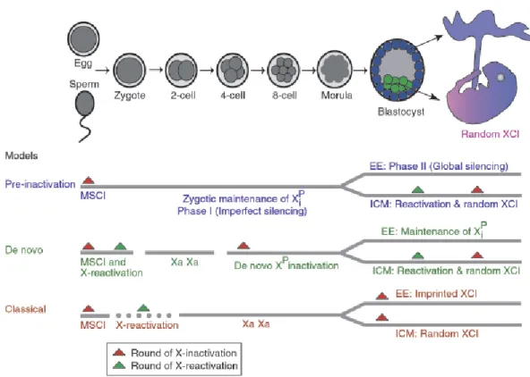
The exact timing of XCI is not known. There are three proposed models for XCI. These models are as shown in Figure 1.3.
The classical model supports that the X chromosomes are transmitted from gametes to embryo fully active and XCI occurs during blastocyst stage coupled with differentiation. This model doesn`t explain how embryo can tolerate twofold imbalance of X-linked gene expression. Paternal X in the sperm is inactived by a process of sex chromosome body in other words XY body inactivation. So another hypothesis called the preinactivation hypothesis defends that inactive X of the sperm is transmitted to the embryo and and more global silencing occurs in the trophoblast while reactivation and random inactivation occur in the epiblast (Huhyn, 2001). It is supported by the finding that in the early cleavage steps of embryogenesis, there is some expression from both Xs and the expression from the paternal X is increasing (Huhyn, 2003; Mak., 2004). The de novo inactivation hypothesis states zygote inherits active X chromosomes and inactivation starts at four-eight cell stage. In the epiblast, reactivation occurs following random XCI. This is based on the observation that two-cell stage embryos have some features of active X (Huhyn, 2004; Migeon 2007).
Imprinted XCI is known to take place in marsupial cells (Cooper, 1971; Namekawa 2007) and placental cells of some eutherians (Takagi, 1975). So it seems likely that XCI evolved as an imprinted phenomenon, which was later modified in eutherians (Graves, 1996).
Figure 1.3 Models in relation to the XCI timing in the embryo a) pre-inactivation
b) de novo inactivation c) classical model (Huhyn, 2004)
1.2.6. Master control region of XCI and its components
Russell was the first to hypothesize that there is a region on X chromosome from which XCI initiates and spreads bidirectionally (Russell, 1963). In both mice (Russell, 1965) and humans (Therman, 1974, 1979), the master control region of XCI was identified observing X-autosome translocations. The X-inactivation center (XIC) was first mapped to band Xq13 in humans and D region of the mouse X chromosome. The region was further defined by transfecting parts of the putative region into mouse embryonic stem cells to see if they could function (Lee, 1999; Heard, 1999: Lee, 1996; Migeon, 1999). XIC is poor in protein-coding genes and it includes many repeats (Ross, 2005). Several genes involved in XCI were identified in XIC (Figure 1.4).
The most crucial gene within is Xist in mouse and XIST in humans. It is larger than 17 kb and it codes for a regulatory RNA transcript. It was identified in many eutherians including mice and humans (Brown, 1992; Brockdorff, 1992).
However it was not identified in monotremes or marsupials (Duret, 2006). The sequence homology among Xist from different species is less than 50% (Chureau, 2002). However genomic organization, promoter region, transcription start sites, tandem repeat and most intron-exon boundries are conserved among species. When male mouse embryonic stem cells were transfected with a YAC transgene containing XIST, it induced random inactivation in male transgenic mice derived from these cells. (Migeon, 1999) This noncoding RNA is expressed from both male and female Xs partially before XCI. The significance of this low expression is not known (Ray, 1997). It continues to be fully expressed after XCI, so it is hypothesized that it can have a function in maintanance of XCI. Its different isoforms exist, which may have a role in regulation (Ma, 2005). Its deletions do not reactivate the inactivated X since there are other mechanisms of maintanance (Brown, 1994). Induced Xist deletions in mouse, spontaneous human XIST mutations such as seen in the cases of people with ring chromosomes and XIC transgenes have shown that role of Xist is XCI.
Another gene within XIC is Tsix which is antisense to Xist. Mouse Tsix is a 40-kb RNA originating 15 kb downstream of Xist and transcribed across the Xist locus (Lee, 1999). Deleting a 65-kb region downstream of Xist results in constitutive Xist expression and X inactivation. Before the onset of X inactivation, Tsix is expressed from both X chromosomes. At the onset of X inactivation, Tsix expression becomes monoallelic, is associated with the future active X and persists until Xist is turned off. Tsix is not found on the inactive X once cells enter the X inactivation pathway. Human TSIX produces a >30-kb transcript that is expressed only in cells of fetal origin. Differences in the structure of human and murine genes indicate that human TSIX was truncated during evolution. Human TSIX can not repress XIST and is coexpressed with it through embryonic development (Migeon, 2001). It was implicated that Tsix and X-inactivation intergenic transcription element (Xite) mutations effect choice and counting (Lee,2005; Migeon, 2002).
A cis element called Xite was found and shown to downregulate Tsix at the onset of XCI. This was suggested as a candidate for X chromosome
controlling element (Xce) (Ogowa, 2003). Before the discovery of Xist, the best candidate for XCI was Xce because it effects the randomness of XCI (Johnston, 1981; Simmler, 1993).
Figure 1.4 Elements of Xic The Xic center that is determined by deletion and
trangene studies. Green parts demonstrate genes that give rise to non-coding transcripts. Grey parts show protein-coding genes with unknown function in relation to XCI. Black parts indicate parts involved in counting and choice determined by targeted deletions. Mapped Xce implicated in choice is also shown (Heard et al, 2004).
1.2.7. Steps of XCI
Steps of XCI include counting, choice, inactivation, spreading and maintenance. In all mammalian cells, all X chromosomes are inactivated but one remains active. The mechanism of XCI seems to depend on determining an active X and repressing all the other X chromosomes by a default pathway. Silencing is chromosome wide. Cis inactivation is mediated by a noncoding RNA molecule transcribed from Xist in XIC. The molecule spreads along the chromosome from which it is expressed and transcriptionally silences it by changing the underlying chromosome.
Counting is the essential first step in general dosage compensation mechanims. A transient interaction is seen between X chromosomes at their Xic at the onset of inactivation in mouse embryonic fibroblasts (Xu, 2006; Bacher, 2006). Whether this transient interaction is a chromosomal cross-talk in relation to XCI and relate to counting is not known (Carrel, 2006). Recently, an X chromosome pairing region was found >100kb upstream of Xist (Augui, 2007).
Choice is the second step. It is hypothesized that inactivation is the default pathway and XIC on the future active X should be repressed somehow.
Xist has the features of a housekeeping gene and silencing X chromosomes
individually is a more complex program than silencing Xist-expressing chromosomes since there is only one active X in each diploid cell. Based on this view, XIC should receive repressory signals for the Xist gene on the future active X. This regulation can be done through DNA elements in the vicinity of Xist acting as enhancers or binding sites for trans-acting factors. The idea of a blocking factor that protects the active X from inactivation was proposed (Lyon 1972). The support for the existence of an autosomal Xist repressor comes from observations that triploid males with 69,XXY chromosomes can have two active Xs in contrast to Klinefelter males (Migeon, 2008).
Another important step in XCI is the spreading of inactivation by modifying chromatin bidirectionally. Spreading and silencing should be distinguished and achieved by different domains of Xist (Wutz, 2002). The specific binding of Xist depends on conserved tandem repeat sequences within its first exon (Beletskii, 2001). These sequences can form stem loop structures which are recognized by binding factors. Silencing occurs only at a critical window of development. This was suggested to depend on the different processing of Xist RNA during development (Ma, 2005).
The observation that inactivation spreading is better in X chromosomes than autosomes have raised the possibility that there are ``way stations`` that enhance spreading (Riggs, 1985; Lyon, 1998). Line 1 (L1) elements were suggested by Mary Lyon as candidates. Studies of X-autosome translocations supported the hypothesis showing the extend of silencing into the autosomes varies. The maximum spreading to autosome observed is 45 Mb from the translocation break point. The spreading can be either continuous or discontinuous so the ability of autosomal parts to maintain the inactive state can be different (Sharp, 2002).
Silencing of X requires its heterochromatinization. After Xist accumulation, transient enrichment of Polycomb-group (PcG) protein complex
around the future inactive X occurs. Than all histone modifications specific to active chromatin are lost. Hypoacetylation occurs, especially globally for H4. During spreading of Xist, histones H3 and H4 undergo lysine modificatons specific to inactive X. Lysine of H2B is ubiquitinated before H3 methylation takes place. The signatures specific to active X are acetylation of H3K9 and di-tri methylation of H3K4. The signatures specific to inactive X are methylation of H3K9 and di-methylation of H3K4. Variants of histone H2 accumulates and is also ubiquitinated. A PcG protein accumulates around future inactive X and have a methylase activity for H3K9 and H3K27 methylation. BRCA1 was shown to localize around inactive X. It has a ubiquitin ligase acitivity, so it is possible that it has a role in H2B ubiquitination (Heard, 2004).
The last event in the silencing is CpG island methylation, which is thought to play a role in the maintanance of XCI. Methylated CpGs bind many methyl binding proteins which bind transcriptional silencing complexes such as methyl CpG binding protein 2 (MeCP2) (Lyon, 2007; Plath, 2002).
1.2.8. Genes escaping XCI
All the genes on the inactive X chromosome are not subject to full inactivation. 15% of the genes escape inactivation based on expression patterns of 95% of X assayable genes in fibroblast systems (Carrel, 2005). These escapees are variable among species. With the exception of pseudoautosomal region, most genes expressed from the inactive X have less than 15% activity. It has been hypothesized that these sequences are skipped by the spreading of inactivation or failure of maintanance (Flippova, 2005 ; Lingenfelter, 1998).
1.2.9.Primary and secondary causes of skewed XCI
The choice of the active X is not dependent on parental origin of the chromosome or its gene content. Exceptions include marsupials and nonhuman placental tissues in which exclusively paternal XCI takes place which is called imprinted XCI. In normal females, approximately half of the cells inactivate their paternal X while the others choose it as active. When the ratio of the two cell populations deviates significantly from a 1:1 distribution, it is called skewed XCI. The causes of skewed XCI is classified depending on whether skewing occurs primary or secondary to the inactivation event. Any alteration in the germline
XIC can hypothethetically cause bias in the initial choice of which X chromosome
will be inactivated in the germline and these causes of skewing are called primary causes (Puck&Willard, 1998). Secondary causes include lethal X-linked mutations, X-autosome translocations, aging, twinning, or monoclonal expansion of cells. In the presence of a deleterious mutation on one of the X chromosomes, the cells in which this X remains active dies. In X-autosome translocations, the cells in which the translocated X is inactivated dies. Skewing was observed to be increased in twins raising several explanations. One of them is twining reduces the number of cells contributing to the embryo increasing the chance of skewing. Skewing can occur as a result of monoclonal expansion of cells with selection for cells that carry the mutant X chromosome (Belmont, 1996; Brown, 2000).
1.3.Self tolerance and Autoimmunity
1.3.1 Immune system
1.3.1.1 Clonal theory of Jerne and Burnett
According to Burnet and Jerne`s theory of clonal selection theory of antibody formation, in a certain point of development, clones of lymphocytes are produced in central lymphoid organs as a result of heritable changes such as a somatic mutation. Lymphocytes have receptors similar to antibodies in structure so antigens stimulate lymphocytes; the corresponding clones of lymphocytes are stabilized in secondary lymphocytic tissue and increase in number. There is a developmental process in the primary lymphoid tissue when any clones of lymphocytes which carry reactive sites corresponding to body determinants are eliminated (Jerne, 1955 ; Burnet, 1957). This theory is important not only for the clonal selection it proposes, but also for the discovery of immunological tolerance.
1.3.1.2 Self tolerance
When antigen binding to T cell receptors (TCR) and B cell receptor (BCR) occur, they turn on cell growth and survival pathways leading to clonal lymphocyte proliferation and immunity. These pathways include nuclear factor kappa B (NF-қB) activation; Myc activation and phosphoinositide 3 phosphate (PI3K) activation of AKT and Ras activation of ERK. The growth induced by antigens is blocked by several mechanisms conferring self-tolerance (Goodnow, 2008). These mechanisms are designated in Figure 1.5.
Mechanisms of self-tolerance can be listed as 1) Autoreactive cells can be deleted with apoptosis. 2) Autoreactive cells can gain a different receptor by receptor editing. 3) The cell can be intrinsically made functionally unresponsive,
which is termed anergy. 4) The cell is extrinsically shut off by limiting the supply of co-stimuli, cytokines etc. The cell can also be suppressed by regulatory T cells. Tolerance mechanisms that take place in generative lymphoid organs when thymocytes are still immature are called central tolerance. The tolerance mechanisms in peripheral lymphoid organs are called peripheral tolerance (Goodnow, 2005).
Figure 1.5 Different mechanisms of self tolerance a) T cell deletion through BIM
induced or FAS activated apoptosis b) Receptor editing by V(D)J recombination or by BCR hypermutation c) Receptor downregulation, role of inhibitory receptors on T cells; role of phosphatases in increasing the threshold of B cell-activation and ubiquitin ligases to tag TCR-CD28 or cytokine receptors to interfere with signalling d) B cell require B cell survival factor (BAFF); self-reactive receptors on T cells require survival factor, IL-7. Costimulation with Toll like receptors (TLRs) for B cells; costimulation through CD28 for T cells; costimulation of B cells with CD40L on T cells are needed for activation (Goodnow et al, 2005).
1.3.2 Autoimmunity
Autoimmune diseases are a diverse group of diseases including more than 80 conditions with an estimated prevalence of 5% to 8% in the United States, effection 14-22 million people. The common feature of autoimmune diseases (AIDs) is that the patients have a defect which leads to the attack of the host`s immune system to self-tissues and organs. There are two subgroups of
autoimmune diseases, which are systemic and organ specific. Autoimmunity is thought to be a complex disease resulting from several genetic sequence variants and environmental triggers including infections and stochastic events. Familial clustering and twin concordance studies have shown that AIDs have a genetic basis. Cooccurence of AIDs in individuals and the occurence of different AIDs in families support the view that AIDs have common predisposing genetic elements and pathogenic pathways (Eaton, 2007; Fox, 2007).
1.3.2.1 Genes associated with autoimmunity
Studies with sib-pairs, isolated populations and comparing patients and controls have shown that there are genetic risk factors for autoimmune diseases (Feltkamp, 1999). In simple diseases, the disease state is determined by a single gene. Common diseases result from a combination of susceptibility alleles. Genome-wide associations, linkage studies and candidate gene based approaches were performed and several MHC and non-MHC genes were found to contribute to predisposition to autoimmunity (Maier, 2008; Welcome Trust Case Control Consortium & Autralo-Angolo-American Spondylitis Consortium, 2007). Mutations in a single gene can cause autoimmunity, but most autoimmune diseases are associated with several sequence variants. Below, there is brief information on some simple genetic traits associated with human autoimmunity (Rioux&Abbas 2005).
Autoimmune regulator (AIRE) was identified as a gene mutated in systemic autoimmune disease, autoimmune polyendocrine syndrome (APS-1). Patients with this syndrome show autoimmunity against endocrine organs, skin and other tissues. AIRE is a transcription factor and normally it is expressed at high levels in several central and peripheral organs and blood cells. Studies with knock out mouse showed that AIRE gene plays a role in the thymic expression of organ specific antigens. This supports a role for AIRE in central tolerance in the thymus.
Cytotoxic T lymphocyte antigen 4 (CTLA-4) is an inhibitory receptor on T cells, which binds to B7-1 and B7-2 on APCs to prevent their binding with CD28 on T cells. This prevents the costimulation necessary for activation of T cells and promotes anergy. CTLA4 knockout mouse develops features of systemic autoimmunity including enlargement of lymphoid organs and organ lympocytic infiltrates. CTLA4 polymorphism was associated with Graves disease, type 1 diabetes (T1D), autoimmune hypothyroidism and other endocrinopathies.
Forkhead box P 3 (FOXP3) encodes a transcription factor of the forkhead family and its expression commits naive T cells to become regulatory T cells that aid in tolerance induction. Mouse studies have shown FOXP3 mutations cause lack of regulatory T cells and lead to systemic autoimmune disease. FOXP3 is also associated with immune dysregulation, polyendocrinopathy, enteropathy and X-linked inheritance (IPEX) in humans. FOXP3 is located within Xp11.23. FOXP3 polymorphisms are associated with autoimmune tyroid disease susceptibility (Ban, 2007).
Protein tyrosine phosphatase, non-receptor type 22 (PTPN22) encodes a lymphoid tyrosine phosphatase which aids in T cell signalling and activation. A polymorphism in PTPN22 was identified in type 1 diabetis and also associated with rheumatoid arthritis and systemic lupus erythematosus.
1.3.2.1.1 X-linked genes associated with immune system
Observation of several immune related pathologies including immunodeficiences and immunoproliferative disease associated with the X chromosome is critical in understanding the role of X chromosome genes in the immune system. These diseases include XLP syndrome (X-linked lymphoproliferative syndrome), X-linked recessive combined immunodeficiency syndrome (SCID), Hyper-IgM syndrome type I (HIGM 1), Wiskott-Aldrich syndrome (WAS) which affect T cells and X-linked agammaglobulinemia (XLA) and X-linked recessive anhydortic ectodermal dysplasia with immunodeficiency (XEDA-ID) which effect B cells (Murchinick, 2006).
As above mentioned, FOXP3, implicated in immune dysregulation syndrome characterized by dysfunction of regulatory T cells, is X-linked. A number of non-MHC loci found to be associated with AID susceptibility also maps to X chromosome (Becker, 1998). Further, there are reports on X chromosome abnormalities implicated in autoimmune diseases (Chagnon, 2006).
1.3.2.2 Molecular Mimicry
It`s observed that there is an association of infectious agents with autoimmune diseases. One of the mechanisms proposed to explain the role of infectious agents in autoimmune etiology is molecular mimicry.
Molecular mimicry is the cross-reactivity of immune reagents with host self antigens and microbial determinants because of shared structures. It was first observed during monoclonal antibody generation that the phosphoprotein of measles virus and a protein of HerpesSimplex virus type 1 (HSV-1) cross-reacted with an human intermediate filament protein (Fujinami,1983). 5% of 800 monoclonal antibodies made against 15 viruses cross-reacted with host self determinants (Bahmanyar, 1987; Oldstone, 1987). By observing symptoms after injecting Hepatitis B virus (HBV) proteins that share sequence similarity with myelin basic protein which is known to cause allergic encephalomyelitis, in vivo
evidence of molecular mimicry was obtained. In this way molecular mimicry was proposed to have a role in autoimmune pathogenesis (Fujinami, 1985). Several diseases were implicated to have a molecular mimicry in their etiology (Oldstone, 1998). Molecular mimicry is demonstrated in animal models but has not been clearly shown in humans (Fairweather, 2004).
There are other mechanisms explaining the role of viruses in the loss of self-tolerance. One of them is epitope spreading, meaning autoimmune response is targeted to a secondary self-antigen after tissue-damage. Another one is the activation of bystander T cells in an antigen-non-specific manner. This enhances the effects of epitope spreading and molecular mimicry. As well as viruses, drugs are also implicated in causing autoimmunity with the drug metabolism forming a neo-self (Christen, 2004; Bach, 2005). Another proposed theory is that microorganisms expose self-antigens to immune system by damaging tissues (Fairweather, 2004).
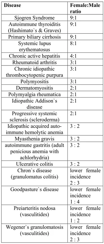
1.3.2.3. Female predominance in autoimmunity
Most of the rheumatic diseases with autoimmune features exhibit higher female incidence. In Table 1.6, AIDs and corresponding female:male ratios are listed. In Figure 1.6, the incidences of selected of AIDs in females and males is designated. The reasons why AID occurs predominantly in women remains to be eluciaded.