UHOD
The Impact of Tumor and Lymph Node
Characteristics on Disease Free Survival
in Squamous Cell Vulvar Cancer
Derya A. CIRIK1, Alper KARALOK1, Isin UREYEN1, Osman TURKMEN1, Tolga TASCI1,
M. Ali NARIN1, Mehmet F. KOSE2, Nurettin BORAN1, Taner TURAN1, Gokhan TULUNAY1
1 Etlik Zübeyde Hanım Women’s Health Training and Research Hospital, Department of Gynecologic Oncology, Ankara
2 Medipol University School of Medicine, Department of Obstetrics and Gynecology, Istanbul, TURKEY
ABSTRACT
We aimed to determine the prognostic significance of tumor and lymph node (LN) characteristics on disease free survival (DFS) in patients who underwent surgery for squamous cell vulvar carcinoma. A total of 94 patients who underwent vulvar surgery and groin dissection were included. The impact of clinicopathologic variables such as age, body mass index, tumor size, tumor depth, total and metastatic lymph node number on DFS were assessed. The estimates of survival were determined with Kaplan-Meier and log rank analysis. In the univariate analysis; age, body mass index, tumor size, tumor location, total LN number, metastatic LN number and adjuvant therapy did not have impact on DFS. The median number of removed LNs was 21. Although removal of higher number of lymph nodes did not improved the DFS, patients who had ≤ 3 metastatic LNs had better 2-year DFS rate compared to those with >3 metastatic LNs (71.8% vs 40.0%; p= 0.042, respectively). In the multivariate analysis, both the depth of tumor invasion and LN involve-ment were the independent predictors of DFS. Additionally, in stage III disease, patients receiving adjuvant therapy had significantly less locoregional recurrence compared to those who did not receive. The presence of LN metastasis and increased tumor depth (> 3 mm) are poor prognostic factors for DFS in squamous cell vulvar cancer. Although the number of lymph nodes removed was not correlated with DFS, patients who had ≤ 3 metastatic LNs had better DFS compared to those with > 3 LNs.
Keywords: Vulvar cancer, lymph node, disease free survival, recurrence
ÖZET
Skuamöz Hücreli Vulva Kanserinde Tümör ve Lenf Nodu Özelliklerinin Hastalıksız Sağkalıma Etkisi
Bu çalışmada, skuamöz hücreli vulva kanseri için cerrahi yapılan hastalarda tümör ve lenf nodu (LN) özelliklerinin hastalıksız sağkalıma (DFS) etkisini incelemek amaçlandı. Vulvar cerrahi ve lenf nodu diseksiyonu yapılan toplam 94 hasta çalışmaya dahil edildi. Yaş, vücut kitle indeksi, tümör boyutu, tümör derinliği, total ve metastatik LN sayısı gibi klinikopatolojik değişkenlerin DFS’ye etkisi araştırıldı. Sağkalım analizleri ise Kaplan-Meier ve log rank analizi ile yapıldı. Univaryant analizine göre, yaş, vücut kitle indeksi, tümör boyutu ve yeri, toplam LN sayısı, metastatik LN sayısı ve adjuvan tedavi DFS üzerine etkili bulunmadı. Ortalama olarak 21 tane LN çıkarıldı. Or-talamadan fazla lenf nodu çıkarılması DFS üzerine etkili olmasa da, 3 ve daha az metastatik LN çıkarılan hastalar, 3’den fazla metasta-tik LN çıkarılanlardan daha iyi 2-yıllık-DFS’ye sahip bulundu (sırasıyla, %71.8 ve %40.0; p= 0.042). Multivaryant analizde, hem tümör derinliği hem de LN tutulumu DFS’nin bağımsız prediktörleri idi. Ek olarak, evre III hastalıkta, adjuvan tedavi alan hastalar almayanlara göre daha az lokal (perineal ve kasık) nüksüne sahip idi. Lenf nodu metastazı ve artmış tümör derinliği (> 3 mm), skuamöz hücreli vulva kanseri için kötü prognostic faktörlerdir. Çıkarılan LN sayısı ile 2-yıllık-DFS korele olmasa da evre III hastalarda, 3 ve daha az metastatik LN çıkarılan hastalarda 3’den fazla LN çıkarılanlara göre daha iyi DFS izlenmektedir.
UHOD
Number: 1 Volume: 26 Year: 2016
INTRODUCTION
Squamous cell vulvar carcinoma (vSCC) is the most common histologic type of vulvar cancer and surgery is the essential step for both staging and treatment. Although new methods for lymph node dissection were introduced, the widely used surgical treatment includes tumor resection with free margins and inguinofemoral lymph node dis-section.1-3 Since two decades, involvement of groin
lymph nodes (LNs) has been reported as the most important prognostic factor for survival.4 The age,
tumor size, tumor location, stage, depth of inva-sion, the LN status and free margins were suggested as the possible predictive factors of recurrence.5-8
However, these risk factors were determined rely-ing on the data generally based on small studies for vSCC. Nearly half of recurrences occur within first two years and when diagnosed, patients with locoregional recurrences have usually the chance of surgical treatment and good prognosis.9,10
There-fore, the clarification of the surgico-pathological factors that facilitate the recurrence were crutial for the patients with vSCC.
In the present study, we assessed the prognostic significance of surgico-pathological variables and LN profile on DFS in patients who underwent sur-gery for vSCC.
PATIENTS AND METHODS
A total of 136 patients with vSCC treated at the Gynecologic Oncology Department of our institu-tion between December 1991 and August 2013. Patients with depth of invasion < 1 mm (n= 7), with advanced stage disease treated with primary chemo- or radiotherapy (RT) (n= 22), who admit-ted with recurrence (n= 1), treaadmit-ted without surgery due to the advanced interstitial lung disease (n= 1) and with non-squamous histology (n= 11) were excluded. Remaining 94 patients who underwent surgery for vSCC were eligible for further analy-sis. This study confirmed the principles outlined in Helsinki Declaration.
After local ethical committee approval, the clini-cal data and pathologic data were collected from the computerized database of the department. A recurrence was defined as the new appearance of a tumor after surgery and at least a 3 months disease
free period. DFS was defined as the time from the date of the surgery to the date of recurrence or last follow-up. Recurrence was diagnosed by clinical examination, biopsy and radiologic imaging. Ac-cording to our protocols; vulvar tumor less than 2 cm and less than 1 mm invasion was resected with at least 1cm tumor free margin via wide lo-cal excision, other tumors were usually resected by radical vulvectomy. All patients scheduled to have bilateral inguinofemoral lymphadenectomy. However one patient had only unilateral lymphad-enectomy due to an orthopedic disease of hip joint and limitation of abduction. Groin dissections were performed with the triple separate incision method. Adjuvant perineal RT was performed when tumor free margin was less than 8 mm and tumor depth >5 mm or when tumor spread into the urethra and anal region. Adjuvant treatment of groins and pelvis was performed via pelvic RT with/without chemotherapy when at least one LN was tumor positive or when tumor size exceeded 4 cm length. Radiotherapy field included the inguinofemoral lymph nodes and at least the lower pelvic nodes. Over 5 weeks, 45 Gy in 25 fractions were given. If there were multiple lymph nodes, up to 60 Gy was given to a reduced volume.
None of the patients took adjuvant chemotherapy. However, adjuvant chemoradiotherapy was given to patients with high risk factors (high number of LN, bigger tumor size, etc) according to the doc-tors’ preferences and experience.
Because most of the recurrence occur in vulva and groin, the diagnosis were usually made with clini-cal examination, biopsy and radiologic imaging when required. When the recurrence was diag-nosed the lower and upper abdominal CT and lung X-ray were performed. Especially in recent years, we also routinely do PET-CT for evaluation. Variables were analyzed by using SPSS software (version 15.0 for Windows, Chicago, IL, USA). For descriptive analyses, categorical variables were defined as numbers and percentages, and nu-meric variables were defined as mean or median. Chi-square and student t-test were used as appro-priate to test the differences between two groups. The estimates of survival were determined using the Kaplan Meier analysis. The survival analysis of categorical variables were examined by log rank
UHOD
Number: 1 Volume: 26 Year: 2016
test and the continuous variables were examined by univariant cox propotional hazard model. The statistical power of the variables were defined by using binary logistic regression and variables with a p value of less than 0.05 analyzed in the multi-variate analysis.
RESULTS
Clinical and Pathological Data
Median age and body mass index (BMI) of the patients were 64.5 years (range: 37-87 years) and 28.4 kg/m2 (range: 18-48 kg/m2), respectively.
De-tails of patients’ characteristics were listed in Table 1. All tumors were primarily confined to the vulva and the median tumor size was 20 mm (range: 2–80 mm) in diameter and median tumor invasion was 3 mm (range: 1-25 mm) in depth. Seventy-nine pa-tients (84%) had radical vulvectomy, 10 (10.6%) had wide local excision and 5 (5.3%) had hemi-vulvectomy. Except for one who had unilateral groin dissection, all had bilateral groin dissection, Of all, 43 (45.7%) had LN metastasis; 10 had uni-lateral and 33 had biuni-lateral LN metastasis in groin. The median number of resected LNs was 21. The number of LNs was ≤ 10 in 8 patients (8.5%) and ≤15 in 19 patients (20.2%). All patients underwent lymphadenectomy and patients were divided into two categories according to the International Fed-eration of Gynecology and Obstetrics (FIGO) stag-ing system. The first group included 51 patients who had no LN metastasis (Stage I and II), and the second group included 43 patients who had LN metastasis (Stage III). There was no patient with stage IV disease.
Table 1. Clinical and pathological characteristics of patients
with vulvar cancer
Parameters n (%) Tumor size [mm] < 10 10 (10.6%) 10-29 48 (51.1%) ≥ 30 36 (38.3%) Tumor location Midline 45 (47.9 %) Right side 18 (19.1%) Left side 31 (33.0 %) Depth of invasion [mm] ≤3 38 (40.4%) > 3 36 (38.3%) Not reported 20 (21.3) Adjuvant therapy No therapy 51 (54.3%) Radiotherapy 34 (36.2%) Radiotherapy + Chemotherapy 9 (9.6%)
Table 2. The sites of first recurrence by stage, localization of tumor and the status of receiving adjuvant therapy
Parameter Sites of recurrence
Perineal, n (%) Inguinal, n (%) Distant, n (%)
Lymph node metastasis (FIGO Stage)
Negative (Stage I-II) 8 (61.5) 4 (30.8) 1 (7.7)
Positive (Stage III) 11 (57.8) 6 (37.5) 2 (10.5)
P 0.307 Localization of tumor Midline 8 (50) 7 (43.7) 1 (6.3) Lateral 11 (68.7) 3 (18.7) 2 (12.6) P 0.829 Adjuvant radiotherapy Not received 12 (60) 7 (35.0) 1 (5) Received 7 (58.3) 3 (25) 2 (16.7) P 0.281
UHOD
Number: 1 Volume: 26 Year: 2016
Patterns of First Recurrence
The median follow-up was 24 months (range: 3-158 months). The estimated 5-year and 10-year DFS were 41% and 28%, respectively. During the follow-up, 32 (34%) patients had the first recur-rence after a median interval of 23.5 months (range: 6-120 months). The distribution of relapses by lo-cation was perineal recurrence in 19 (59.4%) pa-tients, groin recurrence in 10 (31.2%), and distant organ recurrence in 3 (9.4%) patients. Although to-tal recurrence rate was higher in patients with Stage III disease than in those with Stage I&II disease, the difference did not reach statistical significance (44.2% vs 25.5%, p= 0.08, respectively). The sites of recurrences according to the stage were shown
in Table 2. The sites of recurrence were also simi-lar in both groups of patients with LN positivity (Stage III) and negativity (Stage I&II) (p= 0.307). Adjuvant Therapy and Locoregional Recurrence Although LN positivity is an indication for adju-vant therapy in our clinical protocols, 15 (34.9%) of 43 patients with stage III disease did not receive adjuvant therapy after primary surgery due to poor performance status or patients’ preference. Of the patients with Stage III disease, 11 had perineal and 6 had groin recurrences. In stage III disease, pa-tients receiving adjuvant therapy had significantly less locoregional recurrences (perineal and groin) compared to those who did not receive (n= 7/29; 24.1% vs n= 10/15; 66.7%, p= 0.006). Only 2 of Table 3. The factors related to disease free survival in univariate analysis
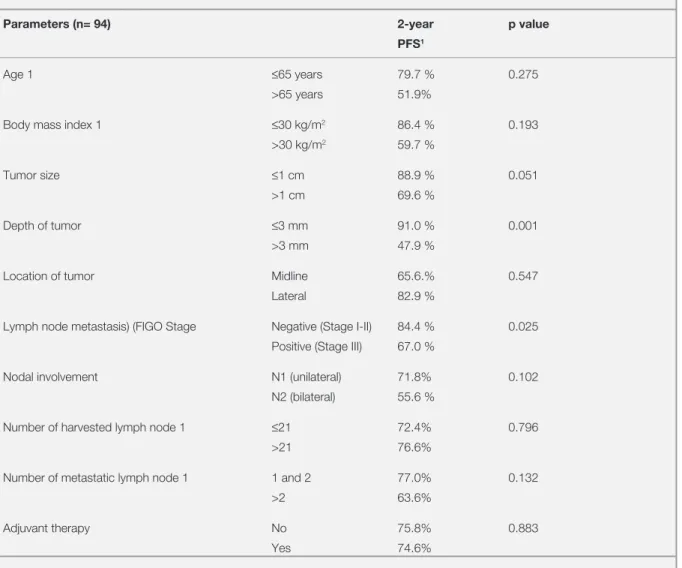
Parameters (n= 94) 2-year p value
PFS1
Age 1 ≤65 years 79.7 % 0.275
>65 years 51.9%
Body mass index 1 ≤30 kg/m2 86.4 % 0.193
>30 kg/m2 59.7 %
Tumor size ≤1 cm 88.9 % 0.051
>1 cm 69.6 %
Depth of tumor ≤3 mm 91.0 % 0.001
>3 mm 47.9 %
Location of tumor Midline 65.6.% 0.547
Lateral 82.9 %
Lymph node metastasis) (FIGO Stage Negative (Stage I-II) 84.4 % 0.025
Positive (Stage III) 67.0 %
Nodal involvement N1 (unilateral) 71.8% 0.102
N2 (bilateral) 55.6 %
Number of harvested lymph node 1 ≤21 72.4% 0.796
>21 76.6%
Number of metastatic lymph node 1 1 and 2 77.0% 0.132
>2 63.6%
Adjuvant therapy No 75.8% 0.883
Yes 74.6% PFS: Disease free survival, 1: Median value
UHOD
Number: 1 Volume: 26 Year: 2016
these 6 patients with groin recurrence received ad-juvant RT and 5 of 11 patients with locoregional recurrences received adjuvant therapy (4 RT and 1 had concomitant chemo-RT after primary surgery). Of the 51 patients with stage I-II disease, 15 re-ceived radiation therapy to perineum. In stage I-II disease, patients receiving adjuvant therapy had similar locoregional recurrence rates compared to those who did not receive (n= 3/14; 21.4% vs n= 9/36; 24%, p= 1.0). A total of 4 groin and 8 per-ineal recurrences developed in patients with stage I-II disease. Of the 4 patients with groin metastasis, only 1 had stage II disease with urethral involve-ment and had received vulvar RT. Of the 8 patients with perineal recurrences, only 2 had received vul-var RT after surgery.
Clinicopathologic Factors and DFS
In the univariate analysis, depth of tumor invasion and LN status were found to be associated with DFS in vSCC (Table 3). The tumor size was also tending
to be associated with DFS. On the other hand, the number of removed LNs was not correlated with improvement in DFS. The 2-year DFS was 72.4% for patients whose ≤ 21 LNs were removed and 76.6% for those whose >21 LNs were removed (p= 0.796). Median number of metastatic LN in Stage III disease was 2 in the current study. Patients who had ≤ 2 metastatic LNs had similar 2-year DFS rate compared to those with >2 metastatic LNs (71.4% vs 63.6%; p= 0.38, respectively). On the other hand, when the patients were grouped according to the number of metastatic lymph nodes; patients who had ≤ 3 metastatic lymph nodes had better 2-year DFS compared to those who had > 3 meta-static lymph nodes (71.8 % vs 40.0 %; p= 0.042). In both groups of patients who received and did not receive adjuvant therapy, 2-year DFS was also similar for patients with ≤ 2 metastatic LNs and with more metastatic LNs (79.3% vs 66.7%; p= 0.289 and 75.4% vs 66.7%; p= 0.301, respective-ly). Two-year DFS was 55.6% for patients with Tablo 4. Multivariate analysis of selected clinicopathological variables regarding disease free survival
Parameter p value HR 95% CI
Tumor size (≤ 10 mm vs >10 mm) 0.093 2.928 0.835 - 10.267
Depth of tumor (≤ 3 mm vs > 3mm) 0.016 3.022 1.227 - 7.442
FIGO stage (stage 1 and 2 vs stage 3) 0.049 2.243 1.003 - 5.019
Body Mass Index (≤ 30 kg/m2 vs > 30 kg/m2) 0.056 0.401 0.157 - 1.025
Figure 1. Disease free survival according to the depth of tumor
invasion
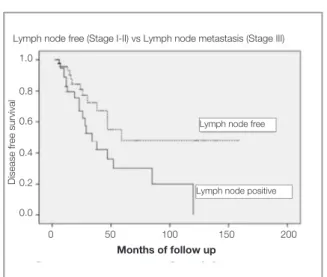
Figure 1. Disease free survival according to the lymph node
status
Months of follow up Months of follow up
Lymph node free (Stage I-II) vs Lymph node metastasis (Stage III)
Disease free survival
Depth of tumoral invasion, ≤ 3 mm vs > 3 mm
Disease free survival
0 50 100 150 200 0 50 100 150 200 1.0 0.8 0.6 0.4 0.2 0.0 1.0 0.8 0.6 0.4 0.2 0.0 ≤ 3 mm > 3 mm
Lymph node free
UHOD
Number: 1 Volume: 26 Year: 2016
bilateral positive LNs while patients with unilat-eral positive LNs had a DFS of 71.8% (p= 0.102). Among 32 patient who had recurrence, 10 were died due to their disease during follow-up. Two-year overall survival was estimated as 91.4 % for the entire cohort.
In the multivariate analysis, depth of tumor inva-sion and LN involvement (stage I-II vs III) were found as the independent prognostic factors for DFS (Figure 1 and 2). Patients with stage III dis-ease were 2.24 times more prone for developing recurrence than those with stage I and II disease. Patients with > 3 mm depth of invasion had 3.022 times more risk for regarding DFS compared to those who had less invasion (Table 4).
DISCUSSION
In the current study, a relatively large cohort (n= 94) who underwent lymphadenectomy for vSCC were analyzed. All patients underwent primary surgical treatment with tumor resection and lym-phadenectomy. The estimated 5-year and 10-year DFS were 41% and 28%, respectively and the re-currence rate was 34% (n: 32/94). In accordance to our results, Maggino et al. reported 37.2% recur-rence within 502 patients in their study, the larg-est series in the literature.10 Iacoponi et al. reported
higher recurrence rate (43.6%) in a study of 87 pa-tients with a median age of 73 years and median tumor size of 35.4 mm.7 Higher incidence of
recur-rence might be attributed to the presence of older patients and bigger tumors in that study. On the other hand, Woelber et al. reported the recurrence rate as only 13.6% in a study of 109 patients 56% of whom had tumor less ≤ 2 cm in greatest diam-eter.5 The lower incidence of recurrence might be
explained by the inclusion of the stage 1A disease and the presence of higher percentage of T1 tumor (≤ 2 cm in greatest diameter, confined to the vulva) in these patients. In the current study, the perineal recurrence was the most common recurrence and seen in 20.2% of patients. This rate was also com-parable with the rates (18.8% to 26.2%) in the oth-er soth-eries.5,6 The incidence of groin recurrence was
10.6% in the current study and similar to the rate of 10.1% groin recurrence in a study of 158 patients who were treated with bilateral lymphadenectomy by Baiocchi et al.11
There are studies investigating the predictive fac-tors for DFS or overall survival for vSCC, but they often had heterogeneous group of patients with non-squamous cell cancer or patients having dif-ferent treatment modalities.7-12,13-15 The results of
this study also verified that LN involvement was clearly confirmed as being the most important prognostic factor for both DFS. In the multivari-ate analysis, LN involvement was the independ-ent prognostic factor regarding DFS in the currindepend-ent study. Woelber et al. stated that patients with uni-lateral and biuni-lateral metastasis were estimated to have 5.1 and 16.9 times more recurrence risk com-pared to those with negative LNs, respectively.5
Similarly, Iacoponi et al. also reported that bilateral LN metastasis was also tend to be associated with higher locoregional recurrences rate compared to unilateral metastasis.7
Up to date, the number of optimal lymph node number that should be removed is not clarified yet. Few authors investigated the prognostic sig-nificance of the number of removed LNs in vulvar cancer and conflicting results were reported.10,12-16
In a large population database trial of 1030 pa-tients, Courtney-Brooks et al. reported higher dis-ease specific survival with removal of ≥ 10 LN in patients with LN negative vulvar cancer.14
Similar-ly, a recent trial also reported an improvement on DFS with removal of ≥15 nodes at bilateral lym-phadenectomy.16
Contrary to this, Baiocchi et al. did not find any survival benefit by removing more LNs (≥12) in patients with Stage I&II disease.11 However, they
also analyzed the patients with Stage III disease and found significantly higher PFS in patients who had removal of higher number LN; 2-year PFS for patients with removal of <12 LN and ≥ 12 LNs were 20.8% vs 52.8%, respectively (p= 0.003). In that study, only 7.6% of 158 patients received ad-juvant therapy. On the other hand, median number of harvested LNs was 21 in the current study and removing higher LN counts also did not improved the DFS in the entire cohort. In our study 45.7% of patients had received adjuvant therapy. This re-sult could be explained by the more effective local control of tumor with the use of more frequent ad-juvant therapy in the current study.
UHOD
Number: 1 Volume: 26 Year: 2016
In a recent study of Vismanathan et al., LN involve-ment and positive margins (< 1 cm with the edge of tumor) but not depth of invasion were found as the clinical predictors of recurrence.17 That study
spe-cifically evaluated the relation between adjuvant RT and perineal recurrence and found significantly lower risk of perineal recurrence with the use of RT. On the other hand, patients with > 3 mm depth of invasion were estimated to have 3.022 times more shorter DFS compared to those who had less invasion in the current study.
Locoregional recurrences usually occur by di-rect tumoral spread or lymphatic embolization to regional LNs and they were attributed to the de-fects either in the surgical treatment or in the local control by the adjuvant therapy. In a recent study, prognostic factors for developing locoregional re-currences were investigated in 78 patients and only receiving adjuvant therapy was found to reduce the perineal and groin recurrence significantly.7
Al-though, distinct criteria for indication of adjuvant therapy following primary tumor resection are still under debate, survival advantage of adjuvant RT on patients with two or more metastatic LNs was confirmed in the literature.18 On the other hand, the
status of patients who had one intracapsular LN in-volvement is controversial. In a recent study, Fons et al. could not show a benefit of adjuvant therapy in patients with a single metastatic LN regarding the DFS.19 However, the negative effect of single
LN involvement on DFS was also shown by other researchers.13,20 According to our clinical protocols,
one metastatic LN was sufficient for the indication of adjuvant therapy. All patients with positive LNs were candidates for adjuvant therapy in the current study. The patients with stage III disease who re-ceived adjuvant therapy had significantly less per-ineal and groin recurrence compared to those who did not receive (p= 0.006).
In the present study, metastatic LN number was not related with DFS in the entire cohort. Patients with ≤ 2 metastatic LN had similar DFS to those who had > 2 metastatic LNs.
However, patients who had ≤ 3 metastatic LNs had better DFS compared to those who had > 3 meta-static nodes. Our results were consistent with a re-cent trial of Panici et al16 that stated that patients
who had > 3 metastatic LN had worse OS and DFS
compared to those who have lesser LNs. In the present study, the impact of metastatic LN on the DFS was analyzed in patients receiving and not re-ceiving adjuvant therapy after subgroup analysis. In both groups of patients, number of metastatic LNs had no association with DFS (p= 0.289 and p= 0.301, respectively). In a study of 157 patients, Woelber et al. investigated the prognostic role of LN metastasis on recurrence. Although the num-ber of metastatic LNs was not also shown to have impact on DFS in patients who received adjuvant therapy, metastatic LN number had significant im-pact on prognosis in patients who did not receive adjuvant therapy in that study.20
Strengths of this study include the relatively high number of patients with a rare disease diagnosed in a single center. Limitations were the retrospective nature of the study and the inclusion of patients di-agnosed within 2 decades.
In conclusion, LN metastasis and tumor with >3 mm depth of invasion were independent prog-nostic factors for DFS in patients with vSCC. Although LN metastasis is very crucial for recur-rence, removing more LNs did not improve DFS. Although there is debate on the indications of ad-juvant therapy for vSCC, receiving adad-juvant RT in even one positive LN may decrease the risk of the recurrence.
REFERENCES
1. Siegel R, Ward E, Brawley O, Jemal A. Cancer statistics, 2011: The impact of eliminating socioeconomic and racial disparities on premature cancer deaths. CA Cancer J Clin 61: 212-236, 2011.
2. Hacker NF, Berek JS, Lagasse LD, et al. Individualization of treatment for stage I squamous cell vulvar carcinoma. Obstet Gynecol 63:155-162, 1984.
3. Wu Q, Zhao YB, Sun ZH, et al. Clinical application of endo-scopic inguinal lymph node resection after lipolysis and lipo-suction forvulvar cancer. Asian Pac J Cancer Prev 14: 7121-7126, 2013.
4. Homesley HD, Bundy BN, Sedlis A, et al. Assessment of cur-rent International Federation of Gynecology and Obstetrics staging of vulvar carcinoma relative to factors for survival (a GOG group). Am J Obstet Gynecol 164: 997-1003, 1991. 5. Woelber L, Manher S, Voelker K, et al. Clinicopathological
prognostic factors and patterns of recurrence in vulvar cancer. Anticancer Research 29: 545-552, 2009.
UHOD
Number: 1 Volume: 26 Year: 2016
6. Martinez-Palones JM, Perez-Benavente MA, Gil-Moreno A, et al. Comparison of recurrence after vulvectomy and lymphad-enectomy with and without sentinel node biopsy in early stage vulvar cancer. Gynecol Oncol 103: 865-870, 2006.
7. Iacoponi S, Zapardiel I, Diestro MD, et al. Prognostic factors associated with local recurrence in squamous cell carcinoma of the vulva. J Gynecol Oncol 24: 242-248, 2013.
8. Stankevica J, Macuks R, Baidekalna I, Donina S. Midline in-volvement as a risk factor for vulvar cancer recurrence. Asian Pac J Cancer Prev 13: 5237-5240, 2012.
9. Onnis A, Marchetti M, Maggino T. Carcinoma of the vulva: Criti-cal analysis of survival and treatment of recurrences. Eur J Gy-neacol Oncol 13: 480-485, 1992.
10. Maggino T, Landoni F, Sartori E, et al. Patterns of recurrence in patients with squamous cell cancer of the vulva. A multicenter CTF Study. Cancer 89: 116-122, 2000.
11. Baiocchi G, Cestari FM, Rocha RM, et al. Does the count af-ter inguinofemoral lymphadenectomy in vulvar cancer correlate with outcome? Eur J Surg Oncol 39: 339-343, 2013. 12. Lataifeh I1, Nascimento MC, Nicklin JL, et al. Patterns of
re-currence and disease-free survival in advanced squamous cell carcinoma of the vulva. Gynecol Oncol 95: 701-705, 2004. 13. Oonk MH, de Hullu JA, van der Zee AG. Current controversies
in the management of patients with early-stage vulvar cancer. Curr Opin Oncol 22: 481-486, 2010.
14. Courtney-Brooks M, Sukumvanich P, Beriwal S, et al. Does the number of nodes removed impact survival in vulvar cancer patients with node-negative disease? Gynecol Oncol 117: 308-311, 2010.
15. Le T, Elsugi R, Hopkins L, et al. The definition of optimal ingui-nal femoral nodal dissection in the management of vulva squa-mous cell carcinoma. Ann Surg Oncol 14: 2128-2132, 2007.
16. Panici PB, Tomao F, Domenici L, et al. Prognostic role of ingui-nal lymphadenectomy in vulvar squamous carcinoma: younger and older patients should be equally treated. A prospective study and literature review. Gynecol Oncol 137: 373-379, 2015.
17. Viswanathan AN, Pinto AP, Schultz D, et al. Relationship of margin status and radiation dose to recurrence in post-opera-tive vulvar carcinoma. Gynecol Oncol 130: 545-549, 2013. 18. Mahner S1, Trillsch F, Kock L, et al. Adjuvant therapy in
node-positive vulvar cancer. Expert Rev Anticancer Ther 13: 839-844, 2013.
19. Fons G, Groenen SMA, Oonk MHM, et al. Adjuvant radiothera-py in patients with vulvar cancer and one intra capsular LN me-tastasis is not beneficial. Gynecol Oncol 114: 343-345, 2009. 20. Woelber L, Eulenburg C, Choschzick M, et al. Prognostic role
of LN metastases in vulvar cancer and implications for adjuvant treatment. Int J Gynecol Cancer 22: 503-508, 2012.
Correspondence
Dr. Derya Akdag CİRİK Yeni Etlik Caddesi No: 55 06010 Kecioren/ANKARA Tel: (+90-312) 567 40 00 Fax: (+90-312) 323 81 91 e-mail: [email protected]