2059
http://journals.tubitak.gov.tr/medical/ © TÜBİTAK
doi:10.3906/sag-2002-14
Antioxidative effects of uridine in a neonatal rat model of hyperoxic brain injury
Nevin AL1,*, Ayşen ÇAKIR2, Cansu KOÇ3, Mehmet CANSEV3, Tülin ALKAN2
1Department of Nursing, Istanbul Medipol University, Institute of Health Sciences, Pediatric Nursing Doctorate Program, Istanbul, Turkey 2Department of Physiology, Bursa Uludağ University Faculty of Medicine, Bursa, Turkey
3Department of Pharmacology, Bursa Uludağ University Faculty of Medicine, Bursa, Turkey
* Correspondence: [email protected] 1. Introduction
Premature birth is a major problem all over the world and preterm infants have increased the risk of mortality and morbidity despite recent advances in neonatal medicine [1]. Preterm infants are vulnerable to several complications including respiratory distress syndrome, bronchopulmonary dysplasia, intestinal injury, compromised immune system, and cardiovascular disorders as well as hearing/vision and neurological problems [2].
Most preterm infants are exposed to supraphysiological oxygen therapy during perinatal period in the neonatal intensive care unit (NICU). Supraphysiological oxygen therapy in the sensitive period of brain development affects the developmental processes as a result of hyperoxia [3,4]. Preterm neonates are especially susceptible to
deterioration caused by reactive oxygen species (ROS) due to the fact that endogenous radical scavenging systems are not fully mature [3]. Although ROS is a function of signal molecules at low concentrations [5,6], its overproduction causes oxidative stress due to the imbalance between oxidative and antioxidative systems [7]. As a result, reactive oxygen radicals are formed, which contribute to oxidative changes in proteins, lipids, and nucleic acids [8], and negatively impact the survival of neuronal cells during development [4,9,10]. In the clinical setting, up to 50% of surviving preterm infants have been reported to exhibit cognitive deficits or behavioral problems during the later stages of development [11].
The level and activity of the most-relevant antioxidant enzymes, such as superoxide dismutase, catalase (CAT), and glutathione peroxidase (GPX) change dynamically
Background/aim: Premature birth is a major problem that results in an increased risk of mortality and morbidity. The management of
such infants consists of supraphysiological oxygen therapy, which affects brain development due, in part, to the deterioration caused by reactive oxygen species (ROS). We showed previously that exogenously administered uridine provides neuroprotection in a neonatal rat model of hyperoxic brain injury. Hence, the aim of the present study was to investigate the effects of uridine on ROS in the same setting.
Materials and methods: Hyperoxic brain injury was induced by subjecting a total of 53 six-day-old rat pups to 80% oxygen (the
hyperoxia group) for a period of 48 h. The pups in the normoxia group continued breathing room air (21% oxygen). Normoxia + saline or hyperoxia + saline or hyperoxia + uridine 100 mg/kg or hyperoxia + uridine 300 mg/kg or hyperoxia + uridine 500 mg/kg was injected intraperitoneally (i. p.) 15 min prior to the hyperoxia procedure. The pups were decapitated and the brains were homogenized to analyze superoxide dismutase (SOD), glutathione peroxidase (GSH-Px), myeloperoxidase (MPO), and malondialdehyde (MDA) enzymes as well as DJ-1 (protein deglycase DJ-1) — an oxidative stress-sensitive protein.
Results: Hyperoxia-induced may cause overproduction of oxygen radicals and the oxidant/antioxidant balance may be disturbed in the
brain. Brain MPO and MDA levels were significantly increased in saline-receiving pups exposed to hyperoxia. Brain SOD and GSH-Px levels were significantly decreased in saline-receiving pups exposed to hyperoxia. Our results showed that uridine administration prevented the hyperoxia-induced decrease in SOD and GSH-Px while counteracting the hyperoxia-induced increase in MPO and MDA in a dose-dependent manner. Uridine also increased the DJ-1 levels in brains of rat pups subjected to hyperoxia.
Conclusion: These data suggest that uridine exhibits antioxidative properties which may mediate the protective effects of uridine in a
neonatal rat model of hyperoxic brain injury.
Key words: Uridine, hyperoxia, antioxidative, neonatal rat, DJ-1
Received: 03.02.2020 Accepted/Published Online: 31.05.2020 Final Version: 17.12.2020
during development and mature stage in the last weeks of gestation, preparing the fetus for lung respiration [12– 14]. They are essential for preserving cells from exposure to oxidative damage [15]. On the other hand, the brain levels of oxidative stress markers myeloperoxidase and malondialdehyde increased significantly after exposure to hyperoxia in animals and humans [16,17]. MPO is a highly potent oxidative enzyme that is capable of inducing both oxidative and nitrosative stress in vivo [18] and MDA is a lipid peroxidation product that is one of the most frequently used markers of oxidative stress [19]. Hence, treatments that raise the levels of antioxidants and lower those of oxidative stress markers have been associated with neuroprotective effects [20].
Uridine is the principal circulating pyrimidine nucleoside in human circulation [21,22], a constituent of breast milk [23], and a precursor of brain membrane phospholipids via the Kennedy pathway [24]. We showed previously that exogenously administered uridine provides neuroprotection in neonatal rat models of hypoxic-ischemic encephalopathy (HIE) [25–27] and hyperoxic brain injury [28]. The neuroprotection by uridine has been shown to be mediated by its antiapoptotic [25] and epigenetic effects (i.e., reducing histone deacetylase activity) [27] in the neonatal HIE model.
Hence, for the first time, the present study aimed to investigate the mechanism by which uridine exhibited neuroprotection in the neonatal rat model of hyperoxic brain injury [28] with regard to hyperoxia-induced oxidative stress. For this purpose, the study analyzed the levels of most relevant oxidative/antioxidative enzymes and DJ-1 protein (protein deglycase DJ-1, also known as Parkinson disease protein 7 [PARK7]) [29], an oxidative stress-sensitive protein which is known to maintain mitochondrial functions by scavenging reactive oxygen species (ROS) [29].
2. Materials and methods
2.1. Animals and experimental design
A total of 7 dated pregnant Sprague-Dawley rats were housed in individual cages at 22 °C with 12h light/dark cycle with free access to food and water. Dams (mothers of newborn rats) delivered 6 to 13 pups and the day of birth was considered postnatal day 1 (P1). A total of 53 pups were included in the study regardless of gender and their weights were checked daily. The pups were pooled in wood shavings, divided randomly, and delivered back to nursing dams.
2.2. Induction of hyperoxic brain injury
The 6-day-old pups were randomly divided into 5 groups as follows: normoxia + saline (n = 10), hyperoxia + saline (n = 11), hyperoxia + uridine 100mg/kg (n = 13), hyperoxia + uridine 300mg/kg (n = 9), and hyperoxia + uridine 500mg/ kg (n = 10).
From P6 to P8 (48 h), the pups in the normoxia group were subjected to room air (21% oxygen) while those in the hyperoxia groups were subjected to 80% continuous oxygen, as has been described previously [9,17–20,29–31]. The preference for studying P6 pups depended on a precise calculation obtained at the web site [32], which showed that brain growth and neurogenesis in developing rat brain on postconceptional (PC) 27 days correspond to those in human brain on PC 204 days (29.1 weeks).
Continuous oxygen was maintained in a plexiglas chamber with 60 × 20 × 20 cm (width × depth × height) dimensions by a flow-through system. Saline (0.9% NaCl; 0.1 ml/10 g body weight) or uridine (100 mg/kg, 300 mg/ kg, or 500 mg/kg; dissolved in saline) was administered intraperitoneally (i. p.) once 15 min before the onset of atmospheric air or oxygen exposure. Doses of uridine administered in this study were selected from our previous studies reporting neuroprotective effects in neonatal rat models of hypoxic-ischemic encephalopathy [25–27] and hyperoxic brain injury [28]. The oxygen level inside the chamber was monitored continuously with a MiniOX 3000 oxygen analyzer (Ohio Medical Corporation, Gurnee, IL, USA) to maintain 80% oxygen saturation. Nursing dams were switched every 24 h to prevent respiratory complications. On completion of 48-hour hyperoxic insult (at P8), the pups were decapitated under deep anesthesia. Then, the brains were removed and chilled in liquid nitrogen immediately and analyzed for biochemical parameters and DJ-1 protein by the Western Blotting [33].
2.3. Biochemical analyses
The brain tissues were homogenized in ice-cold phosphate-buffered saline (PBS, pH 7.4). The homogenates were utilized to analyze the levels of GSH-Px, SOD, MDA, and MPO using commercially available ELISA kits (Sunredbio Technology Co. Ltd., Shanghai, China) according to the kit procedure.
DJ-1 protein analyses:
Homogenates were boiled in Laemmli buffer [34] and loaded onto Sodium Dodecyl Sulfate Polyacrylamide Gel Electrophoresis (SDS-PAGE; Mini Protean II, Bio-Rad, Hercules, CA, USA) at equal protein content analyzed by the Lowry method [35]. Each sample was run electrophoretically and protein bands were transferred to polyvinylidene fluoride (PVDF) membranes (Millipore, Billerica, MA, USA). The membranes were blocked with 5% nonfat dry milk dissolved in tris buffered saline (TBST) and incubated overnight with the primary antibody, DJ-1 (1:1000, Cell Signaling Technology, Danvers, MA, USA). On the following day, the membranes were incubated with HRP (horse radish peroxidase) bound rabbit anti IgG secondary antibody (1:5000, Cell Signaling Technology, Danvers, MA, USA) for 1 h and then visualized by incubation with enhanced chemiluminescence solution
(Millipore, Billerica, MA, USA). The protein bands were scanned by Licor CDigit scanner (LI- COR Biotechnology, Lincoln, NE, USA), and the density of the bands was compared using the software of Licor CDigit system. Following this procedure, the membranes were cleared with stripping buffer (Thermo Fisher Scientific, Rockford IL, USA) and then incubated with mouse anti-β-III-tubulin antibody (Millipore, Temecula, CA, USA) used as a house-keeping protein and the procedure was repeated with the appropriate secondary antibody.
2.4. Statistical analysis
Statistical analyses were performed using the Sigma Plot 12.0 software. The data were expressed as mean ± standard error of means (SEM). The Shapiro-Wilk test was used for determining the normality of the data. When the data showed a normal distribution and passed the normality test, the difference between groups was determined by the parametric One-Way ANOVA followed by the post hoc Tukey test. On the other hand, when the data failed the normality test, the difference between the groups was determined by the nonparametric Kruskal-Wallis H test followed by the post hoc Dunn’s test with Bonferroni correction. P < 0.05 was considered statistically significant.
3. Results
3.1. Biochemical analyses
Compared with the normoxia + saline group, the brain MDA levels of the pups in the hyperoxia + saline group were significantly increased (P < 0.001) (Figure 1A, Table 1). Uridine treatment at 300 mg/kg and 500 mg/kg in the pups exposed to hyperoxia reduced brain MDA levels significantly (P < 0.01 and P < 0.001, respectively), while uridine at 100 mg/kg did not.
Similarly, the brain MPO levels were increased in saline-receiving pups exposed to hyperoxia significantly (P < 0.001) (Figure 1B, Table 1) and compared with the normoxic group. The enhanced brain MPO levels were reduced significantly in hyperoxic pups treated with uridine at 300 mg/kg and 500 mg/kg (P < 0.0 and P < 0.001, respectively) doses, while no change was observed with 100 mg/kg uridine.
On the contrary, the brain SOD levels of the pups in the hyperoxia + saline group were significantly decreased (P < 0.001) (Figure 1C, Table 1) compared to those in the normoxia + saline group. Treatment with uridine at 300 mg/kg and 500 mg/kg in the pups exposed to hyperoxia increased brain SOD levels significantly (P < 0.01 and P < 0.001, respectively), but uridine at 100 mg/kg was not effective.
In good accord with data derived in SOD analyses, the levels of GSH-Px in the brains of pups in the hyperoxia + saline group were significantly decreased (P < 0.001) (Figure 1D, Table 1) compared to those in the normoxia
+ saline group, and uridine increased the brain GSH-Px levels at 300 mg/kg and 500 mg/kg in the pups exposed to hyperoxia significantly (P < 0.05 and P < 0.01, respectively). Uridine administered at 100 mg/kg dose was again not effective.
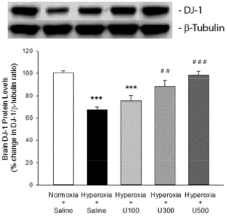
3.2. Levels of DJ-1 protein
The brain DJ-1 protein levels (expressed as % change in DJ-1/ β-tubulin ratio) of pups in the hyperoxia + saline group were significantly (P < 0.001) reduced by about 33% compared to those in the normoxia + saline group (Figure 2, Table 2). Uridine treatment at 300 mg/kg and 500 mg/kg in the pups exposed to hyperoxia prevented the decrease in the brain DJ-1 protein levels significantly (P < 0.01 and P < 0.001, respectively), but not at 100 mg/kg dose (Figure 2, Table 2).
4. Discussion
These data show that uridine exhibits antioxidative properties in a neonatal rat model of hyperoxic brain damage suggesting one mechanism by which uridine provides neuroprotection in such model [28]. We showed, for the first time, that a single dose of uridine injected to P6 pups prevented the hyperoxia-induced decreases in SOD and GSH-Px levels as well as the hyperoxia-induced increases in MDA and MPO levels, in a dose-dependent manner. In addition, uridine treatment also enhanced the decreased levels of DJ-1, an oxidative stress-sensitive protein.
Preterm birth is a major health problem for neonates and their caregivers due to the lack of maturity of organs and systems, including the brain. Survival rates of preterm neonates have been increasing with efforts in modern neonatal intensive care units (NICUs) while the same is not true for morbidity rates [36,37]. In addition to the complications observed due to prematurity itself, the fact that most premature infants receive supraphysiological oxygen therapy for conditions like cardiac resuscitation and respiratory distress during their NICU stay [38] contributes to the morbidities associated especially with tissues like the intestines [39], lungs [40], retina [41,42], and the brain [3,10,43].
The fact that extrauterine environment (PaO2 100 mm Hg) is richer in oxygen compared to the intrauterine environment (PaO2 20–25 mm Hg) renders birth an oxidative challenge for the newborn. Hence, the fetal to neonatal transition exposes the newborn to an oxygen-rich world [3], creating a considerably high risk for oxidative stress at birth. This risk is additionally exacerbated by the low efficiency of natural antioxidant systems in preterm newborns [44,45] and the supraphysiological oxygen therapy that they receive.
Pathological hallmarks of supraphysiological oxygen treatment in the brain include enhanced inflammation,
Figure 1. Brain levels of (A) MDA, (B) MPO, (C) SOD, and (D) GSH-Px in neonatal rats subjected to hyperoxic brain damage.
Hyperoxia+Saline group, *P < 0.05, **P < 0.01 and ***P < 0.001 compared to Normoxia+Saline group. Normoxia+Saline group, #P < 0.05, ##P < 0.01 and ###P < 0.001 compared to Hyperoxia+Saline group.
Table 1. Brain levels of MDA, MPO, SOD, and GSH-Px in neonatal rats subjected to hyperoxic brain damage.
Normoxia + Saline Hyperoxia + Saline Hyperoxia + U100 Hyperoxia + U300 Hyperoxia + U500
MDA (nmol/g protein) 4.35 ± 0.1 5.75 ± 0.2*** 5.45 ± 0.2*** 4.85 ± 0.1## 4.47 ± 0.1###
MPO (ng/g protein) 3.19 ± 0.1 4.83 ± 0.1*** 4.57 ± 0.2*** 4.04 ± 0.1**# 3.70 ± 0.1###
SOD (ng/g protein) 7.51 ± 0.2 5.54 ± 0.3*** 6.30 ± 0.2 7.09 ± 0.3## 7.39 ± 0.3###
GSH-Px (ng/g protein) 17.66 ± 0.6 13.23 ± 0.5*** 14.67 ± 0.7* 16.28 ± 0.5# 17.06 ± 0.7##
Hyperoxia+Saline group, *P < 0.05, **P < 0.01 and ***P < 0.001 compared to Normoxia+Saline group. Normoxia+Saline group, #P < 0.05, ##P < 0.01 and ###P < 0.001 compared to Hyperoxia+Saline group.
oxidative stress, and matrix metalloproteinase activity accompanied by autophagy, increased apoptotic cell death, and reduced neuro-glial development in the brain [17– 20,31,46,47]. Hyperoxic insult to the brain is associated with increased levels of IL-1 b and IL-18 mRNA, oxidized glutathione (GSSG), MDA [17,47], thiobarbituric acid reactive substances, and hydrogen peroxide [20]. In addition, hyperoxia changes the balance of the reactive oxygen species (ROS)-dependent thioredoxin/ peroxiredoxin system and reduces the level of DJ-1, a hydroperoxide-responsive protein [29].
Various neuroprotective strategies have been investigated experimentally to reduce hyperoxic brain damage in preterm neonates including caffeine [20],
erythropoietin [17,47,48], dexmedetomidine [31], topiramate [49], acetylcholinesterase (AChE) inhibitors [50], and, recently, mesenchymal stem cells [51] and fingolimod [52].
Uridine is the principal circulating pyrimidine nucleoside in humans [21,22] and a precursor of membrane phospholipids [24].
We showed previously that exogenously administered uridine provides neuroprotection and improves long-term cognitive deficits [26] by reducing apoptotic cell death [25] and inhibiting histone deacetylase activity [27] in a rat model of HIE. In addition, our previous findings showed, in the hyperoxia model, that uridine administration for 5 consecutive days during continuous hyperoxic insult
Table 2. Levels of DJ-1 protein in brains of neonatal rats subjected to hyperoxic damage.
Normoxia + Saline Hyperoxia + Saline Hyperoxia + U100 Hyperoxia + U300 Hyperoxia + U500
% Change in DJ-1/β-tubulin ratio 100 ± 2 67.2 ± 2.6*** 75.4 ± 4.8*** 88.2 ± 5.5## 98.7 ± 3.2###
Hyperoxia+Saline group, ***P < 0.001 compared to Normoxia+Saline group. Normoxia+Saline group, ##P < 0.01 and ###P < 0.001 compared to Hyperoxia+Saline group.
Figure 2. Levels of DJ-1 protein in brains of neonatal rats subjected to hyperoxic
damage. Hyperoxia+Saline group, ***P < 0.001 compared to Normoxia+Saline group. Normoxia+Saline group, ##P < 0.01 and ###P < 0.001 compared to Hyperoxia+Saline group.
reduced apoptosis in pups’ brains and protected against long-term cognitive deficits [28]. Although uridine has been shown to exhibit antiinflammatory effects in models of pulmonary diseases [53,54] or colitis [55], the effect of uridine on oxidative stress has not yet been studied.
Hence, the aim of the present study was to investigate whether uridine prevents oxidative injury as a mechanism of action in the benefit it provides in neonatal hyperoxic brain damage. We showed, for the first time, that uridine exhibits antioxidative properties by preventing the hyperoxia-induced decreases in SOD and GSH-Px levels and the increases in MDA and MPO levels, as well as by enhancing the decreased levels of an oxidative stress-sensitive protein, DJ-1. These data provide the first evidence that the neuroprotective effect of uridine in a neonatal rat model of hyperoxic brain injury depends, at least in part, on uridine’s action on oxidative stress parameters.
Our data on SOD, GSH-Px, MDA, and MPO are in good accord with previous studies reporting that antioxidative strategies provide neuroprotection in the hyperoxia-induced brain injury in neonatal animals [17–20,31,47]. In addition, we found, in accordance with previous studies [29], that the levels of DJ-1, an oxidative stress-sensitive protein, were reduced following supraphysiological oxygen treatment. DJ-1 functions as a redox-sensitive chaperone
to protect tissues against oxidative stress and cell death [56–59]. Hence, in cases where DJ-1 levels are decreased, oxidative stress is triggered, which in turn contributes to cell death, as shown in a neuronal cell line [60], or in the immature brain following hyperoxic insult [29]. Therefore, treatments that raise the expression of DJ-1 protein may prevent oxidative stress and provide benefit in terms of neuroprotection. In the present study, we showed that uridine treatment significantly restored DJ-1 levels in a dose-dependent manner and this effect may comprise one mechanism by which uridine prevents brain cell death in the hyperoxic setting [28].
In conclusion, our data show, for the first time, that uridine prevents oxidative damage by preventing the hyperoxia-induced decreases in SOD and GSH-Px levels and the increases in MDA and MPO levels, as well as by enhancing the decreased levels of DJ-1 protein. These data provide a deeper mechanistic insight into the neuroprotective effect of uridine in neonatal hyperoxic brain injury.
Acknowledgments
This study was supported by the funds from Bursa Uludağ University Scientific Research Projects Council (ID: HDP(T)-2017/24). The authors declare no conflicts of interest with regard to the present study.
References
1. Manuck TA, Rice MM, Bailit JL, Grobman WA, Reddy UM et al. Preterm neonatal morbidity and mortality by gestational age: a contemporary cohort. American Journal of Obstetrics & Gynecology 2016; 215 (1): 103-114.
2. Institute of Medicine (US) Committee on Understanding Premature Birth and Assuring Healthy Outcomes, Mortality and acute complications in preterm infants Behrman RE, Butler AS, 2007. Preterm Birth: Causes, Consequences, and Prevention. 2007 National Academies Press Washington 313-345.
3. Reich B, Hoeber D, Bendix I, Felderhoff-Mueser U. Hyperoxia and the immature brain. Developmental Neuroscience 2016; 38: 311-330.
4. Yis U, Kurul SH, Kumral A, Cilaker S, Tuğyan K et al. Hyperoxic exposure leads to cell death in the developing brain. Brain Development 2008; 30 (9): 556-562.
5. Irani K, Xia Y, Zweier JL, Sollott SJ, Der CJ et al. Mitogenic signaling mediated by oxidants in Ras- transformed fibroblasts. Science 1997; 275 (5306): 1649-1652.
6. Lander HM. An essential role for free radicals and derived species in signal transduction. The FASEB Journal 1997; 11: 118-124.
7. Finkel T, Holbrook NJ. Oxidants, oxidative stress and the biology of ageing. Nature 2000; 408: 239-247.
8. Fridovich I. Fundamental aspects of reactive oxygen species, or what’s the matter with oxygen? Annals of the New York Academy of Sciences 1999; 893: 13-18.
9. Bendix I, Schulze C, Haefen C, Gellhaus A, Endesfelder S et al. Erythropoietin modulates autophagy signaling in the developing rat brain in an in vivo model of oxygen-toxicity. International Journal of Molecular Sciences 2012; 13: 12939-12951.
10. Felderhoff-Mueser U, Bittigau P, Sifringer M, Jarosz B, Korobowicz E et al. Oxygen causes cell death in the developing brain. Neurobiology of Disease 2004; 17: 273-282.
11. Volpe JJ. Perinatal brain injury: from pathogenesis to neuroprotection. Mental Retardation and Developmental Disabilities Research Reviews 2001; 7: 56-64.
12. Frank L, Sosenko IRS. Prenatal development of lung antioxidant enzymes in four species. Journal of Pediatrics 1987; 110 (1): 106-110.
13. Friel JK, Friesen RW, Harding SV. Roberts LJ. Evidence of oxidative stress in full-term healthy infants. Pediatric Resource 2004; 56: 878-882.
14. Tiina MA, Kari OR, Mika S, Vuokko LK. Expression and development profile of antioxidant enzymes in human lung and liver. American Journal of Respiratory Cell and Molecular Biology 1998; 19: 942-949.
15. Zaghloul N, Nasim M, Patel H, Codipilly C, Marambaud P et. al. Overexpression of extracellular superoxide dismutase has a protective role against hyperoxia-induced brain injury in neonatal mice. FEBS Journal 2012; 279: 871-881.
16. Perrone S, Tataranno ML, Stazzoni G, Buonocore G. Biomarkers of oxidative stress in fetal and neonatal diseases. The Journal of Maternal-Fetal & Neonatal Medicine 2012; 25: 2575-2578. 17. Sifringer M, Brait D, Weichelt U, Zimmerman G, Endesfelder
S et al. Erythropoietin attenuates hyperoxia-induced oxidative stress in the developing rat brain. Brain, Behavior, and Immunity 2010; 24: 792-799.
18. Chisolm GM, 3rd Hazen SL, Fox PL, Cathcart MK. The oxidation of lipoproteins by monocytes-macrophages. Biochemical and biological mechanisms. Journal of Biological Chemistry 1999; 274: 25959-25962.
19. Gutteridge JMC. Lipid peroxidation and antioxidants as biomarkers of tissue damage. Clinical Chemistry 1995; 41 (12):1819-1828.
20. Endesfelder S, Makki H, von Haefen C, Spies CD, Buhrer C et al. Neuroprotective effects of dexmedetomidine against hyperoxia-induced injury in the developing rat brain. PLoS One 2017;12 (2): 1-20.
21. Cansev M. Uridine and cytidine in the brain: Their transport and utilization. Brain Resource 2006; 52 (2): 389-397.
22. Wurtman RJ, Regan M, Ulus IH, Yu L. Effect of oral CDP-choline on plasma CDP-choline and uridine levels in humans. Biochemical Pharmacology 2000; 60: 989-992.
23. Thorell L, Sjoberg LB, Hernell O. Nucleotides in human milk: sources and metabolism by the newborn infant. Pediatric Research 1996; 40 (6): 845-852.
24. Kennedy EM, Weiss SB. The function of cytidine coenzymes in the biosynthesis of phospholipids. Journal of Biological Chemistry. 1956; 222: 193-214.
25. Cansev M, Minbay Z, Goren B, Yaylagul EO, Cetinkaya M et al. Neuroprotective effects of uridine in a rat model of neonatal hypoxicischemic encephalopathy. Neuroscience Letters. 2013; 542: 65-70.
26. Goren B, Cakir A, Ocalan B, Kocoglu SS, Alkan T, Cansev M. Long-term cognitive effects of uridine treatment in a neonatal rat model of hypoxic- ischemic encephalopathy encephalopathy. Brain Resource 2017; 1659: 81-87.
27. Koyuncuoğlu T, Türkyılmaz M, Gören B, Cetinkaya M, Cansev M et al. Uridine protects against hypoxic-ischemic brain injury by reducing histone deacetylase activity in neonatal rats. Restorative Neurology and Neuroscience 2015; 33 (5): 777-784. 28. Goren B, Cakir A, Sevinc C, Kocoglu S, Ocalan B et al. Uridine treatment protects against neonatal brain damage and long-term cognitive deficits caused by hyperoxia. Brain Research 2017; 1676: 57-68.
29. Bendix I, Weichelt U, Strasser K, Serdar M, Endesfelder S et al. Hyperoxia changes the balance of the thioredoxin/ peroxiredoxin system in the neonatal rat brain. Brain Resource 2012; 1484: 68-75.
30. Endesfelder S, Zaak I, Weichelt U, Bührer C, Schmitz T. Caffeine protects neuronal cells against injury caused by hyperoxia in the immature brain. Free Radical Biology and Medicine 2014; 67: 221-234.
31. Sifringer M, von Haefen C, Krain M, Paeschke N, Bendix I et al. Neuroprotective effect of dexmedetomidine on hyperoxia-induced toxicity in the neonatal rat brain. Oxidative Medicine and Cellular Longevity 2015; 1-10.
32. Clancy B, Darlington RB, Finlay BL. Translating developmental time across mammalian species. Neuroscience 2001; 105 (1): 7-17.
33. Ritter J, Schmitz T, Chew LJ, Bührer C, Möbius W et al. Neonatal Hyperoxia Exposure Disrupts Axon–Oligodendrocyte Integrity in the Subcortical White Matter. Journal of Neuroscience 2013; 33 (21): 8990-9002.
34. Laemmli UK. Cleavage of structural proteins during the assembly of the head of bacteriophage T4. Nature 1970; 227: 680-689.
35. Lowry OH, Rosebrough NJ, Farr AL, Randall RJ. Protein measurement with the folinphenol reagents. Journal of Biological Chemistry 1951; 193: 265-275.
36. Marlow N, Wolke D, Bracewell MA, Samara M, Group ES. Neurologic and developmental disability at six years of age after extremely preterm birth. New England Journal of Medicine 2005; 352: 9-19.
37. Wood NS, Marlow N, Costeloe K, Gibson AT, Wilkinson AR. Neurologic and developmental disability after extremely preterm birth. EPICure Study Group. New England Journal of Medicine 2000; 343: 378-384.
38. Sola A, Golombek SG, Montes Bueno MT, Lemus-Varela L, Zuluaga C et al. Safe oxygen saturation targeting and monitoring in preterm infants: can we avoid hypoxia and hyperoxia? Acta Paediatrica 2014; 103 (10): 1009-1018. 39. Giannone PJ, Bauer JA, Schanbacher BL, Reber KM. Effects
of hyperoxia on postnatal intestinal development. Biotech Histochem 2007; 82 (1): 17-22.
40. Gien J, Kinsella JP. Pathogenesis and treatment of bronchopulmonary dysplasia. Current Opinion in Pediatrics 2011; 23: 305-313.
41. Perrone S, Vezzosi P, Longini M, Marzocchi B, Paffetti P et al. Biomarkers of oxidative stress in babies at high risk for retinopathy of prematurity. Frontiers in Bioscience 2009; 1: 547-552.
42. Saugstad OD. Oxygen and retinopathy of prematurity. Journal of Perinatology 2006; 26: 46-50.
43. Collins MP, Lorenz JM, Jetton JR, Paneth N. Hypocapnia and other ventilation-related risk factors for cerebral palsy in low birth weight infants. Pediatric Resource 2001; 50: 712-719. 44. Kumar VH, Patel A, Swartz DD, Wang H, Wynn KA et al.
Exposure to supplemental oxygen and its effects on oxidative stress and antioxidant enzyme activity in term newborn lambs. Pediatric Resource 2010; 67: 66-71.
45. Patel A, Lakshminrusimha S, Ryan RM, Swartz DD, Wang H et al. Exposure to supplemental oxygen downregulates antioxidant enzymes and increases pulmonary arterial contractility in premature lambs. Neonatology 2009; 96:182-192.
46. Brehmer F, Bendix I, Prager S, van de Looij Y, Reinboth, BS et al. Interaction of inflammation and hyperoxia in a rat model of neonatal white matter damage. PLoS One 2012; 7: 1-13. 47. Sifringer M, Genz K, Brait D, Brehmer F, Löber R et al.
Erythropoietin attenuates hyperoxia-induced cell death by modulation of inflammatory mediators and matrix metalloproteinases. Developmental Neuroscience 2009; 31: 394-402.
48. Yis U, Kurul SH, Kumral A, Tugyan K, Cilaker S et al. Effect of erythropoietin on oxygen-induced brain injury in the newborn rat. Neuroscience Letters 2008; 448: 245-249.
49. Kurul SH, Yis U, Kumral A, Tuğyan K, Cilaker S et al. Protective effects of topiramate against hyperoxic brain injury in the developing brain. Neuropediatrics 2009; 40: 22-27.
50. Sifringer M, Bendix I, von Haefen C, Endesfelder S, Kalb A et al. Oxygen toxicity is reduced by acetylcholinesterase inhibition in the developing rat brain. Developmental Neuroscience 2013; 35: 255-264.
51. Kim YE, Park WS, Sung DK, Ahn SY, Sung SI et al. Intratracheal transplantation of mesenchymal stem cells simultaneously attenuates both lung and brain injuries in hyperoxic newborn rats. Pediatric Research 2016; 80: 415-424.
52. Serdar M, Herz J, Kempe K, Lumpe K, Reinboth BS et al. Fingolimod protects against neonatal white matter damage and long-term cognitive deficits caused by hyperoxia. Brain, Behavior, and Immunity 2016; 52: 106-119.
53. Cicko S, Grimm M, Ayata K, Beckert J, Meyer A et al. Uridine supplementation exerts anti-inflammatory and anti-fibrotic effects in an animal model of pulmonary fibrosis. Respiratory Research 2015; 16: 105-115.
54. Evaldsson C, Ryden I, Uppugunduri S. Anti-inflammatory effects of exogenous uridine in an animal model of lung inflammation. International Immunopharmacology 2007; 7: 1025-1032.
55. Jeengar MK, Thummuri D, Magnusson M, Naidu VGM, Uppugunduri S. Uridine Ameliorates Dextran Sulfate Sodium (DSS)-Induced Colitis in Mice. Scıentıfıc Reports 2017; 7: 3924, 1-10.
56. Bonifati V, Rizzu P, van Baren MJ, Schaap O, Breedveld GJ et al. Mutations in the DJ-1 gene associated with autosomal recessive early-onset parkinsonism. Science 2003; 299: 256-259. 57. Jin J, Meredith GE, Chen L, Zhou Y, Xu J et al. Quantitative
proteomic analysis of mitochondrial proteins: relevance to Lewy body formation and Parkinson’s disease. Molecular Brain Research 2005; 134:119-138.
58. Lev N, Ickowicz D, Melamed E, Offen D. Oxidative insults induce DJ-1 upregulation and redistribution: implications for neuroprotection. Neurotoxicology 2008; 29: 397-405.
59. Lev N, Ickowicz D, Barhum Y, Lev S, Melamed E et al. DJ-1 protects against dopamine toxicity. Journal Neural Transmission. 2009; 116: 151-160.
60. Yokota T, Sugawara K, Ito K, Takahashi R, Ariga H et al. Down regulation of DJ-1 enhances cell death by oxidative stress, ER stress, and proteasome inhibition. Biochemical and Biophysical Research Communications 2003; 312: 1342-1348.