Original investigation
Limitations of Cross-Talk Between Osteosarcoma and Bone Marrow-Derived
Mesenchymal Stem Cells
Yasemin Basbinar
1, Tugba Uysal
1, Caner Karaca
2, Ezgi Daskin
1, Hanife Ecenur Meco
1, Ahu Pakdemirli
3,
Turkan Yigitbasi
4, Hulya Ellidokuz
51Dokuz Eylul University, Institute of Oncology, Translational Oncology Department, Izmir, Turkey 2Dokuz Eylul University, Institute of Health Sciences, Department of Oncology, Izmir, Turkey
3Dokuz Eylul University, Vocational School of Health Services, Department of First and Emergency Aid, Izmir, Turkey 4Istanbul Medipol University, Faculty of Medicine, Department of Biochemistry, Istanbul, Turkey
5Dokuz Eylul University Oncology Institute Department of Preventive Oncology, School of Medicine Department of Biostatistics and Medical Informatics, Izmir, Turkey.
Address for Correspondence: Yasemin Basbinar, E-mail: [email protected] Received: 12.04.2019201; Accepted: 27.04.2019; Available Online Date: 28.05.2019
©Copyright 2019 by Dokuz Eylül University, Institute of Health Sciences - Available online at www.jbachs.org
Cite this article as: Basbinar Y, Uysal T, Karaca C, Daskin E, Meco HE, Pakemirli A, Yigitbasi T, Ellidokuz H. Limitations of Cross-Talk Between Osteosarcoma and Bone Marrow-Derived
Mesenchymal Stem Cells. J Basic Clin Health Sci 2019; 3:83-88. https://doi.org/10.30621/jbachs.2019.611
Abstract
Objectives: Metastasis is a multi-step process which leads the tumor cells to escape from primer tumor region due to their need to gain malign phenotypes. While the effect of bone marrow-derived mesenchymal stem cells upon metastasis is not certain, some studies point out bone marrow-derived mesenchymal stem cells (BM-MSCs) to have this ability due to cell-cell interaction, released cytokines, and organization with the extracellular matrix in the micro-environment. Cross-talk via soluble factors also shifts the metastatic character.
Patients and Methods: In this study, the effects of mesenchymal stem cells on tumor behavior by creating different microenvironments in 3-dimensional (3D) in vitro cancer model is analyzed. The BM-MSCs and osteosarcoma cells were co-cultured via hanging-drop modeled 3D structure in normoxic and hypoxic conditions, and the cross-talk was modeled to measure their chemoattractant effects. The invasion and migration rates were measured with xCELLigence DP real-time cell analysis system. Mann Whitney U Test was used to compare independent samples. All P-values <0.05 were considered statistically significant.
Results: In this study, the most effective chemoattractant that increases the rate of migration in the osteosarcoma cell line under both normoxic (P 0.02) and hypoxic (P 0.004) conditions have been found to be the chemoattractant obtained from the BMSC culture.
Conclusion: Soluble factors secreted by BM-MSCs to micro-environment are highly effective chemoattractants for osteosarcoma, nevertheless the stem cells that have been co-cultured with the MG-63 decrease this behavior. These results could provide a new scientific approach to downregulate the metastasis induced by the effect of BM-MSCs.
Keywords: Osteosarcoma, MG-63, BM-MSCs, Metastasis, Cross-Talk
Osteosarcoma is the most common type of bone cancer and the second leading cause of cancer-related deaths in children and young adults (1, 2). Although the origin of the disease remains unclear, the most likely candidates are seen as mesenchymal stem cells (MSCs) or osteoprogenitor cells (3). MSCs are characterized by their ability to differentiate into mesenchymal cell types such as osteoblasts, chondrocytes, adipocytes, muscle cells, pericytes, reticular fibroblasts, and even neural cells. They also represent a heterogeneous population of cells consisting of multipotent and progenitor cells and provide micro-environmental regulation that controls the dormancy and proliferation of hematopoietic stem cells in bone marrow and migrate to damaged areas in many pathological conditions such as inflammation, tissue repair, and neoplasia (4, 5). Recent studies show that these cells take important roles in tumor behavior.
The tumor cells communicate with neighbouring and distant cells to attract allies and reprogram micro-environment to benefit tumor growth, invasion, and metastasis. This cross-talk mechanisms involved in the collection of MSCs in tumor site are very similar to the activation and migration processes of inflammatory cells (6). The tumor cells secrete soluble factors and pro-inflammatory proteins to attract MSCs to the region (7–9). Microvesicles and exosomes also take an important role to transfer materials between cells (10). Furthermore, this interaction is double-sided. While primer and metastatic tumor cells attract MSCs from bone marrow, the stem cells also attract them into themselves (11–13).
The cross-talk not only influence the attraction of cells but, also modulate the phenotype and functional characteristics, therefore
INTRODUCTION
tumor progression (14, 15). In this dynamic micro-environment, soluble factors and extracellular vesicles reprogram the MSCs. In turn, the stem cells secrete pro-tumorogenic molecules and bodies (16). As a result, the tumor gets more aggressive in character while MSCs also changes to cancer stem-cell like phenotype (17). Bone marrow-derived mesenchymal stem cells (BM-MSCs) are locally adjacent to tumor tissues. In addition, the cross-talk influences metastasis character of both cells. This study is aimed to investigate the effect of the cross-talk between Osteosarcoma (OS) and BM-MSCs in both hypoxic and normoxic conditions. Since this cross-talk double-sided, the results might suggest new approaches to limit metastasis to bone marrow and also prevent the migration.
MATERIALS AND METHODS
Cell CultureHuman Umbilical Vein Endothelial Cells (HUVEC) (ATCC®
CRL-1730™) was obtained from American Type Culture Collection
(Rockville, CT, USA) and cultured in Dulbecco’s Modified Eagle Medium low-glucose containing 10% fetal bovine serum (FBS,
Cegrogen Biotech GmbH, Stadtallendorf, Germany) and 1%
penicillin/streptomycin (Biochrom GbmH, Berlin, Germany). Cells were incubated in 5% CO2 incubator at 37°C in humidified air. Bone Marrow-Derived Mesenchymal Stem Cell Culture
Human BM-MSCs (ATCC® PCS-500–012™) were cultured in
Mesenchymal Stem Cell Basal Medium for Adipose, Umbilical and Bone Marrow-derived MSCs (ATCC® PCS-500–030™)
supplemented with 7% FBS, 15 ng/mL rh IGF-1, 125 pg/mL Rh FGF-b, and 2.4 mM L-Alanyl-L-Glutamine. Cells were incubated in 5% CO2 incubator at 37°C in humidified air. After 72 hours, the culture media of the cells were collected and maintained at -80°C to use in invasion/migration experiments as chemoattractant. In Vitro Co-culture Models
The same amount of MG-63 and BM-MSCs were seeded into 25 cm2 flasks in DMEM low glucose (Cat no: F1218, Biochrom GmbH,
Germany) with 10% FBS and 1% penicillin-streptomycin.
Hanging-drop plates were used for preparing the 3D cell culture model. The volume of each droplet was calculated as 50 μl as BM-MSCs and BM-MSCs + MG-63 co-culture. The number of cells to be resuspended in each drop was optimized to 2.5×10⁴ cells. 3 ml
of PBS was added for moisture in the chamber, located on the side of the plate. Cell incubated in 37°C, 5% CO2 for 72 hours. After 72 hours, the culture media of the cells were collected and maintained at-80ºC to use in invasion/migration experiments as chemoattractants.
Normoxic and Hypoxic Conditions
Normoxic conditions were provided in 5% CO2, 20% O2 and 37° C incubators, and hypoxic conditions in 5% CO2, 1% O2 and 37°C incubators.
Measuring the Effect of BM-MSCs on Invasion and Migration Under Normoxic and Hypoxic Conditions
Osteosarcoma cells and BM-MSCs were cultured both as separate
and co-cultures under normoxic and hypoxic conditions. xCELLigence RTCA System (ACEA, Biosciences, San Diego, CA) and CIM-Plate 16 were used to determine the effects of these conditions on invasion and migration capacities of osteosarcoma cells. Matrigel was used to determine invasion of cells.
To prepare the xCELLigence System, 1% and 20% FBS, BM-MSCs and BM-MSCs + MG-63 cultured in both normoxic and hypoxic conditions, that are seeded as a chemoattractant in the lower wells of the plates separately. 4x10⁴ cells cultured in both conditions are
seeded to the upper wells as their culture conditions match with the chemoattractants. The measurements were made real time for 48 hours.
Identifying the Shifting in Metastatic Phenotype of OS with Fluorescent Immunohistochemistry
The group to which 20% FBS was administered in MG-63 cells was accepted as a positive group in which invasion and migration were induced. Cells were incubated for 48 hours in normoxic and hypoxic conditions in Chambered Cell Culture Slides (Falcon,
USA). The culture media was aspirated and the cells were washed
with PBS. Subsequently, cells were fixed by incubation with 4% paraformaldehyde (Electron Microscopy Sciences, Hatfield, Pa.,
USA) for 20 min at room temperature. After the fixation, cells
were incubated overnight at 4°C with N-cadherin (dilution 1:500) antibody diluted in staining buffer (1% bovine serum albumin in PBS and 0.3% Triton X-100). After overnight incubation, the cells were washed twice with PBS to avoid residual primary antibody. After washing steps, the cells were incubated with Alexa Fluor 568 (dilution 1:500) secondary antibodies for 2 hours at room temperature under mild agitation. After application of the secondary antibodies, DNA was incubated with nucleotide 4’, 6-Diamidino-2-Phenylindole, Dihydrochloride (DAPI) (dilution 1:1000) for 5 min at room temperature. After all staining steps, the cells were carefully washed and viewed with a fluorescence microscope after selection of the appropriate wavelength (568 nm for N-fader).
Statistical analysis
Analyzing the data, SPSS V. 15.0 (SPSS, Chicago, Illinois, USA) was used. For numerical values found in the figures and also text, the mean ± the standard error (SE) was used. A non-parametric Mann Whitney U test was used to determine the statistical significance. All P-values <0.05 were considered statistically significant.
RESULTS
MG-63 and BM-MSCs Cross-Talk Effect on BM-MSCs
In the migration model, measurements were made real time for 48 hours. As a chemoattractant, 1% FBS medium was used as a negative control and 20% FBS medium as a positive control. The total and time-dependent effects (rates) of chemoattractants were obtained from BM-MSCs and the co-culture between MG-63 and the stem cells by xCelligenceDP real-time cell analysis instrument. It is observed that the total effect of normoxic microenvironment on cell migration in 3D cell culture that 20% FBS, BM-MSCs and
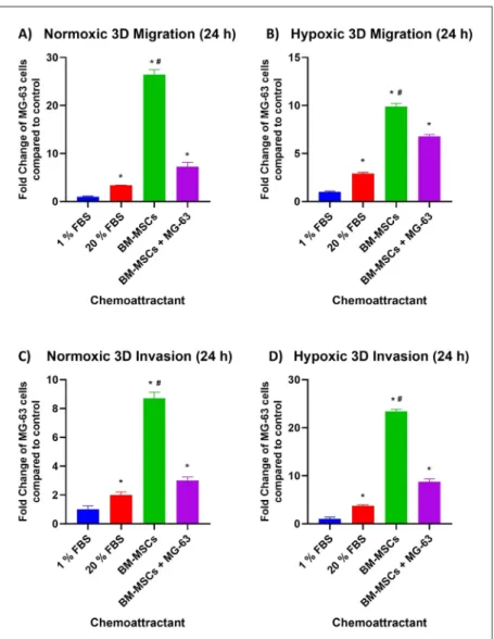
BM-MSCs + MG-63 chemoattractants increased the migration 1.83, 16.11 and 4.42 fold; on the other hand in hypoxia the chemoattractants increased by 3.04, 10.38 and 7.11 fold respectively, compared to 1% FBS at the end of 12 hours. At the 24th hour, these chemoattractants increased cell migration 3.38, 26.43 and 7.25 fold in normoxia and 2.90, 9.88 and 6.78 fold in hypoxia and it is also compared to 1% FBS. In addition to migration rates between 12th and 24th hours in MG-63, the chemoattractants increased the rate 0.09, 0.17, 0.12 fold in normal oxygen levels and rate 0.03, 0.26 and 0.13 fold in the hypoxic microenvironment.
Similarly to the migration model, measurements were also made real time for 48 hours and same chemoattractants were used in the invasion assay. However, the major difference between the migration and the invasion models was the matrigel that mimics the structure of basal membrane which leads only the cells having invasive character could pass from upper chamber to a lower one. The assay established that the total effect of normoxic micro-environment on cell invasion in 3D cell culture with 20% FBS, BM-MSCs and BM-MSCs + MG-63 chemoattractants increased
the invasion 2.24, 9.51 and 2.51 fold; and the chemoattractants increased it 3.82, 12.29 and 5.28 fold respectively, compared to 1% FBS at the end of 12 hours in hypoxia. At the 24th hour, the chemoattractants enhanced the cell invasion 1.99, 8.71 and 2.99 fold in normoxia and 3.74, 23.43 and 8.76 fold in hypoxia and it is also compared to 1% FBS. Moreover, invasion rates between 12th and 24th hours in MG-63 cells measured by xCelligence DP RTCA appraised that the chemoattractants raised the rate 0.07, 0.30, 0.17 fold in normal oxygen levels and rate 0.03, 0.26 and 0.13 fold in the hypoxic microenvironment.
The most effective chemoattractant that increases the rate of migration in the osteosarcoma cell line under both normoxic (P 0.02) and hypoxic (P 0.004) conditions has been found to be the chemoattractant obtained from the BM-MSC culture. Moreover, the experiment revealed that when MG-63 cells were co-cultured with BM-MSCs, the capacity of BM-MSC on the migration of osteosarcoma decreases. (P 0.004 for normoxia and hypoxia) A parallel effect is also observed in invasion results. (P 0.004 for both conditions) Also on the invasion, it seems that the cross-talk reduces the effect of BM-MSCs (Figure 1).
Figure 1. A) Fold change graphs of MG-63 cells that migrate
and invade to chemoattractants 1% FBS, 20% FBS, BM-MSCs and co-culture between the stem cells and MG-63 under normoxic (A, C) and hypoxic (B, D) conditions compared to
1% FBS at the 24th hour.
*represents statistically significance compared to 1% FBS and #P=0.004 compared with the co-culture
Effect of Co-culture on Metastatic Phenotype of BM-MSCs
The mesenchymal character representing the metastatic phenotype of BM-MSCs and BM-MSCs + MG-63 co-culture chemoattractants were evaluated with osteosarcoma cells exposed to these BM-MSCs and BM-MSC + MG-63 in the metastasis process. Changes in the mesenchymal phenotype marker N-cadherin (red) expression were demonstrated by fluorescence immunocytochemical techniques. MG-63 cell line under normal growth conditions was used as a control and osteosarcoma cells exposed to BM-MSCs and BM-MSCs + MG-63 co-culture chemoattractants were compared (Figure 2).
To obtain quantitative data of N-cadherin expression, pixel analysis was performed on the obtained fluorescence immunocytochemical images by an image analysis program. As a result of the generated graphs and structured statistical analysis with Mann Whitney U tests, when the expressions in the control group were compared with the MG-63 cells incubated with the BM-MSCs chemoattractant, the increases in the N-cadherin expressions were found to be statistically significant (P 0.003 for co-culture and P 0.002 for BM-MSCs). A decrease in N-Cadherin
expression of the MG-63 exposed to the co-culture between BM-MSCs and MG-63 is observed, compared to the stem cell condition. (P 0.015) This result is similar with earlier findings of this study in the migration and the invasion assays.
DISCUSSION
The present study investigates the effectiveness of BM-MSCs on migration and invasion of MG-63 osteosarcoma cell lines. In the migration and the invasion models, the results revealed that the BM-MSCs is the most effective chemoattractant in both normoxic and hypoxic microenvironment. Even though the MG-63 and the stem cell co-culture has also a positive chemoattractant effect on tumor cells, its effect is lower than the effect of BM-MSCs alone. The altered expressions of N-cadherin point out that there is a related shift in metastatic phenotype of the MG-63 exposed to BM-MSCs and the culture conditions. As a consequence, it is concluded that the co-culture limits the effectiveness of BM-MSCs on metastatic behavior. Several studies have found the presence of stem cells which facilitate migration and growth in the environment (18–20). In
Figure 2. MG-63 osteosarcoma cells’ interaction with respectively; (A) Control, (B) BM-MSCs and (C) BM-MSCs
+ MG-63 co-culture chemoattractants. (D) Density graph determined by Image-J processing of N-cadherin
expressions with fluorescence immunocytochemistry in MG-63 cells.
osteosarcoma, Huang J et. al showed that MSCs promotes both growth and metastasis (21). Other studies also point an increase in the metastasis via cross-talk between OS and BM-MSCs (22, 23). The metastasis increases by 10-fold considering the impact of BM-MSCs in the literature (24, 25). The data obtained from the results in the present study are also similar. Additionaly, it has been observed that a direct co-culture between OS and BM-MSCs as an attractant limits the capacity to both migrate and invade.
Contrary to the results in osteosarcoma, Sarah M. Ridge et al. recorded that treatment with condition mediums of some metastatic prostate cancer to BM-MSCs has enhanced the capacity on the migration of PC3 cell line compared to untreated MSCs. However, such an effect is not observed for 22Rv1 or DU145 prostate cancer cells, which suggests that PC3 may modulate the behavior of MSCs that is beneficial specifically to PC3 cells (12) The source of BM-MSCs investigated in the study is bone marrow aspirates from Pca patients and healthy volunteers. Furthermore, the exposure time of MSCs to the condition mediums of Pca cell lines is much longer when the time is 30 days in the study whereas we treated BM-MSCs in co-culture with osteosarcoma for 72 hours. The extended time would probably cause differentiation of BM-MSCs and researchers studied the changes in both character and phenotype of differentiated BM-MSCs. Moreover, the direct co-culture would affect BM-MSCs and the secreted soluble factors which change the metastasis of indirect co-cultured MG-63 s via paracrine effect in the study. The phenotypes of cancer cells are essential for the examination of metastatic characteristics. N-Cadherin is a marker which increases with metastasis in many types of cancers such as breast and colon cancers, neuroblastoma, and osteosarcoma (26–29). In addition, H. Rai and J. Ahmed point out the increase in N-cadherin which is related to the mesenchymal transition in tumor progression since it mediates a less stable cell-cell adhesion (30). N-cadherin, a mesenchymal marker, was examined by fluorescent immunocytochemistry. The staining was performed to demonstrate the effect of the BM-MSCs chemoattractant group and the BM-BM-MSCs + MG-63 co-culture chemoattractant group on the invasion and migration to demonstrate that the metastasis process is induced by cross-talk in BM-MSCs and BM-MSCs + MG-63 co-culture group.
Therefore, the increase in N-cadherin expression might explain the enhanced migration capacity in the OS exposed to BM-MSCs whereas the lower expression observed in the co-culture treated MG-63, consistent with the results in migration and invasion models of this study.
This study investigates the effects of mesenchymal stem cell on tumor behaviour in 3-dimensional (3D) in vitro cancer model mimicking in vivo tissue features best with creating different microenvironments (31). It allows the cell to make cell-cell interaction without polarization, an organization with the extracellular matrix in addition to growth factors, chemokines, and cytokines. Moreover, real-time measurements made it possible to observe the process (32, 33). The decreased shifting in
N-cadherin expression of co-culture compared to BM-MSCs also support the results for migration and invasion models. The study could be expanded with an animal model since in vivo studies mimic microenvironments more accurately and could provide some conditions which are difficult to predict.
In the co-culture conditions, BM-MSCs and MG-63 cross-talk by releasing cytokines and chemokines which contribute to tumor invasion and migration. It is revealed that this interaction limits the effect of BM-MSCs on metastasis. In future studies, it is suggested to research the role of the cytokines and chemokines in this manner. This could provide a new scientific approach to downregulate the metastasis by decreasing the effect of BM-MSCs.
Acknowledgement
Special thanks to Gizem Çalıbaşı-Koçal, Mahdi Akbarpour, Ece Çakıroğlu, and Kağan Dağdeviren, for their professional and technical support on this project.
Funding Statement
This work was supported by Dokuz Eylül University Scientific Research Coordination Unit (Project Number: 2017.KB.SAG.003) and Tailor of Science Biotechnology Innovation I.C. (Project Number: TSCI.Project.001)
Informed Consent: Written informed consent was obtained from patient who
participated in this study.
Peer-review: Externally peer-reviewed.
Author Contributions: Concept - TU; Design - YB; Supervision - YB; Fundings - TY;
Materials - AP; Data Collection and/or Processing - TU; Analysis and/or Interpretation - HCM; Literature Search - CK; Writing Manuscript - ED; Critical Review - HE
Conflict of Interest: No conflict of interest was declared by the authors.
Financial Disclosure: The authors declared that this study has received no financial
support.
REFERENCES
1. Siclari VA, Qin L. Targeting the osteosarcoma cancer stem cell. J Orthop Surg Res BioMed 2010;5:78. [CrossRef]
2. Longhi A, Errani C, De Paolis M, Mercuri M, Bacci G. Primary bone osteosarcoma in the pediatric age. State of the art. Cancer Treat Rev 2006;32:423–436. [CrossRef]
3. Tang N, Song WX, Luo J, Haydon RC, He TC. Osteosarcoma development and stem cell differentiation. Clin Orthop Relat Res 2008;466:2114–2130. [CrossRef]
4. Caplan AI. The mesengenic process. Clin Plast Surg 1994;3:429–435. 5. Wagner W, Ho AD. Mesenchymal Stem Cell Preparations-Comparing
Apples and Oranges. Stem Cell Rev 2007;3:239–248. [CrossRef]
6. Robado de Lope L, Alcíbar OL, Amor López A, Hergueta-Redondo M, Peinado H. Tumour-adipose tissue crosstalk: fuelling tumour metastasis by extracellular vesicles. Philos Trans R Soc Lond B Biol Sci 2018;373:20160485. [CrossRef]
7. Peinado H, Zhang H, Matei I, et al. Pre-metastatic niches: organ-specific homes for metastases. Nat Rev Cancer 2017;5:302–317.
[CrossRef]
8. Mishra PJ, Mishra PJ, Humeniuk R, et al. Carcinoma-associated fibroblast-like differentiation of human mesenchymal stem cells. Cancer Res 2008;68:4331–4339. [CrossRef]
9. Hass R, Otte A. Mesenchymal stem cells as all-round supporters in a normal and neoplastic microenvironment. Cell Commun Signal 2012;10:26. [CrossRef]
10. Becker A, Thakur BK, Weiss JM, Kim HS, Peinado H, Lyden D. Extracellular Vesicles in Cancer: Cell-to-Cell Mediators of Metastasis. Cancer Cell 2016;30:836–848. [CrossRef]
11. Simmons PJ, Torok-Storb B. Identification of stromal cell precursors in human bone marrow by a novel monoclonal antibody, STRO-1. Blood 1991;78:55–62.
12. Ridge SM, Bhattacharyya D, Dervan E, et al. Secreted factors from metastatic prostate cancer cells stimulate mesenchymal stem cell transition to a pro-tumourigenic ‘activated’ state that enhances prostate cancer cell migration. Int J Cancer 2018;142:2056–2067.
[CrossRef]
13. Jones E, McGonagle D. Human Bone Marrow Mesenchymal Stem cells in vivo. Rheumatology (Oxford) 2008;47:126–131. [CrossRef]
14. Théry C, Zitvogel L, Amigorena S. Exosomes: composition, biogenesis and function. Nat Rev Immunol 2002;2:569–579. [CrossRef]
15. Colombo M, Raposo G, Théry C. Biogenesis, Secretion, and Intercellular Interactions of Exosomes and Other Extracellular Vesicles. Annu Rev Cell Dev Biol 2014;30:255–289. [CrossRef]
16. Alfranca A, Martinez-Cruzado L, Tornin J, et al. Bone microenvironment signals in osteosarcoma development. Cell Mol Life Sci 2015;72:3097–3113. [CrossRef]
17. El-Badawy A, Ghoneim MA, Gabr MM, et al. Cancer cell-soluble factors reprogram mesenchymal stromal cells to slow cycling, chemoresistant cells with a more stem-like state. Stem Cell Res Ther 2017;8:254. [CrossRef]
18. Tu B, Du L, Fan QM, Tang Z, Tang TT. STAT3 activation by IL-6 from mesenchymal stem cells promotes the proliferation and metastasis of osteosarcoma. Cancer Lett 2012;325:80–88. [CrossRef]
19. Zhang P, Dong L, Yan K, et al. CXCR4-mediated osteosarcoma growth and pulmonary metastasis is promoted by mesenchymal stem cells through VEGF. Oncol Rep 2013;30:1753–1761. [CrossRef]
20. Bian ZY, Fan QM, Li G, Xu WT, Tang TT. Human mesenchymal stem cells promote growth of osteosarcoma: Involvement of interleukin-6 in the interaction between human mesenchymal stem cells and Saos-2. Cancer Sci 2010;101:2554–2560. [CrossRef]
21. Zhang P, Dong L, Long H, et al. Homologous mesenchymal stem cells promote the emergence and growth of pulmonary metastases of the rat osteosarcoma cell line UMR-106. Oncol Lett 2014;8:127–132.
[CrossRef]
22. Avnet S, Di Pompo G, Chano T, et al. Cancer-associated mesenchymal stroma fosters the stemness of osteosarcoma cells in response to intratumoral acidosis via NF-κB activation. Int J Cancer 2017;140:1331–1345. [CrossRef]
23. Mandel K, Yang Y, Schambach A, Glage S, Otte A, Hass R. Mesenchymal Stem Cells Directly Interact with Breast Cancer Cells and Promote Tumor Cell Growth In Vitro and In Vivo. Stem Cells Dev 2013;22:3114–3127. [CrossRef]
24. Zhang T, Lee YW, Rui YF, Cheng TY, Jiang XH, Li G. Bone marrow-derived mesenchymal stem cells promote growth and angiogenesis of breast and prostate tumors. Stem Cell Res Ther 2013;4:70. [CrossRef]
25. Hazan RB, Phillips GR, Qiao RF, Norton L, Aaronson SA. Exogenous expression of N-cadherin in breast cancer cells induces cell migration, invasion, and metastasis. J Cell Biol 2000;148:779–790. [CrossRef]
26. Lammens T, Swerts K, Derycke L, et al. N-Cadherin in Neuroblastoma Disease: Expression and Clinical Significance. PLoS One 2012;7:e31206. [CrossRef]
27. Zhuo H, Jiang K, Dong L, et al. Overexpression of N-cadherin is correlated with metastasis and worse survival in colorectal cancer patients. Chinese Sci Bull 2013;58:3529–3534. [CrossRef]
28. Rai H, Ahmed J. N-Cadherin: A Marker of Epithelial to Mesenchymal Transition in Tumor Progression. Internet J Oncol 2014;10. Available at: https://print.ispub.com/api/0/ispub-article/14796
29. Fang S, Yu L, Mei H, et al. Cisplatin promotes mesenchymal-like characteristics in osteosarcoma through Snail. Oncol Lett 2016;12:5007–5014. [CrossRef]
30. Duval K, Grover H, Han L-H, et al. Modeling Physiological Events in 2D vs 3D Cell Culture. Physiology (Bethesda) 2017;32:266–277.
[CrossRef]
31. Rahim S, Üren A. A real-time electrical impedance based technique to measure invasion of endothelial cell monolayer by cancer cells. J Vis Exp 2011;50. [CrossRef]
32. Liu Y, Feng Y, Liu H, Wu J, Tang Y, Wang Q. Real-time assessment of platinum sensitivity of primary culture from a patient with ovarian cancer with extensive metastasis and the platinum sensitivity enhancing effect by metformin. Oncol Lett 2018;16:4253–4262.