Background: Bruxism is a parafunctional habit, usually performed in sleep, by rhythmic and involuntary teeth being squeezed or squeaked. The most common methods of treatment are the use of occlusal splints. Aims: The aim of this study was to compare the efficacy of occlusal splinting with botulinum toxin administration in the treatment of TMJ pain. Subjects and Methods: For this purpose, 40 patients with bruxism were divided into two groups and one group was treated with occlusal splint and the other group received masseter muscle botulinum toxin injection. Then, the participants in both groups were evaluated in terms of pain, functional movement, and maximum bite force change at 2 weeks, 6 weeks, 3 months, and 6 months. Mann–Whitney U test was used to examine the differences between two independent groups. While Friedman test was used for differences between dependent groups, the Wilcoxon test was used for the differences between two repetitive measurements. Chi‑square test was used to examine the relationship between categorical variables. Results: When pain was evaluated, both methods were effective in pain reduction, botulinum toxin injection was found to be less effective in reducing pain but no difference was found between the two methods. The maximum bite force decreased in the 2nd and 6th weeks and increased in the 3rd and 6th months in patients receiving botulinum toxin. In patients using occlusal splints, there was no change until the 3rd month and an increase was seen in the 6th month. In this study, it was observed that low dose BTX‑A and occlusal splint use were effective in eliminating bruxism‑related pain but not superior to each other. Conclusions: According to these results, low dose botulinum toxin can be considered as an alternative treatment in patients who cannot use occlusal splint for various reasons.
Keywords: Bite force, botulinum toxin, bruxism
Botulinum Toxin Treatment of Temporomandibular Joint Pain in
Patients with Bruxism: A Prospective and Randomized Clinical Study
DI Kaya, H Ataoğlu1
Address for correspondence: Dr. DI Kaya, Parsana Mah, Beyhekim Cad, No. 3, Konya, Turkey. E‑mail: [email protected]
during sleep and characterized by involuntary rhythmic or spasmodic nonfunctional grinding and clenching of teeth. In patients with bruxism, as a result of overloading on the stomatognathic structures, headache, neck pain, limitation in functional movement of mandibula, pain and spasm in masticatory muscles may occur.[2,3]
The most recent hypothesis about the etiology of bruxism support the role of central and autonomic
Introduction
T
emporomandibular disorder is examined in two subgroups: muscle originated disorders and temporomandibular joint disorders.[1] Temporomandibular disorders include internal structure irregularities, or mismatch disorders involving Temporomandibular joint muscles and surrounding tissues. In patients with intraarticular irregularities; masticatory muscle sensitivity or hyperactivity may be seen. Muscle hyperactivity is sometimes seen as the cause of intraarticular disorder and sometimes as a factor accompanying intraarticular disorder. Bruxism is a parafunctional activity usually seenDepartment of Oral and Maxillofacial Surgery, Konya Oral and Teeth Health Hospital, Konya, 1Department
of Oral and Maxillofacial Surgery, Medipol University, Istanbul, Turkey
Abstract
This is an open access journal, and articles are distributed under the terms of the Creative Commons Attribution‑NonCommercial‑ShareAlike 4.0 License, which allows others to remix, tweak, and build upon the work non‑commercially, as long as appropriate credit is given and the new creations are licensed under the identical terms.
For reprints contact: [email protected]
How to cite this article: Kaya DI, Ataoğlu H. Botulinum toxin treatment of temporomandibular joint pain in patients with bruxism: A prospective and randomized clinical study. Niger J Clin Pract 2021;24:412-7.
Access this article online Quick Response Code:
Website: www.njcponline.com DOI: 10.4103/njcp.njcp_251_20 Received: 11-May-2020; Revision: 30-May-2020; Accepted: 03-Jul-2020; Published: 15-Mar-2021
nervous systems in the formation of oromandibular activity during sleep. More specifically, sleep‑related mechanisms under the influence of brain chemicals and maintenance of airway patency during sleep may increase motor activity underlying the genesis of sleep bruxism and rhythmic masticatory muscle activity, the motor manifestation of sleep bruxism preceding tooth grinding during sleep.[4]
Botulinum toxin is the metabolite of bacterium Clostridium botulinum. Botulinum toxin, which prevents signal transduction in neuromuscular junction and is a highly effective neurotoxin, began to be used in modern medicine by understanding its effect mechanism. Botulinum Toxin Type A (BTX‑A) inhibits the release of acetylcholine by directly affecting the neuromuscular junction and other cholinergic synapses. Thus, it prevents transmission between two neurons. This causes paralysis of muscle and blocks its function. Because of this feature, Botulinum Toxin A has been used for many years in medicine as well as for the prevention of hyperactivity seen in the masseter muscles of bruxism patients.[5] The most common treatment option in the treatment of bruxism is the use of stabilization splints. Studies have reported that parafunctional habit and the damage caused by this habit are reduced in patients using stabilization splints.[6‑9] In the light of this literature information, the hypothesis of the study is that BTX‑A is more effective than occlusal splint applications in the treatment of myofascial pain.
Subjects and Methods
Selection of patient
The presented research was performed on 40 patients (7 males, 33 females) who are aged between 18 and 45 years (mean 26.333). They were referred to The Oral and Maxillofacial Surgery Clinic, with a history of bruxism and pain complaint in the maxillofacial area. The criteria of the study were determined for the patients aged between 18 and 65 years old, without a systemic disease, with bruxism‑originated Myofascial pain syndrome and pain in masseter muscle during palpation. Patients taking systemic steroids or immunosuppressive drugs, non‑steroidal anti‑inflammatory drugs (NSAID) users within the last 7–10 days, occlusal splints users within the last 6 months, having removable or fixed prosthesis, having missing teeth, being performed Temporomandibular Joint (TMJ) surgery, the ones who received physiotherapy for TMJ within the last 6 months and patients with occlusal etching, pregnant and nursing women were excluded from the study. For this study, approval was obtained from the clinical research ethics committee of Selçuk University Faculty of Medicine. Approval date and number:23.11.2017/37.
To make a clinical diagnosis of Myofascial pain syndrome five major and at least one minor criteria are needed.[10]
The patients were informed about the procedures to be performed and the possible complications and they were asked to sign the Informed Consent Form. In 20 of the patients included in the study, 24 units of BTX‑A were applied on one side of the masseter muscle, and an occlusal splint was applied in the other 20 patients for at least 8 h a day. The inclusion of patients in the groups was performed randomly. One patient in the occlusal splint group was excluded from the study because he did not participate in the control sessions. Measurements were performed in the second week, sixth week, third month and sixth month to evaluate pain and bite force in the patients included in the study.
Assessment of maximum bite force
In this study, a modular system with low cost, precise, and accurate measurement has been developed to measure the bite force. This modular system consists of two main structures. The first one is a microprocessor which collects data and the second one is a sensor system design for a precise and accurate measurement. In the sensor system, FlexiForce Pressure Sensor (Tekscan ‑ USA), a commercially purchased sensor, was used to measure compressive force.
The sensor has a sensing zone of 9.53 mm diameter. For accurate measurement, it is important to apply forces only to this zone in the sensor. Metal washers are stick on both surfaces of the sensing zone of the sensor to provide this. Two supportive components made of PE 1,000 plastic are used to provide straight forces on the washers and to prevent damage to other parts of the sensor.
The system, developed based on sensor system design, consists the units of sensor, sensor supply and voltage divider circuit, microcontroller board and imaging. When the force is applied to the sensor via the sensor supply in second row and voltage divider circuit, alternating signal depending on the applied force (analog voltage) was produced. This analogue signal was converted to digital values with the Arduino UNO microprocessor development board and it was provided to be operable in computer environment. Afterwards, the acquired values were transferred to imaging units.
Analog signals from the sensor circuit (DC voltage, 0–5 V) are converted to digital values (ADC, 0‑1023) by means of the hardware features of the development board (10‑bit ADC/Analog to Digital Converter). These
digital values were processed by the software prepared for being installed on the developer card and force values were acquired. The acquired results can be both seen instantly on the LCD monitor connected to the system and can be viewed and stored on a computer via USB connection. In order to obtain force values from digital data from the sensor and to test the accuracy of the system, specific loads were applied to the sensor system design by INSTRON brand tension/compression testing machine. The digital values read from the LCD screen obtained as a result of gradually increasing loading from 25N to 450N were saved. This loading series were applied repeating 10 ten times. The mean and standard deviation of the values obtained for each load stage were calculated. Standard deviation values decreased as load increased. The operating range in the measurement of compressive force is usually between 100 and 250 N. In this range, the standard deviation value varies between 3.734 and 0.707. It is seen that these values are at the desired level for the designed measurement system.
The maximum bite force of the participants was measured by using prepared bite force measuring device. Measurements were made by placing a bite device on the occlusal surface of the first molar tooth in the mandibula. When measuring, the patients were asked to bite the device in their mouths as strongly as possible for 5 s. This measurement was repeated 4 times for the right side and 4 times for the left side and the highest values were recorded. After a 3‑minute resting, the whole measurement was repeated by starting from the opposite side and the highest values were recorded. Out of a total of 16 measurements, the highest value were recorded.
Assessment of pain
Pain assessment of patients was carried out by Visual Analogue Scale (VAS). For the VAS evaluation method, on a 10‑unit line, the values were explained to the patient as follow: no pain, 0; the most severe pain, 10; moderate pain, 5. Before being included in the working group, the patient was asked to determine a value for the pain felt. According to the treatment group, this measurement was repeated after 2 weeks, 6 weeks, 3 months, and 6 months after starting treatment.
Statistical analysis
The acquired data were evaluated with SPSS 21.0 package program. Mann–Whitney U test was used to examine the differences between two independent groups. While Friedman test was used for differences between dependent groups, the Wilcoxon test was used for the differences between two repetitive measurements.
Chi‑square test was used to examine the relationship between categorical variables. The significance level was found to be 0.05.
Results
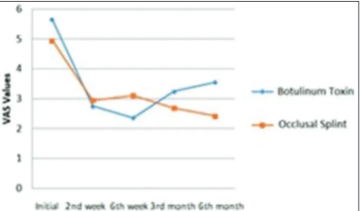
Pain‑related measurements in patients using BTX‑A and occlusal splints are shown [Figure 1].
The mean VAS values obtained at each control session in patients applied BTX‑A and occlusal splint are given in the Table 1. Accordingly, there was a statistically significant decrease in pain during the control periods after application for both methods.
Both methods were found to be effective in reducing pain in the group analysis. However, according to the Mann–Whitney U test performed to compare both methods, it is seen that the methods are not superior to each other [Table 2].
Measurements of maximum bite force in patients using BTX‑A and occlusal splints are shown [Figure 2].
The mean maximum bite force value obtained in each control session in patients treated with BTX‑A is given in the Table 3. There was a statistically significant decrease in the maximum bite force assessment in the 2nd and 6th weeks of BTX‑A application, whereas there was no difference in 3rd and 6th month measurements with initial data.
Figure 1: Pain‑related measurement graph
Table 1: The mean VAS values of BTX-A and Occlusal splint patients
Mean SD P
BTX-A OS BTX-A OS BTX-A OS
0‑2w 2.9 2.0 0.496 0.315 0.000* 0.000* 0‑6w 3.3 1.8 0.492 0.420 0.000* 0.003* 0‑3m 2.4 2.2 0.406 0.482 0.000* 0.001* 0‑6m 2.1 2.5 0.552 0.441 0.003* 0.001* 0‑2w: Difference between initial and 2nd week, 0‑6w: Difference
between initial and 6th week, 0‑3m: Difference between initial
and 3rd month, 0‑6m: Difference between initial and 6th month,
BTX‑A: Botulinum Toxin, OS: Occlusal Splint, SD: Standart Deviation, p<0.05, *: significant result
bite force during the first 3 follow‑up sessions. However, maximum bite force increased significantly at the end of the 6th month.
As a result of analysis of variance (Repeated Measures ANOVA); measurements differ significantly in itself (F = 13.409, P = 0.001). However, there was no statistically significant difference between the applied methods (F = 0.119, P = 0.732).
Discussion
In the consensus recommendations of Ahn et al.,[11] in 2013 on the clinical applications of BTX‑A, a total of 3‑point injections as to be 8–10 units per point in the masseteric muscle were successful for one side. To ensure the standardization of the quantity applied in the study, a total of 24 units of BTX‑A injections were administered for each muscle 8 units per point at a minimum concentration.
In the study conducted by Conti et al.[12] evaluating the efficacy of oral devices used in patients with bruxism, control measurements were performed on the patients at the second week, the sixth week, the third month, and the sixth month. Also in the present study, periodic follow‑up was performed at similar time intervals in order to closely monitor the effect of the treatments applied on the changes in the parameters examined in the patients.
In their study carried out on 24 patients with bruxism with myofascial pain, Jadhao et al.[13] divided the patients into three groups. Accordingly, in one group, 30 units of BTX‑A were administered to the masseter muscle and 20 units to the anterior temporal muscle. Another group was injected with sterile saline solution. Another group was evaluated as a control group and no injections were made. According to the maximum bite force measurements made in the first week, the third month and the sixth month, there was a significant decrease in the BTX‑A group compared to the placebo and control groups. In the present study, on the other hand, in the BTX‑A administered group, while there was a significant decrease in maximum bite force in the second and the sixth week, no statistically significant difference was found in the third and sixth month measurements. In the occlusal splint used group, there was a significant increase in the sixth month measurements. However, it was understood that the difference between the two methods was not statistically significant. The reason for this difference may be the fact that while we, in our study, injected the masseter muscle only, Jadhao et al.[13] injected both the masseter muscle and anterior temporal muscle in their study. In the same study, pain status during rest and chewing was evaluated with VAS. In Table 2: Comparison of BTX-A and occlusal splint for
pain
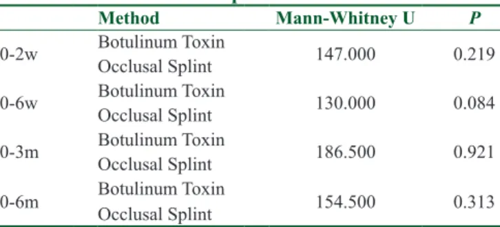
Method Mann‑Whitney U P
0‑2w Botulinum Toxin Occlusal Splint 147.000 0.219 0‑6w Botulinum Toxin Occlusal Splint 130.000 0.084 0‑3m Botulinum Toxin Occlusal Splint 186.500 0.921 0‑6m Botulinum Toxin Occlusal Splint 154.500 0.313 0‑2w: Difference between initial and 2nd week, 0‑6w: Difference
between initial and 6th week, 0‑3m: Difference between initial and
3rd month, 0‑6m: Difference between initial and 6th month, p<0.05,
*: significant result
Figure 2: Measurements of maximum bite force
Table 3: The mean bite force values of BTX-A
Mean SD P 0‑2w 57.700 14.718 0.001* 0‑6w 66.600 16.395 0.001* 0‑3m 13.700 17.392 0.441 0‑6m ‑40.150 21.858 0.082 0‑2w: Difference between initial and 2nd week, 0‑6w: Difference between initial and 6th week, 0‑3m: Difference between initial and 3rd month, 0‑6m: Difference between initial and 6th month,
SD: Standart Deviation, p<0.05, *: significant result
Table 4: The mean bite force values of occlusal splint
Mean SD P 0‑2w ‑8.789 17.655 0.625 0‑6w ‑43.894 22.583 0.068 0‑3m ‑34.578 22.631 0.144 0‑6m ‑52.526 23.529 0.039* 0‑2w: Difference between initial and 2nd week, 0‑6w: Difference
between initial and 6th week, 0‑3m: Difference between initial
and 3rd month, 0‑6m: Difference between initial and 6th month,
SD: Standard Deviation, p<0.05, *: significant result
The mean maximum bite force value obtained in each control session in patients treated with occlusal splint is given in the Table 4. There was no significant change in
the measurements, it was seen that while the pain level of the patients in the BTX‑A injected group decreased, the pain remained at the same level in the placebo and control groups. In this study, it was observed that pain levels in BTX‑A administered group and occlusal splinted used group were significantly reduced compared to initial data, however, no statistically significant difference was found in the comparison of two methods. Therefore, as a result of this study, it can be said that the use of occlusal splints was as effective as BTX‑A application in pain control.
In a study of 30 patients with bruxism, Zhang et al.[14] divided the patients into three groups. While 50 units of BTX‑A were administered to the masseter muscle in one group, in another group, sterile saline solution was administered to the masseter muscle. No injection were made to the other group. Maximum bite force was measured in all patients at the first month, the third month, and the sixth month. According to the study, it was seen that the maximum bite force was significantly reduced in the BTX‑A administered group compared to the placebo and control groups. On the other hand, there was no significant difference between placebo and control groups. The average maximum bite force value of BTX‑A administered group in the sixth month decreased compared to the initial data, however, this change was not statistically significant. Also in this study, while the maximum bite force value at second week 2 and sixth week decreased significantly, there was no statistically significant difference in the third and sixth month measurements compared to the initial measurement. While Zhang et al.[14] injected 50 units of BTX‑A injection to each masseter muscle, we, in our study, administered 24 units of BTX‑A injection to each masseter muscle. This dose difference may also have made a difference in the medicine efficiency duration. Studies comparing the groups administered different doses of BTX‑A would clarify this issue.
Yurttutan et al. divided the patients into 3 groups in a study conducted with 73 patients. The patients in the first group were given occlusal splints, the patients in the second group were treated with masseter muscle botulinum toxin, and the patients in the third group were treated with botulinum toxin with occlusal splints. At the end of 6 months, pain and questionnaire evaluation showed a significant decrease in the complaints of the patients in the second and third groups. This result leads to questioning the use of occlusal splints in patients with bruxism.[15] In the study conducted by Goiato et al.,[16] 30 female patients with bruxism with myofascial pain were examined. Patients included in the study used occlusal splints for 30 days. Patients using occlusal splints were
evaluated for pain and bite force on initial stage, 7th day, and 13th day. According to the results, it was seen that the bite force increased significantly after 30 days. Pain level, on the other hand, decreased significantly after 30‑day period. These two data are consistent with the measurements of our study in the same time period. As a result of VAS evaluations of patients using occlusal splints, while a decrease was seen in patients’ pain, there was an increase in their maximum bite force. In this sense, the study seems to be consistent with the study of Goiato et al.[16] There may be a relationship between the feeling pain during biting with maximum force and the increase in maximum bite force with decreasing pain over time. However, when Goiato et al.[16] examined the correlation between the increase in bite force and pain reduction parameters, they found that there was no strong connection. This statement refutes the idea that there is a decrease in clenching force due to pain in patients with bruxism.
Fernandez‑Nunez examined 68 studies and stated that botulinum toxin applications are a safe and effective treatment option in patients with bruxism. He also underlined that botulinum toxin applications give better results than traditional treatment options. He stated that botulinum toxin applications should be included in daily clinical practice, especially for patients with severe bruxism.[17] Agren examined 311 studies and showed that there is insufficient evidence that botulinum toxin treatments can treat bruxism. However, individual studies have shown promising results. This dilemma suggests that more studies are needed to evaluate the effects of botulinum toxin.[18]
The easy application of the occlusal splint, its low cost, and the ability to terminate the treatment at any time. In clinical practice of dentistry, it has become the first choice for treatment of bruxism. Use of occlusal splints; it causes nausea reflex and creates a feeling of having a foreign object in the mouth. Furthermore, the fact that the occlusal splint is very difficult to use 24 h a day is a disadvantage compared to the use of BTX‑A in the treatment of bruxism. BTX‑A is a costly procedure that requires repeated dosing and is more invasive than splint applications. According to the results of this clinical study, low doses of BTX‑A may be considered as an alternative treatment in patients who cannot use occlusal splints for various reasons.
To better understand the efficacy of Botulinum Toxin Type‑A in the treatment of patients with bruxism; more clinical trials with more participants and comparing different dosages are needed.
Declaration of patient consent
The authors certify that they have obtained all appropriate patient consent forms. In the form the patient(s) has/have given his/her/their consent for his/her/their images and other clinical information to be reported in the journal. The patients understand that their names and initials will not be published and due efforts will be made to conceal their identity, but anonymity cannot be guaranteed.
Financial support and sponsorship
Selcuk University Scientific Research Projects Coordination, Konya. Project no: 17202055.
Conflicts of interest
The authors declare that they have no conflict of interest.
References
1. McNeill C. Management of temporomandibular disorders: Concepts and controversies. J Prosthet Dent 1997;77:510‑22. 2. Akamatsu Y, Minagi S, Sato T. A new method for recording
mandibular position during nocturnal bruxism. J Oral Rehabil 1996;23:622‑6.
3. Kataoka K, Ekuni D, Mizutani S, Tomofuji T, Azuma T, Yamane M, et al. Association between self‑reported bruxism and malocclusion in university students: A cross‑sectional study. J Epidemiol 2015;25:423‑30.
4. Klasser GD, Rei N, Lavigne GJ. Sleep bruxism etiology: The evolution of a changing paradigm. J Can Dent Assoc 2015;81:f2. 5. Kane MAC, Sattler G. Illustrated Guide to Aesthetic Botulinum
Toxic Injections: Basics, Indications, Uses. Quintessence; New Malden, United Kindom. 1st edition. 2013.
6. Bourbon B. Craniomandibular Examination and Treatment. In: Myers RS, editor. Philadelphia: W.B. Saunders Co; 1995. p. 669‑715.
7. Okeson JP. Management of Temporomandibular Disorders and Occlusion. Mosby; St. Louis,United States.4th edition. 1998.
8. Yengin E. Temporomandibular rahatsızlklarda teşhis ve tedavi. İstanbul: Dilek Matbaacılık; 2000.
9. Marbach JJ. Temporomandibular pain and dysfunction syndrome. History, physical examination, and treatment. Rheum Dis Clin North Am 1996;22:477‑98.
10. Travell JG, Simons DG. Myofascial Pain and Dysfunction. The Trigger Point Manual. Baltimore: Williams and Wilkins 1983. 2nd edition. p. 5‑201.
11. Ahn BK, Kim YS, Kim HJ, Rho NK, Kim HS. Consensus recommendations on the aesthetic usage of botulinum toxin type A in Asians. Dermatol Surg 2013;39:1843‑60.
12. Conti PC, Corrêa AS, Lauris JR, Stuginski‑Barbosa J. Management of painful temporomandibular joint clicking with different intraoral devices and counseling: A controlled study. J Appl Oral Sci 2015;23:529‑35.
13. Jadhao VA, Lokhande N, Habbu SG, Sewane S, Dongare S, Goyal N. Efficacy of botulinum toxin in treating myofascial pain and occlusal force characteristics of masticatory muscles in bruxism. Indian J Dent Res 2017;28:493‑7.
14. Zhang LD, Liu Q, Zou DR, Yu LF. Occlusal force characteristics of masseteric muscles after intramuscular injection of botulinum toxin A(BTX‑A) for treatment of temporomandibular disorder. Br J Oral Maxillofac Surg 2016;54:736‑40.
15. Yurttutan ME, Tütüncüler Sancak K, Tüzüner AM. Which treatment is effective for bruxism: Occlusal splints or botulinum toxin? J Oral Maxillofac Surg 2019;77:2431‑8.
16. Goiato MC, Zuim PRJ, Moreno A, Dos Santos DM, da Silva EVF, de Caxias FP, et al. Does pain in the masseter and anterior temporal muscles influence maximal bite force? Arch Oral Biol 2017;83:1‑6.
17. Fernández‑Núñez T, Amghar‑Maach S, Gay‑Escoda C. Efficacy of botulinum toxin in the treatment of bruxism: Systematic review. Med Oral Patol Oral Cir Bucal 2019;24:e416‑e24. 18. Ågren M, Sahin C, Pettersson M. The effect of botulinum toxin
injections on bruxism: A systematic review. J Oral Rehabil 2020;47:395‑402.