One-stage surgical correction of congenitalthoracic lordosis--report of 2 cases
Tam metin
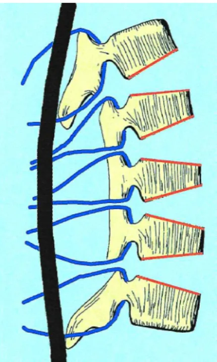
Şekil

Benzer Belgeler
The revised conceptual framework theorises that, Contact (CON), Fulfilment (FUL), Privacy (PRV) and Responsiveness (RSP) act as determinants of customer
We did not detect a difference of the age of presentation for both the study and the control groups; thus, we claim that intraspinal defects might not have a
The true axial ima- ges were analyzed by measuring chord length, trans- verse pedicle width and transverse pedicle angle of concave and convex pedicles.. Measurements were
The length of DFS varied according to the histopathologi- cal type of tumor: the mean for epithelial tumors was 40.1 months (range: 9–75.2 months), while it decreased to 28.2
Multiple lung cancers are classified as synchronous when more than 1 type of lung cancer is detected at the same time, and as metachronous tumors if the second tumor is detected
To overcome such problems besides pedicled harvesting technique of the ITA, skeletonization is described by Keeley, which improves length and blood flow of the
The main strategy of coronary artery bypass grafting (CABG) is based on grafting the left internal thoracic artery (LITA) to left anterior descending artery (LAD) and using
All of the patients were evaluated with a preoperative posteroanterior chest X-ray, and those who required supracostal access along with those suspected of
