926
The onset of labor is associated with several events that include the appearance of gap junctions between the smooth muscle cells in the myometrium.1Gap junctions are made up from a family of proteins, called connexins, that can be classified according to their molecular weights.2
It has been shown that at least three members of the family of connexins (Cx43, Cx45, and Cx26) are associ-ated with various stages of pregnancy in rat myo-metrium.3, 4The appearance of Cx43 in the myometrium coincides with the onset of labor in several species, in-cluding women.5The presence of the Cx43 protein and immunohistochemically detectable gap junctions have also been shown in some human leiomyomas from pre-menopausal nonpregnant women but not in the autolo-gous myometrium.6 The timing of expression and
ap-pearance of Cx43 shows similarities between rats7 and human subjects,8but the existence of a variety of con-nexin expressions has not been shown in the latter.
The aim of our study was to examine the presence of Cx43 and Cx26 proteins and the distribution of gap junc-tions in cell cultures prepared from myometrial tissues obtained from the upper and lower uterine segments of nonpregnant and pregnant women and from leiomyoma tissues.
Material and methods
Cell culture. Strips of myometrium from nonpregnant women and myoma tissue were obtained at hysterectomy and from the upper and lower uterine segments of the pregnant uterus at cesarean delivery. Tissues obtained from the myomas were prepared separately: one tissue specimen dissected from the tumor tissue itself and the other, from the analogous myometrium. The specimen from the tumor tissue was taken 1 cm below the surface of the myoma.
The protocol of the study was approved by the institu-tion’s ethics committee, and the patients were informed and gave consent to participate in the study. The tissues were minced into very small pieces that were rinsed with phosphate-buffered sodium choride solution (PBS) re-peatedly and then digested overnight with collagenase 2.5 mg/mL (Sigma Chemical Co, St Louis, Mo), hyal-From the Department of Women’s and Children’s Health, Section of
Obstetrics and Gynecology,aand the Department of Biochemistry,b Uppsala University.
Supported by funds from the Swedish Medical Research Council and the Swedish Society of Medicine.
Received for publication March 15, 1999; revised September 14, 1999; accepted November 4, 1999.
Reprint requests: H. Nadir Çiray, MD, PhD, Kadir Has University, Faculty of Medicine, Vefabey sokak No 5, TR-80810 Gayrettepe-Istanbul, Turkey.
Copyright © 2000 by Mosby, Inc.
0002-9378/2000 $12.00 + 0 6/1/104235 doi:10.1067/mob.2000.104235
derived from myometrial tissues from nonpregnant and pregnant
women and from leiomyomas
H. Nadir Çiray, MD, PhD,aXin Fu, MD, PhD,aMatts Olovsson, MD, PhD,aGöran Ahlsen,b
Cynthia Shuman,bBo Lindblom, MD, PhD,aand Ulf Ulmsten, MD, PhDa Uppsala, Sweden
OBJECTIVE: Our objective was to study the appearance and distribution of connexins 43 and 26 in various human myometrial cell cultures.
STUDY DESIGN: Scrape loading, Western blotting, and immunohistochemical techniques were applied to cultured cells derived from myometrial tissues obtained from nonpregnant and pregnant women (upper and lower uterine segments) and from leiomyomas (tumor and analogous myometrial tissues).
RESULTS: Scrape loading revealed the presence of metabolic coupling in all tissues. Indirect immunohisto-chemical studies showed membrane localization of connexin 43 in all myometrial cultures. Western blots and indirect immunohistochemical studies showed the presence and localization of the connexin 26 protein and associated gap junctions in tissues from myomas and from nonpregnant and pregnant women except for those derived from the upper segment of the pregnant uterus.
CONCLUSION: These results show that human myometrial cultures express various gap junction proteins and that there are regional differences in expression of connexins in tissues from pregnant women. (Am J Obstet Gynecol 2000;182:926-30.)
donkey antirabbit horseradish-peroxidase, conjugated (Amersham Pharmacia Biotech) to a final dilution of 1:1000 [wt/vol] of bovine serum albumin in Tris-buffered saline solution. Streptavidin-horseradish-peroxi-dase conjugate 1:1000 [wt/vol] (Amersham Pharmacia Biotech) was added for the detection of molecular weight markers. All rinses between incubations were done with Tris-buffered saline solution containing 0.1% [vol/vol] Tween-20. Blots were developed with the Enhanced Chemiluminescence Kit (Amersham Pharma-cia Biotech) according to the manufacturer’s instruc-tions. For documentation, a Biomax (Eastman Kodak, Rochester, NY) film was used.
Indirect immunofluorescence. The cells were rinsed with PBS (pH 7.4) at 37°C and then fixed with 4% buffered formalin for 15 minutes at room temperature and in methanol for 5 minutes at –20°C. The cells were washed with PBS and incubated in blocking solution (2% human serum albumin, 0.2% Triton X-100 in PBS) for 15 minutes. They were then stained with either an anti-Cx43 mouse monoclonal antibody (Zymed Laboratories) 1:200 diluted in 0.2% Triton X-100 in PBS or an anti-Cx26 rabbit polyclonal antibody (Zymed), 1:100 diluted in 0.2% Triton X-100 in PBS. Incubations were carried out for 30 minutes at room temperature. After being rinsed, the cells were incubated with a sec-ondary antibody, fluorescein isothiocyanate–conjugated rabbit antimouse antibody (Serotec Ltd, Oxford, United Kingdom) 1:200 in PBS and tetramethylrhodamine isothiocyanate (Zymed). Negative controls were cul-tured cells that were incubated according to the proto-col described here but with an irrelevant immunoglobu-lin G2αas primary antibody. Nonspecific staining was not detected. Examination was then conducted with an in-verted Nikon Diaphot microscope equipped with a re-flected light fluorescence system for application of fluo-rescein isothiocyanate, and photographs were taken (Tmax 400 ASA, Eastman Kodak).
Results
Scrape loading illuminated the layer of cells at the im-mediate vicinity of the scraped tissue in all cultures. The adjacent cells (second layer) were also illuminated, and the intensity of fluorescence was diminished gradually on the axis perpendicular to the scrape line (Fig 1).
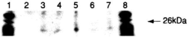
Western blot analysis showed that an immunopositive band with an apparent molecular weight of approximately 23 kd could be detected in tissues obtained from leiomy-oma, analogous myometrium, and myometrium of the lower uterine segment from both pregnant and nonpreg-nant women, but it could not be detected in the upper seg-ment of the myometrium from pregnant women (Fig 2). The negative staining in the upper segment did not de-pend on low total protein concentration, which was con-firmed by the silver-stained gel (data not shown).
uronidase 200 g/mL (Life Technologies, Inc, Grand Island, NY), and deoxyribonuclease 50 g/mL (Life Technologies) at 37°C in Dulbecco modified Eagle medium (Life Technologies). The cells were cultured in the Dulbecco medium supplemented with 5% fetal bovine serum (Life Technologies), penicillin G 100 IU/mL (Gibco), and streptomycin sulfate 50 g/mL (Life Technologies) in multiwell dishes. Incubation was car-ried out in 5% carbon dioxide at 37°C. The viability of the cells was >85%, as assessed by trypan blue dye exclu-sion.
Scrape loading. Scrape loading was used to study meta-bolic coupling qualitatively between myometrial cells, as described by El-Fouly et al.9Briefly, the cells were rinsed with PBS, and 0.05% lucifer yellow CH in PBS was added. The bottom of the well was scraped with a razor blade, and the dye was left for 2 minutes. Then the cells were rinsed with PBS and the culture medium was added. Dye transfer was examined under a fluorescent microscope, and photographs were taken.
Sodium dodecyl sulfate–polyacrylamide gel elec-trophoresis and Western blot analysis. All samples were concentrated about 5-fold in prerinsed Nanosep 10 k (Pall-Filtron; Pall Medical, Ann Arbor, Mich) centrifugal concentrators and then diluted in loading buffer (10-mmol/L Tris hydrochloride, 1-(10-mmol/L ethylenedi-aminetetraacetic acid, 2.5% sodium dodecyl sulfate [wt/vol], and 5% β-mercaptoethanol [vol/vol]; pH 8.0).
Protein separation for silver staining and Western blot analysis were performed on a Phast (Amersham Pharma-cia Biotech, Uppsala, Sweden) system by means of two precast 8% to 25% acrylamide gradient gels, running in parallel for 80 ampere volt hours. One of the gels con-tained biotin-conjugated molecular weight markers (Amersham Pharmacia Biotech). Marker proteins were treated as described here before loading.
One of the gels was silver stained according to the manufacturer’s instructions (Pharmacia Development Technique File 210). The other gel was electroblotted onto Immobilon P (Millipore Corp, Bedford, Mass) blot-ting membrane by means of semidry transfer, according to Pharmacia Development Technique File 221. Transfer buffer consisted of 0.7-mol/L glycine and 25-mmol/L Tris hydroxyacetate (pH 7.7), and blots were performed for 30 minutes at 25 mA. All incubations of blotting membrane were carried out at 37°C under gentle agita-tion for 30 to 60 minutes. Nonspecific binding was blocked in 3% [wt/vol] bovine serum albumin (Cohn fraction V, Sigma) in 25-mmol/L Tris hydrochloride and 140-mmol/L sodium chloride (pH 8.0; Tris-buffered saline solution) and was subsequently treated with poly-clonal rabbit antisera raised against Cx26 (Zymed Corp, San Francisco) to a final dilution of 1:2000 in 20-mmol/L phosphate, 140-mmol/L sodium chloride, and 0.2% [vol/vol] Triton X-100. The secondary antibody was
The characteristic pattern of punctual staining by Cx43 was observed in all cultures (Fig 3, a to e). The lo-calization of the fluorescent spots was in accordance with the plasma membrane. The pattern and intensity of Cx43 staining varied slightly between the cultures; tissues from the upper segment of the pregnant uterus showed longi-tudinal and intense staining (Fig 3, c); nonpregnant, lower-segment, and analogous myometrial tissues did not display any particular pattern, and the frequency of spots was lower than in the upper segment (Fig 3, a, b, and e); and myoma tissues showed a wheatfield-like appearance and intense staining (Fig 3, d). When the cultures were stained by Cx26 antibody, fluorescent spots were de-tected in line with the plasma membrane of the cells, as in Cx43, except from those obtained from the upper seg-ment of the myometrium from pregnancy, in which no staining could be detected (Fig 3, h). The staining inten-sity of Cx26 was weaker than that of Cx43 in all cultivated tissues (Fig 3, f, g, i, and j).
Comment
The cultivated myometrial tissues in our study exhibit (1) metabolic coupling as shown by scrape loading, (2) Cx26 protein in all tissues except in myometrium from the upper uterine segment in pregnant women, and (3) immunohistochemically detectable Cx43 and Cx26 gap junctions between the cells of pregnant, non-pregnant, myomatous, and analogous myometrium, ex-cept for Cx26, which was not present in the tissues from
the upper uterine segment in pregnancy. Therefore these results indicate that (1) functional coupling of cul-tivated human myometrial cells is established through gap junctions made up of at least 2 different connexin proteins and (2) the presence and appearance of con-nexins display regional differences in human myo-metrium in pregnancy.
It has previously been shown by electron microscopy10 and electrophysiologic examination11-14 that the fre-quency of gap junctions increases significantly during labor. The expression of Cx43 is also increased with the onset of labor8and is accompanied by the appearance of immunohistochemically detectable Cx43 gap junctions in the myometrium.5, 15Recently, patch-clamp experi-ments showed that Cx43 is the major gap junction pro-tein in the human myometrium.16Taken together, these findings indicate that Cx43 gap junctions may contribute to the synchronization of uterine contractions during labor by establishing an extensive intercellular communi-cation between myometrial cells. The extensive presence of Cx26 protein and gap junctions among cultivated myo-metrial tissues in our study indicates that gap junctional intercellular communication may not be solely responsi-ble for a functionally syncytial tissue in labor. It was previ-ously shown in myometrium from pregnant rats that tem-poral expressions of Cx43 and Cx26 differed: Cx26 was elevated before labor, whereas Cx43 expression peaked on the day of labor.4 The regulation of expression also varied; Cx26 was up-regulated by progesterone4 and Cx43 was up-regulated by estradiol.17Another gap junc-tion protein, Cx45, which has not yet been identified in human subjects, was expressed at early gestation and in postpartum rat myometrial tissues.3It is likely, when the myometrium is in quiescence, that intercellular commu-nication is established by gap junctions formed by several members of the connexin family, but the need to enhance the coupling of the cells during labor is main-tained by an increase in Cx43 expression and subsequent gap junction formation. We previously showed the pres-ence of metabolic18and electrical19, 20coupling between human myometrial cells before labor. It has been re-ported that when the cytoplasmic tail of Cx43 in human cardiac gap junctions was shortened with site-directed Fig 1. Scrape-loading photomicrograph of human myometrial
cultures. Illumination of cells adjacent to scrape line (lower right corner) is seen. Intensity of fluorescence gradually decreases away from first layer of cells. Axis of propagation is from lower left to upper right corner of figure.
Fig 2. Western blots of Cx26 from various human myometrial
cultures. Contrast of image is enhanced. Left to right, Molecular weight marker, upper segment—pregnancy, lower segment— pregnancy, myoma, lower segment—nonpregnancy, analogous myometrium, upper segment—nonpregnancy, and molecular weight marker. Lack of band in upper segment of myometrium from pregnancy is seen.
mutagenesis to the length of Cx26 the unitary conduc-tance of the channels was decreased to 50 ps from 60 and 100 ps.21Our previous results on studies of pregnant human myometrium before labor showed that dye cou-pling was more extensive when a fluorescent probe with a smaller molecular mass was injected intracellularly than when a probe with a higher mass was used.18If the lower conductance of Cx26 gap junction channels is caused by a smaller pore size, then the observed difference between fluorescent probes in metabolic coupling in nonlabor
human tissues may be a result of the dominance of Cx26 gap junctions. This hypothesis can be confirmed indi-rectly by quantitative analysis of Cx26 protein and im-munohistochemically detectable gap junctions in these tissues.
The absence of Cx26 protein and gap junctions in cul-tures obtained from the upper segment of myometrium during pregnancy indicates regional differences in the connexin expression in human myometrium. We served the presence of Cx26 gap junctions when we ob-Fig 3. Indirect immunohistochemical results of human myometrial cultures with Cx43 (a-e) and Cx26 (f-j). Cultures
obtained from myometrium of nonpregnancy (a and f), from lower segment (b and g) and upper segment (c and h) of myometrium during pregnancy, from myoma (d and i), and from analogous tissue (e and j) are seen.
tained biopsy specimens from regions that were close to the upper segment in myometrium from nonpregnant subjects. However, these segments are not clear, as in the tissue from pregnancy, and it is likely that the enlarge-ment of the myometrium during pregnancy is accompa-nied by regional variances in protein expression so that functional activity can be maintained during labor.
Our study is, to our knowledge, the first to demon-strate the presence of Cx26 protein and gap junctions in both the tumor tissue and the analogous myometrium. The presence of Cx43 gap junctions was previously shown in leiomyomas, but they were absent in the analo-gous tissue when the tissue was devoid of 17β-estradiol.6 Our results are in contrast to the latter finding, and the difference may arise in preparation and maintenance of the tissue.
In future studies researchers need to clarify the tempo-ral expression and regulation of Cx26 protein so that the significance of various gap junction proteins in human myometrium during pregnancy and in myomas can be understood.
We acknowledge Margareta Nordling for maintenance of cell cultures.
REFERENCES
1. Garfield RE, Blennerhassett MG, Miller SM. Control of myome-trial contractility: role and regulation of gap junctions. Oxf Rev Reprod Biol 1998;10:436-90.
2. Beyer EC, Paul DL, Goodenough DA. Connexin family of gap junction proteins. J Membr Biol 1990;116:187-94.
3. Albrecht JL, Tadros PN, Orsino A, Lye SJ, Sadovsky Y, Beyer EC. Rat uterine myometrium contains the gap junction protein con-nexin 45, which has a differing temporal expression pattern from connexin 43. Am J Obstet Gynecol 1996;175:853-8. 4. Orsino A, Taylor CV, Lye SJ. Connexin-26 and connexin-43 are
differentially expressed and regulated in the rat myometrium throughout late pregnancy and with the onset of labor. Endocrinology 1996;137:1545-53.
5. Tabb T, Thilander G, Grover A, Hertzberg E, Garfield RE. An immunochemical and immunocytochemical study of the in-crease in myometrial gap junctions (and connexin 43) in rats and humans during pregnancy. Am J Obstet Gynecol 1992;167:559-67.
6. Andersen J, Grine E, Eng CLY, Zhao K, Barbieri RL, Chumas JC, et al. Expression of connexin 43 in human myometrium and leiomyoma. Am J Obstet Gynecol 1993;169:1266-76.
7. Lye SJ, Nicholson BJ, Mascarenhas M, MacKenzie L, Petrocelli T. Increased expression of connexin-43 in the rat myometrium during labor is associated with an increase in the plasma estro-gen/progesterone ratio. Endocrinology 1993;132:2380-6. 8. Chow L, Lye SJ. Expression of the gap junction protein
connexin-43 is increased in the human myometrium toward term and with the onset of labor. Am J Obstet Gynecol 1994;170:788-95. 9. E1-Fouly MH, Trosko JE, Chang CC. Scrape-loading and dye
transfer: rapid and simple technique to study gap junctional in-tercellular communication. Exp Cell Res 1987;168:422-30. 10. Çiray HN, Guner H, Håkansson H, Roomans GM, Ulmsten U.
Morphometric analysis of gap junctions in nonpregnant and term pregnant human myometrium. Acta Obstet Gynecol 1995;74:497-504.
11. Miyoshi H, Boyle MB, MacKay LB, Garfield RE. Voltage-clamp studies of gap junctions between uterine muscle cells during term and preterm labor. Biophys J 1996;71:1324-34.
12. Sims SM, Daniel EE, Garfield RE. Improved electrical coupling in uterine smooth muscle is associated with increased numbers of gap junctions at parturition. J Gen Physiol 1982;80:353-75. 13. Miller SM, Garfield RE, Daniel EE. Improved propagation in
myometrium associated with gap junctions during parturition. Am J Physiol 1989;256(1 Pt 1):C130-41.
14. Cole WC, Garfield RE, Kirkaldy JS. Gap junctions and direct in-tercellular communication between rat uterine smooth muscle cells. Am J Physiol 1985;249(1 Pt 1):C20-31.
15. Sakai N, Tabb T, Garfield RE. Studies of connexin 43 and cell-to-cell coupling in cultured human uterine smooth muscle. Am J Obstet Gynecol 1992;167:1267-77.
16. Miyoshi H, Boyle MB, MacKay LB, Garfield RE. Gap junction currents in cultured muscle cells from human myometrium. Am J Obstet Gynecol 1998;178:588-93.
17. Lefebvre DL, Piersanti M, Bai XH, Chen ZQ, Lye SJ. Myometrial transcriptional regulation of the gap junction gene, connexin-43. Reprod Fertil Dev 1995;7:603-11.
18. Çiray HN, Persson BE, Roomans GM, Ulmsten U. Dye-coupling between term pregnant human myometrial cells before labor: carboxyfluorescein versus lucifer yellow. Cell Biol Int 1995; 19:609-17.
19. Çiray HN, Bäckstrom T, Ulmsten U, Roomans GM. Steroid hor-mone effects on intercellular communication between term pregnant human myometrial cells before labor. Biol Reprod 1996;55:379-85.
20. Çiray HN, Bäckstrom T, Ulmsten U. Ineffectiveness of oxytocin on intercellular communication between human myometrial cells before labor. Am J Obstet Gynecol 1998;178:855-61. 21. Fishman G, Moreno A, Spray D, Leinwand L. Functional analysis
of human cardiac gap junctional channel mutants. Proc Natl Acad Sci U S A 1991;88:3525-9.