Annals of Medical Research
DOI: 10.5455/annalsmedres.2020.06.578
Prognostic significance of poorly differentiated cluster
grading system in intestinal type gastric adenocarcinoma
Semra Gurunluoglu1, Emine Samdanci2, Ahmet Kadir Arslan3, Nusret Akpolat2, Nurhan Sahin4, Hasan Gokce2
1Department of Medical Pathology, Malatya Turgut Ozal University Training and Research Hospital, Malatya, Turkey 2Department of Medical Pathology, Faculty of Medicine, Inonu University, Malatya, Turkey
3Department of Biostatistics and Medical Informatics, Faculty of Medicine, Inonu University, Malatya, Turkey 4Department of Medical Pathology, Faculty of Medicine, Bezmialem University, Istanbul, Turkey
Copyright © 2020 by authors and Annals of Medical Research Publishing Inc.
Abstract
Aim: Gastric carcinoma is the fourth most common carcinoma worldwide. The relationships established between the tumor
morphology and the prognosis have not been very effective until recent times. Poorly Differentiated Clusters (PDCs); are structures those can be easily identified in H&E sections. A new grading system based on PDC count has been studied in colorectal carcinoma; and found to be associated with prognosis. We have aimed to investigate this concept in gastric carcinoma.
Material and Methods: Our study included 80 cases, consisting of 16 females and 64 males having gastric carcinoma with intestinal
morphology. For each case; conventional grade, PDC grade and prognostic parameters to be applied in the study were determined, and statistically compared.
Results: Significant discrepancy was found between the two grading systems. PDC tumor grades were statistically related to; median
metastatic lymph node counts, metastatic lymph node ratios, lymph node stages (pN) and the presence of perineural invasion.
Conclusion: In stomach carcinoma; PDC grading system was found to be related to a significant part of the expected parameters
and may have a prognostic value.
Keywords: Poorly differentiated clusters; gastric carcinoma; histologic grade
Received: 08.06.2020 Accepted: 06.08.2020 Available online: 24.08.2020
Corresponding Author: Semra Gurunluoglu , Clinic of Pathology Laboratory, Malatya Education and Research Hospital, Malatya, Turkey, E-mail: [email protected]
INTRODUCTION
Gastric carcinoma is the fourth most common carcinoma (1, 2) and it is the fifth most common cause of cancer-related deaths in men and the fourth most common cause in women all over the world (3). Although the diagnosis and treatment have been improved to an important degree in recent years, the five-year survival rate is still around 29.7%. It is very important to predict which patients will have rapid progression and poor prognosis; in terms of choosing patients who will benefit from aggressive treatment methods (1).
Although traditional histopathological variables such as tumor size, depth of invasion (tumor stage) are used effectively and widely in gastric carcinoma, they do not provide sufficient prognostic information. Therefore, it is necessary to reveal new prognostic factors those are easy to use (1).
The aim of this study is to investigate the prognostic value of poorly differentiated cluster (PDC) concept and grading system in gastric carcinoma.
MATERIALS AND METHODS
Study Design and ParticipantsWe started this study, after the approval of the Inonu University scientific research and publication ethics board; numbered as 2017/27-11. The cases were selected according to the inclusion and exclusion criteria from the available data of the hospital records.Cases who have recieved preoperative neoadjuvant chemo-radiotherapy, and/or have got multiple primary cancers such as synchronous and metachronous tumors were excluded (4). At the beginning 120 gastric carcinoma cases were included; those underwent total or subtotal gastrectomy and regional lymphadenectomy at Inonu University, Turgut Ozal Medical Center.
Pathology reports, paraffin block and H-E stained slides, taken from the archive of the Pathology Department, were evaluated retrospectively by two pathologists which were unaware of each other's evaluation results.
From those120 cases, which had the diagnosis between
the years 2011-2017 in one center, 38 cases were excluded because of the tumor morphology (ten cases were diagnosed as mucinous carcinoma and 3 cases were diagnosed as diffuse carcinoma, and 25 had diffuse carcinoma component). Eighty-two cases with gastric carcinoma of intestinal morphology were evaluated in this study.
All the H&E stained sections, belonging to the patients, with tumor were examined; the presence and intensity of PDCs were determined.
Two cases were excluded from the study; because no PDCs were detected in those. As a result we continued the study with 80 patients which were diagnosed in our center between the years 2011-2017. The age of the cases ranged from 38 to 86; 16 of them were females, 64 of them were males.
Prognostic parameters
Prognostic parameters to be applied in the study were determined by reviewing the criteria of the World Health Organisation (WHO) and the literature created on this issue. Determined parameters are; gender, patient age, patient age groups, tumor localization, tumor size, tumor size groups (4), tumor stage (depth of invasion), number of metastatic lymph nodes, metastatic lymph node ratio, lymph node stage (pN), presence of lymphovascular invasion, presence of perineural invasion (5).
Since there were no cases under the age 30, patient age groups were determined as; <=39, 40-49, 50-59, 60-69 and >70 years; since there were no cases covering entire stomach tumor site groups were determined as proximal third, middle third, distal third, and other localization. Tumor size groups were determined as <40mm and 41-60 mm, 61-80 mm and >80mm (7).
Tumor stage (the depth of tumor invasion) groups are created according to WHO (2010) criteria; tumors showing invasion of lamina propria or muscularis mucosa are grouped as T1a, tumors showing submucosal invasion are grouped as T1b, tumors invading muscularis propria are grouped as T2, tumors invading subseroza are grouped as T3, tumors perforating visceral periton are grouped as T4a, tumors invading the surrounding tissues are grouped as T4b (5).Since the number of cases were small; T1a, T1b, T4a, T4b groups were included in the two groups as T1a-T1b and T4a-T4b; by checking the compliance with the literature (4).
The lymph node number of cases in the study varies between 4-71. The lymph node stage (pN) for each case was determined according to WHO (2010) criteria. Patients with no metastatic lymph nodes N0, patients with 1-2 lymph node metastases N1, patients with 3-6 lymph node metastases N2, patients with 7-15 lymph node metastases N3a, patients with lymph node metastase number equal to or more than 16 staged as N3b (5). Cases were divided into five groups according to lymph node stage as; N0, N1, N2, N3a and N3b.
For the data on prognostic parameters, pathology reports and the clinical information in HIMS hospital information management system are taken as basis (Table 1).
Histological Grading
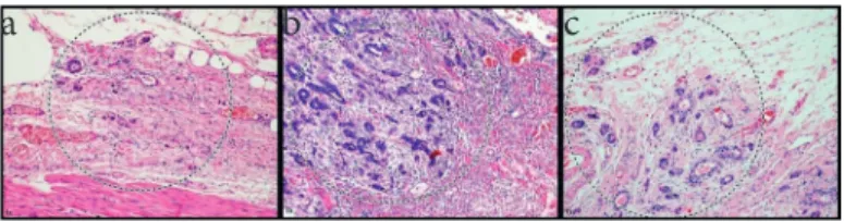
Histopathological examination was performed with an Olympus BX51 brand microscope; bearing 10x ocular and 4x, 10x, 20x, 40x and 100x objective. In determining the PDCs degree; previously determined criteria for this system for colorectal carcinoma were used (6,7). Cell groups consist of five or more neoplastic cells, which do not form gland structures, are looked for in the sections of all the tumor containing blocks and the regions where they are located are marked with 4x and 10x objective; the most intense area was chosen; it was counted in a magnification area of a 20X objective lens (1 mm diameter microscopic area). Tumor having a PDC count below 5, graded as 1 (Figure 1a); Tumor having a PDC count between 5-9, graded as 2 (Figure 1b); tumor having a PDC count more than 9 graded as 3 (7) (Figure 1c).
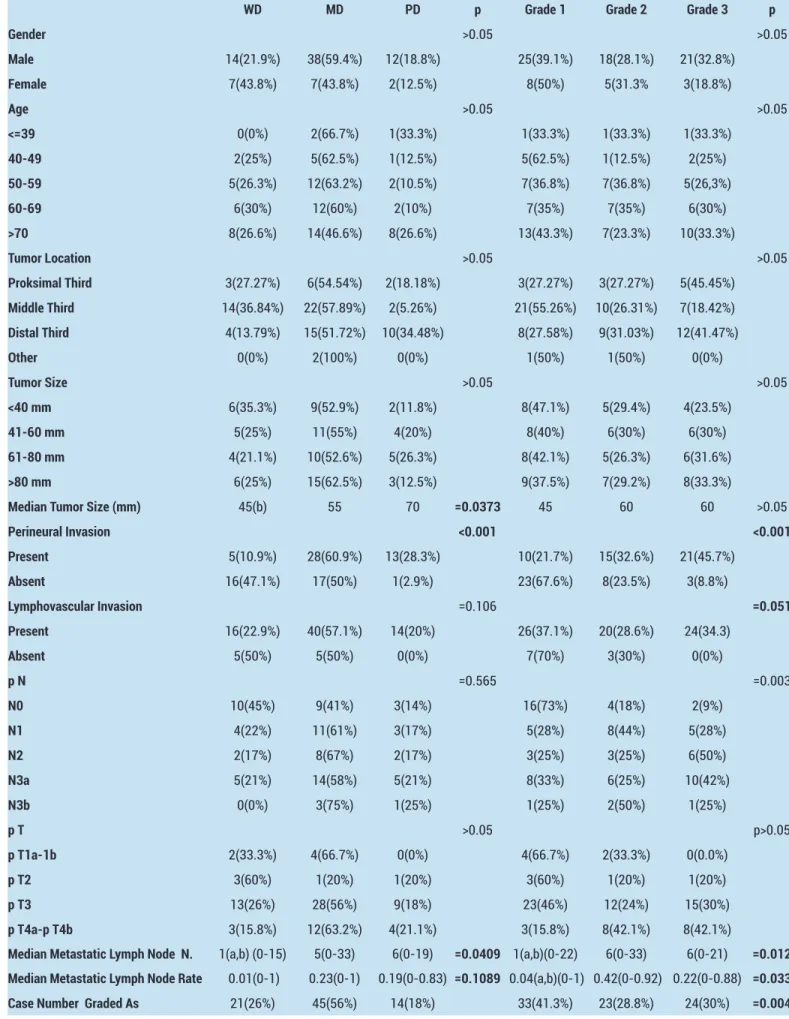
Conventional grading was made by applying the WHO criteria (2010).Tumors composed of well formed glands were graded as well differentiated (WD) (Figure 2a); Tumors showing a morphology between well and poorly differentiated tumors, were graded as moderately differentiated (MD) (Figure 2b); tumors those consist of highly irregular glands, those could be hardly noticed, were graded as poorly differentiated (PD) (Figure 2c) (5). The PDC degrees and conventional degrees were recorded at separate times for each case (Table 1). All data were organized and statistical analysis was performed.
Figure 1. H&E stained sections of gastric adenocarcinoma: a)
PDC Grade 1, b) PDC Grade 2, c) PDC Grade 3 (X100)
Figure 2. H&E stained sections of gastric adenocarcinoma; a)
Well differentiated (WD), b) Moderately differentiated (MD), c) Poorly differentiated (PD), (X100)
Statistical Analyses
In the statistical power analysis performed before starting the study, the minimum sample size to be included in this study was calculated as 40.
The quantitative data used in the study are summarized
a
b
c
b
c
as mean ± standard deviation or median (min-max) and qualitative data are summarized as numbers (percentage). Relevance of quantitative data on group basis to normal distribution was investigated by Shapiro-Wilk test. Whether there is a statistical difference between the independent groups in terms of quantitative variables providing parametric test assumptions was examined by one-way variance analysis (ANOVA). Whether there is a statistical difference between independent groups in terms of quantitative variables that do not provide parametric test assumptions was examined by Kruskal-Wallis H test. After variance analysis, multiple comparisons were made with Tukey and Tamhane's T2 tests, respectively, depending on whether the assumption of the homogenety of the variances was provided. After the Kruskal-Wallis H test, multiple comparisons were performed with Conover testing. Whether there was a statistical difference between qualitative independent groups was examined by Pearson chi-square test. Whether there was a statistical difference between qualitatively dependent groups was examined by McNemar-Bowker test. p<0.05 is accepted as a level of statistical significance.
Table 1. Patient demographics and findings
Parameters Case Number
Gender Female 16 Male 64 Age <=39 3 40-49 9 50-59 19 60-69 20 >70 30 Tumor Location Proximal third 11 Middle third 38 Distal third 29 Other 2 Tumor Size <40mm 17 41-60mm 20 61-80mm 19 >80mm 24 Perineural Invasion Present 46 Absent 34 Lymphovascular Invasion Present 70 Absent 10 p N N0 22 N1 18 N2 12 N3a 24 N3b 4 p T T1a-1b 6 T2 5 T3 50 T4a-4b 19 Conventional Grade WD 21 MD 45 PD 14 PDC Grade Grade 1 33 Grade 2 23 Grade 3 24
RESULTS
According to the PDC grading system: 33 of the 80 cases (41.3%) were graded as 1; 23 of them (28.8%) graded as 2; 24 of them (30%) are graded as 3.According to the conventional grading system, 21 of the 80 cases were graded as WD; 45 of them were graded as MD; 14 of them were graded as PD. A statistically significant discrepancy was found in terms of the number of cases included in PDC tumor degrees and conventional tumor degrees, (p=0.004) (Table 2).
Tumor Stage
Cases were divided into four groups, according to the depth of tumor invasion, as T1a-b; T2; T3; T4a-b. T1a-b group included 6; T2 group included 5; T3 group included 50 and T4a-b group included 19 cases.
There was no statistically significant relationship between tumor stage (depth of invasion) and conventional tumor grade variables (p>0.05)(Table 2). T1a-b tumor stage was associated with a low PDC grade and a T4a-b tumor stage with a high PDC grade. However, there was no statistically significant relationship between the tumor stage (depth of invasion) and the PDC tumor grade (p> 0.05) (Table 2). Tumor Size
The distribution of cases to tumor size groups, and the number of the cases in each group of grade, has been as follows: In <40mm group; 6 cases WD, 9 cases MD 2 cases PD; 8 cases grade 1, 5 cases grade 2, 4 cases grade 3. In 41-60 mm group; 5 cases WD, 11 cases MD, 4 cases PD; 8 cases grade 1, 6 cases grade 2, 6 cases grade 3. In 61-80 mm group; 4 cases WD, 10 cases MD, 5 cases PD; 8 cases grade 1, 5 cases grade 2, 6 cases grade 3. In >80mm group; 6 cases WD, 15 cases MD, 3 cases PD; 9 cases grade 1, 7 cases grade 2, 8 cases grade 3. There were no statistically significant difference between tumor size groups in terms of conventional grades or PDC grades (Table 2).
Median tumor size increased inversely with the degree of tumor differentiation (45 mm in WD cases; 55 mm in MD cases; 70 mm in PD cases); this increase made a statistically significant difference between WD and PD tumors (p<0.05) (Table 2).
Median tumor size was 45 mm for PDC Grade 1 tumors; 60 mm for PDC Grade 2 and Grade 3 tumors. There was no statistically significant difference between the PDC grades in terms of median tumor size (p> 0.05) (Table 2). Median Metastatic Lymph Node Count and Ratio
Median metastatic lymph node count was detected as 1 for the WD tumors; 5 for the MD tumors; and 6 for the PD tumors. It was significantly lower for WD tumors than those were for MD and PD tumors, and the difference between was found to be statistically significant (p <0.05). Median metastatic lymph node ratio was found to be 0.01 for the WD tumors; 0.23 for the MD tumors; and 0.19 for the PD tumors.There was no statistically significant relationship between median metastatic lymph node ratio and conventional tumor grade (p> 0.05) (Table 2).
Table 2. The Results of statistical analysis
WD MD PD p Grade 1 Grade 2 Grade 3 p
Gender >0.05 >0.05 Male 14(21.9%) 38(59.4%) 12(18.8%) 25(39.1%) 18(28.1%) 21(32.8%) Female 7(43.8%) 7(43.8%) 2(12.5%) 8(50%) 5(31.3% 3(18.8%) Age >0.05 >0.05 <=39 0(0%) 2(66.7%) 1(33.3%) 1(33.3%) 1(33.3%) 1(33.3%) 40-49 2(25%) 5(62.5%) 1(12.5%) 5(62.5%) 1(12.5%) 2(25%) 50-59 5(26.3%) 12(63.2%) 2(10.5%) 7(36.8%) 7(36.8%) 5(26,3%) 60-69 6(30%) 12(60%) 2(10%) 7(35%) 7(35%) 6(30%) >70 8(26.6%) 14(46.6%) 8(26.6%) 13(43.3%) 7(23.3%) 10(33.3%) Tumor Location >0.05 >0.05 Proksimal Third 3(27.27%) 6(54.54%) 2(18.18%) 3(27.27%) 3(27.27%) 5(45.45%) Middle Third 14(36.84%) 22(57.89%) 2(5.26%) 21(55.26%) 10(26.31%) 7(18.42%) Distal Third 4(13.79%) 15(51.72%) 10(34.48%) 8(27.58%) 9(31.03%) 12(41.47%) Other 0(0%) 2(100%) 0(0%) 1(50%) 1(50%) 0(0%) Tumor Size >0.05 >0.05 <40 mm 6(35.3%) 9(52.9%) 2(11.8%) 8(47.1%) 5(29.4%) 4(23.5%) 41-60 mm 5(25%) 11(55%) 4(20%) 8(40%) 6(30%) 6(30%) 61-80 mm 4(21.1%) 10(52.6%) 5(26.3%) 8(42.1%) 5(26.3%) 6(31.6%) >80 mm 6(25%) 15(62.5%) 3(12.5%) 9(37.5%) 7(29.2%) 8(33.3%)
Median Tumor Size (mm) 45(b) 55 70 =0.0373 45 60 60 >0.05
Perineural Invasion <0.001 <0.001 Present 5(10.9%) 28(60.9%) 13(28.3%) 10(21.7%) 15(32.6%) 21(45.7%) Absent 16(47.1%) 17(50%) 1(2.9%) 23(67.6%) 8(23.5%) 3(8.8%) Lymphovascular Invasion =0.106 =0.051 Present 16(22.9%) 40(57.1%) 14(20%) 26(37.1%) 20(28.6%) 24(34.3) Absent 5(50%) 5(50%) 0(0%) 7(70%) 3(30%) 0(0%) p N =0.565 =0.003 N0 10(45%) 9(41%) 3(14%) 16(73%) 4(18%) 2(9%) N1 4(22%) 11(61%) 3(17%) 5(28%) 8(44%) 5(28%) N2 2(17%) 8(67%) 2(17%) 3(25%) 3(25%) 6(50%) N3a 5(21%) 14(58%) 5(21%) 8(33%) 6(25%) 10(42%) N3b 0(0%) 3(75%) 1(25%) 1(25%) 2(50%) 1(25%) p T >0.05 p>0.05 p T1a-1b 2(33.3%) 4(66.7%) 0(0%) 4(66.7%) 2(33.3%) 0(0.0%) p T2 3(60%) 1(20%) 1(20%) 3(60%) 1(20%) 1(20%) p T3 13(26%) 28(56%) 9(18%) 23(46%) 12(24%) 15(30%) p T4a-p T4b 3(15.8%) 12(63.2%) 4(21.1%) 3(15.8%) 8(42.1%) 8(42.1%)
Median Metastatic Lymph Node N. 1(a,b) (0-15) 5(0-33) 6(0-19) =0.0409 1(a,b)(0-22) 6(0-33) 6(0-21) =0.012 Median Metastatic Lymph Node Rate 0.01(0-1) 0.23(0-1) 0.19(0-0.83) =0.1089 0.04(a,b)(0-1) 0.42(0-0.92) 0.22(0-0.88) =0.033
The median metastatic lymph node count was detected as; 1 for PDC Grade 1 tumors, 6 for PDC Grade 2 tumors, 6 for PDC Grade 3 tumors.The median metastatic lymph node ratio was detected as; 0.04 for PDC Grade 1 tumors, 0.42 for PDC Grade 2 tumors ; 0.22 for PDC Grade 3 tumors.Median metastatic lymph node count and median metastatic lymph node ratio were significantly lower for Grade 1 tumors than those were for Grade 2 and Grade 3 tumors, and the differences between were found to be statistically significant (p <0.05) (Table 2).
Lymph Node Stage
According to the lymph node staging system; 22 of the cases were staged as N0, 18 of them were staged as N1; 12 of them were staged as N2; 24 of them were staged as N3a; four of them were staged as N3b; and the lymph node stage groups were builded. There was no statistically significant relationship between the conventional grades and lymph node stages of the cases (p>0.05) (Table 2). High lymph node stages were associated with high PDC grade and this relationship was found to be statistically significant (p<0.05) (Table 2).
Lymphovascular İnvasion
Lymphovascular invasion was observed in 70 cases; 16 of these 70 cases (22.9%) had WD; 40 of them (57.1%) had MD; 14 of them (20%) had PD tumors.Of the 10 patients, those did not have lymphovascular invasion, 5 (50%) had WD; 5 (50%) had MD tumors. Absence of lymphovascular invasion was associated with a lower WHO grade (WD and MD); however, this relation was not found to be statistically significant (p> 0.05) (Table 2). Of the 10 patients, those did not have lymphovascular invasion; 7 (70%) had PDC Grade 1; 3 had PDC Grade 2. As a result, the absence of lymphovascular invasion was associated with a lower PDC grade, however this relation was not found to be statistically significant (p = 0.051) (Table 2).
Perineural Invasion
Perineural invasion was observed in 46 of the cases.Five of these cases (10.9%) were graded as WD; 28 (60.9%) of them were graded as MD; thirteen (28.3%) of them graded as PD. The incidence of perineural invasion was observed to increase with the increase in WHO grade; differences observed between groups were found to be statistically significant (p<0.001) (Table 2). Ten of the cases (21.7%) were graded as PDC Grade 1; 15 (32.6%) of them were graded as PDC Grade 2; 21 (45.7%) of them were graded as PDC Grade 3; the presence of perineural invasion was associated with a high PDC grade; this relationship was found to be statistically significant (p<0.05) (Table 2). Neither the conventional, nor the PDC tumor grade showed statistically significant correlation with; the mean patient age, the patient age groups, the gender andthe tumor location groups (p>0.05) (Table 2).
Evaluation of the Results
High median metastatic lymph node count, median metastatic lymph node ratio, lymph node stages and the presence of perineural invasion were associated with a high PDC grade; and those relationships were found to be statistically significant (p<0.05); with these results one can suggest that PDC grade might have a role in assessing prognosis.
DISCUSSION
The prognosis of gastric carcinoma is determined by many factors associated with the patient and the tumor. Naturally, the ones we are most interested in are; the ones those could be interfered with, such as; the width of the resection and the lymph node dissection. Our main goal is to have the widest possible knowledge, from a prognostic point of view, before starting the treatment.Thus, the most appropriate specific treatment could be planned for each patient (4).
So far, many prognostic factors have been proposed by different authors. Some of them are; the tumor depth, the number of metastatic lymph nodes, the tumor size, presence of the residual residual tumor after resection (8), the lymphovascular invasion, the perineural invasion (4). In malignancies, the tumor grade emerges as an expression of molecular changes, providing a clue to the biological behavior of the tumor; it has been shown to provide prognostic information, independently from the stage, for many malignancy (9). The relationship of tumor grade based on World Health Organization criteria with patient survival has been shown in some studies (10), and not shown in some others (8, 11). We have not found a decisive judgement on this issue in the literature.
Many studies have been conducted to demonstrate the relationship of the histopathological structure of gastric carcinoma to the clinical behavior and new classification schemes have been introduced (12-14).
On the other hand, there is also a histopathological parameter previously studied in colorectal carcinoma, such as tumor budding (TB). TB has been studied many times in the colon; has been associated with lymph node metastasis, distant metastasis and recurrence of the disease (6, 15-17). Later on, in 2014, it was found to be prognostically valuable in gastric carcinoma ina study conducted by Tanaka and et al, and other studies with similar results followed (3, 14, 18).
TB refers to small structures, consisting of less than five tumor cells. These structures are difficult to recognize in H&E sections, especially when neoplastic cells are situated in a desmoplastic stroma or there is inflammatory cell accumulation within the tumor microenvironment, immunohistochemical dye might be needed to detect them (3, 6).
Poorly Differentiated Cluster (PDC); is a structure consisting of five or more tumor cells those do not form a
gland, which could be easily determined in H&E sections and settles at the invasive border of the tumor. A new grading system based on PDC count has been developed (6, 19). In this system the area where PDCs are most dense is counted in the 20x magnification area (1 mm diameter microscopic area). If there are less than 5 clusters the tumor is considered grade 1, 5-9 is considered grade 2, 10 or more is considered as grade 3 (6).
This new concept and system has previously been shown to have a prognostically predictive value in colorectal carcinomas (6, 15, 16, 20-22). Many studies have shown that histological grading in colorectal carcinomas is a prognostic factor independent of the stage; however, there were serious difficulties regarding the reproducibility of grading and its applicability to tumors in different morphology (9, 21). In this situation the concept of PDC has come up, which constitutes a different perspective in evaluating the tumor behaviour (21).
Studies performed in colorectal carcinomas showed that PDC degree has a strong relationship with lymphovascular invasion and lymph node metastases. Moreover it has enabled the prediction of lymph node involvement, with higher specificity and sensitivity compared to the conventional histologic prognostic factors (5, 16, 23). High ADK grade (Grades 2 and Grade 3) has shown a strong association with a short disease-free survival and disease-specific survival.This association was independent of the pTNM stage and histological features such as conventional grade (7, 19, 21, 23-25). It was denoted in colorectal carcinomas that, the reproducibility of the data obtained by this grading system; is higher than it is by conventional grading system (6, 25).
The PDC and TB are two different entities with similar morphologies those do not show gland formation. They have also been reported to be similar in terms of etiopatogenesis (6).
Many different etiopathogenetic mechanisms have been proposed about the formation of PDCs (6). There are in vitro studies showing migration of the tumor cells, separated from the tumor mass individually or in groups, in a desmoplastic stroma; as a result of the epithelial-mesenchymal transformation process (26, 27). Epithelial-mesenchymal transformation is thought to be an effective mechanism for the immigration and the invasion of epithelial cells. (27). In this respect, PDCs were associated with the upregulation of Wnt/beta-catenin signaling pathway; such as metalloproteinase, dysintegrin, and L1-cell adhesion molecule (LICAM); and beta- catenin expression, also associated with the loss of e-cadherin and claudin (6).
These findings suggest that PDCs facilitate tumor spread and metastasis through lymphovascular invasion. Consequently, PDCs; can play an important role in explaining the aggressive behavior of carcinoma and predicting the tumor behavior (6).
In our study, comparing the grading system based on PDCs with the grading system based on WHO criteria, we observed a significant incompatibility. PDC grading system; significantly reduced the number of Grade 2 (grade corresponding to MD) tumors and increased the number of Grade 1 (grade corresponding to WD) tumors. A similar incompatibility has been observed in literature,in studies investigating the grading system based on PDCs in colorectal carcinomas, Barresi’s study being one of them (7, 28).
The number of metastatic lymph nodes in gastric carcinoma is a highly effective variable on survival (29). In the study of colorectal carcinomas; PDC grade has shown a correlation with the increasing number of metastatic lymph nodes in studies performed for colorectal carcinomas (16, 22). Moreover PDC grade showed a strong relationship with the presence of occult lymph node metastases; the same relationship has not been observed between the conventional grading and the parameter in question (7). Based on the results it was commended that the presence and number of PDCs may have an important role in assessing the risk of lymph node involvement (16, 22). Especially in early stage carcinomas; if local excision is planned, in which case limited number of or no lymph nodes would be dissected, this method might be useful in making the decision (6).
In our study, the number of median metastatic lymph nodes of PDC Grade 1 gastric carcinomas were lower than; Grade 2 and Grade 3 tumors and this difference was found to be statistically significant. It can be interpreted that; a lower PDC degree is associated with a lower number of lymph node metastases and therefore might be associated with a better prognosis. According to conventional grading system; median metastatic lymph node numbers weresignificantly lower in WD tumors compared to MD and PD tumors. It could be interpreted that WD tumors tend to have less lymph node metastasis and therefore are associated with a better prognosis. In our study, lymph node stages of the cases were correlated with PDC tumor grade i.e; low lymph node stage (pN) was observed in low-grade tumors (p<0.05). This correlation has made a statistically significant difference between all PDC grades. Lymph node stage is one of the most important prognostic factors in gastric carcinoma and is a part of the TNM staging system (5). This condition has been interpreted as; PDC gradig system was closely related to the prognosis of the patient. A similar relationship was not observed between the conventionalgrade and the lymph node stage.
Metastatic lymph node ratio has been accepted as an effective factor in predicting prognosis in literature (4, 8, 10). This factor can be explained as the ratio of the tumor positive lymph node number to the total number of lymph nodes dissected; in fact, it is a simple measure of the efficacy of the lymphadenectomy (5, 7). Adequate
lymph node dissection is important both for ensuring not to leave any tumor residue and for the correct staging (5). In a comprehensive study of 1654 cases conducted by J.R. Siewerd et al, metastatic lymph node ratio was determined as the most important independent prognostic factor in patients without residual tumors (4). In our study, metastatic lymph node ratio was found to be low in PDC Grade 1 tumors; compared to Grade 2 and Grade 3 tumors and this difference was statistically important; conventional grading system has not recorded a similar relationship.These results have been interpreted as PDC grade might be an important prognostic marker.
In the study conducted by Barresi et al, the PDC grade provided more information, about the anatomical spread of the disease and the biological properties of the tumor, than conventional grading system did. A high number of PDC in biopsy speciment has been highly decisive for histological features associated with aggressive tumor behavior; such as infiltrative tumor border, tumor budding, lymphovascular invasion, perineural invasion; in resection speciment (16).
The presence of lymphovascular invasion in gastric carcinomas varies between 5.4-86 % and its presence has been associated with low survival rates and aggressive tumor behaviour (30). In our study, although low conventional grade was associated with the lack of lymphovascular invasion, this association did not show a istatistically significant difference (p=0.106).
PDC grade showed a strong relationship with lymphovascular invasion in colorectal carcinomas (5, 16, 23). In our study, the absence of lymphovascular invasion was combined with a low PDC grade; however, this relationship was not found to be statistically significant (p=0.051). This may be related to the scarcity of the sample number.
Perineural invasion plays an important role in the local spread of the tumor (31). Studies investigating the prognostic importance of perineural invasion in gastric carcinomas in the literature, showed that the perineural invasion is common and its incidence increases with the stage of the disease (31). Moreover its presence was associated with worsened prognosis (32, 33,34).
In our study, the number of perineural invasion positive cases increased in correlation with the increase in tumor grade and this correlation was statistically significant (p<0.05).As tumor differentiation decreases the risk of perineural invasion increases; therefore, well differentiated tumors are associated with good prognosis.
PDC degrees in colorectal carcinomas have been decisive for histological features associated with aggressive tumor behavior such as perineural invasion (16). In our study, we have seen an increase in the incidence of perineural invasion with an increase in PDC grade. This correlation was found to be statistically significant (p<0.001). The
presence of perineural invasion supported by many studies as an important prognostic factor (31-34); its correlation with the high PDC grade can be interpreted as the PDC grading system could be a valuable method for predicting the prognosis.
Many studies have been associated the large tumor size with poor prognosis in gastric carcinomas (4, 35, 36). Saito H. et al concluded that the tumors above 8 cm were associated with a poor prognosis, in the study evaluating the limit value of tumor size as 8 cm from a prognostic point of view(36). In the literature, there are studies linking an increase in tumor size with a decrease in differentiation (35, 37).
In our study, when the patients were divided into groups according to the tumor size, there was no significant difference between groups in terms of PDC and conventional grades. However, as conventional grade increased, the median tumor size increased also. This situation has made a statistically significant difference between WD tumors and PD tumors. This result can be interpreted as the tendency of less differentiated tumors to reach larger sizes, as in other studies in the literature (35, 37).
In literature we did not encounter any correlation of PDC grade with tumor size, perusing the studies of colorectal carcinomas. In our study, no significant correlation was found between PDC grade and tumor size groups either. Among the prognostic factors tumor depth accepted as a factor determining the course of the disease and has become part of UICC staging system (5, 8). Neither the PDC nor the conventional grade was associated with the tumor depth in our study. This relationship was not observed in some of the studies investigating PDCs in the CRCs either (7, 20), and in one of them it is commended that; when evaluated in biopsy specimens; PDC grade might be predictive for an advanced tumor stage (16). On the other hand, PDC tumor grade is a prognostic factor independent of the tumor stage, this is a prominent result of the studies conducted in CRC. Because PDC grade was particularly effective in determining cases with a stage I tumor and a poor prognosis in CRCs.In studies, cases being TNM stage I and rich in PDCs were associated with a prognosis equivalent to and even worse than stage III cases in terms of total survival (7, 23, 38). Since chemotherapy after resection is a controversial practice in stage I and stage IIA CRC cases, this has been valuable information (7, 16). Because progression occurs in 10 to 25% of low-stage CRCs during the clinical follow-up those do not receive adjuvant chemotherapy (15).
PDC tumor grade in colorectal carcinomas was associated with the course of the disease; it was observed that the patients who showed progression five years after diagnosis had a higher PDC grade than those who did not have progression. Same studies did not exhibit such a
relationship with conventional grade and the survival (7). This type of analysis was not carried out in our study. The limitations of this study are the low sample number and the lack of analysis of the patient survival rates.
CONCLUSION
In our study; Based on the relationship between median tumor size, number of metastatic lymph nodes and presence of perineural invasion, it can be interpreted that conventional degree has a place in predicting prognosis. Most of the gastric carcinomas of the intestinal type have PDCs in different densities. In gastric carcinomas of the intestinal type; PDC grading system is not compatible with the conventional grading system.
In gastric carcinomas of the intestinal type, the grading system based on PDC density was associated with the presence of lymphovascular invasion, but this relationship was not statistically significant.In studies conducted in colorectal carcinomas, a statistically significant relationship was observed between PDC grade and the presence of lymphovascular invasion. The lack of a similar result in our study might be related to the scarcity of the sample number.
PDC grading system, in intestinal type gastric carcinomas, has been associated with proven parameters of prognostic importance; such as the number of metastatic lymph nodes, the metastatic lymph node ratio, the lymph node stage and the presence of perineural invasion.
The presence of PDC and the grading system based on it are associated with an important portion of the identified prognostic parameters. They might have a prognostic significance for intestinal type gastric carcinoma. This judiciary needs to be supported by broader studies.
Conflict of interest: The authors declare that they have no conflict of interest.
Financial Disclosure: There are no financial supports.
Ethical approval: The approval of the Inonu University scientific research and publication ethics board; numbered as 2017/27-11.
REFERENCES
1. Zhou ZH, Ji CD, Zhu J, et al. The prognostik value and pathobiologic significans of Glasgow microenvironment score in gastric cancer. J Cancer Res Clin Oncol. 2017;143:883-94.
2. McLoughlin JM. Adenocarcinoma of the stomach: a review. Proc (Bayl Univ Med Cent) 2004;17:391–9. 3. Olsen S, Jin L, Fields RC, et al. Tumor budding in
intestinal-type gastric adenocarcinoma is associated with nodal metastasis and recurrence. Hum Pathol 2017;68:26-33.
4. Siewert JR, Böttcher K, Stein HJ, et al. Relevant prognostic factors in gastric cancer: ten-year results of the German Gastric Cancer Study. Ann Surg. 1998;228:449-61.
5. Lauwers GY, Carneiro F, Graham DY, et al. WHO classification of tumours of the digestive system. 4th ed. Lyon: IARC Press 2010:45-80.
6. Bonetti LR, Barresi V, Betteli S, et al. Poorly differentiated clusters (PDC) in colorectal cancer: what is and ought to be known. Diagn Pathol 2017;11:31.
7. Barresi V, Bonetti LR, Branca G, et al. Colorectal carcinoma grading by qualifying poorly differentiated clusters is more reproducible and provides more robust information than conventional grading. Virchovs Arch 2012;461:621-8.
8. Dhar DK, Kubota H, Tachibana M, et al. Long-term survival of transmural advanced gastric carcinoma following curative resection: multivariate analysis of prognostic factors. World J Surg 2000;24:588-94. 9. Chandler I, Houlston RS. Interobserver agreement
in grading of colorectal cancers-findings from a nationwide web-based survey of histopathologists. Histopathology 2008;52:494-9.
10. Bouliaris K, Rachiotis G, Diamantis A, et al. Lymph node ratio as a prognostic factor in gastric cancer patients following D1 resection. Conparison with the current TNM staging system. EJSO 2017;43:1350-6. 11. Zhu Z, Sun X, Sun Z, et al. Histopathology-based
prognostic score is independent prognostic factor of gastric carcinoma. BMC Cancer 2014;11: 663.
12. Carnerio F, Seixas M, Sobrinho-Simoes M. New elements for an updated classification of the carcinomas of the stomach. Pathol Res Pract 1995;191: 571-84.
13. Songun I, van de Velde CJ, Arends JW, et al. Classification of gastric carcinoma using the Goseki system provides prognostic information additional to TNM staging. Cancer 1999;85:2114-8.
14. Gulluoglu M, Yegen G, Ozluk Y, et al. Tumor budding is independently predictive for lymph node involvement in early gastric cancer. Int J Surg Pathol 2015;23:349-58.
15. Barresi V, Bonetti LR, Ieni A, et al. Poorly differentiated clusters: clinical impact in colorectal cancer. Clin Colorectal Cancer 2017;16:9-15.
16. Barresi V, Bonetti LR, Ieni A, et al. Histopathologic grading based on counting poorly differentiated clusters in preoperative biopsy predicts nodal involvement and pTNM stage in colorectal cancer patients. Hum Pathol 2014;45:268-75.
17. Wang L, Kevans D, Mulcahy H, et al. Tumor budding is a strong and reproducible prognostic marker in T3N0 colorectal cancer. Am J Surg Pathol 2009;33:134-41. 18. Tanaka K, Shimura T, Kitajima T, et al.
Tropomyosin-related receptor kinase B at the invasive front and tumour cell dedifferentiation in gastric cancer. Br J Cancer 2014;110: 2923-3910.
19. Ueno H, Kajiwara Y, Shimazaki H, et al. New criteria for grading of colorectal cancer. Am J Surg Pathol 2012;36:193-201.
20. Barresi V, Reggiani Bonetti L, Ieni A, et al Prognostic significance of grading based on the counting of poorly differentiated clusters in colorectal mucinous adenocarcinoma. Hum Pathol 2015;46: 1722-9.
21. Kim JW, Shin KM and Kim BC. Clinicopathologic impacts of poorly differentiated cluster-based grading system in colorectal carcinoma. J Korean Med Sci 2015;30:16-23.
22. Machado I, Valera- Alberni M, Martínez de Juan F, et al. Histological factors predicting loco-regional lymph node metastasis in early invasive colorectal adenocarcinoma pT1.Gastroenterologia Hepatologia (English Edition) 2016;39: 1-8
23. Barresi V, Branca G, Ieni A, et al. Poorly differentiated clusters (PDCs) as a novel histological predictor of nodal metastases in pT1 colorectal cancer. Virchows Arch 2014;464:655-62.
24. Ueno H, Hase K, Hashiguchi Y, et al. Novel risk factors for lymph node metastasis in early invasive colorectal cancer: a multi-instution pathology review. J Gastroenterol 2014;49:1314-23.
25. Ueno H, Hase K, Hashiguchi Y, et al. Site-specific tumor grading system in colorectal cancer: multicenter pathologic review of the value of quantifying poorly differentiated clusters. Am J Surg Pathol 2014;38: 197-204.
26. Friedl P, Wolf K. Tumour-cell invasion and migration: diversity and escape mechanisms. Nat Rev Cancer 2003;3:362-74.
27. Ueno H, Shino E, Kajiwara Y, et al. Prognostic impact of histological categorisation of epithelial- mesenchimal transition in colorectal cancer. Br J Cancer 2014;111: 2082-90.
28. Jurescu A, Văduva A, Tăban S, et al. Poorly differentiated clusters: prognostic significance in colorectal carcinomas. Pol J Pathol 2019;70:235-45. 29. Roder JD, Böttcher K, Busch R, et al. Clasification
of regional lymph node metastasis from gastric carcinoma. Cancer 1998;82:621-31.
30. Dicken BJ, Graham K, Hamilton SM, et al. Lymphovascular Invasion is associated with poor survival in gastric canceran application of gene-expression and tissue array techniques. Ann Surg 2006; 243: 64-73
31. Duraker N, Şişman S, Can G. The significance of perineural invasion as a prognostic factor in patients with gastric carcinoma. Surg Today 2003;33:95-100. 32. Tanaka A, Watanabe T, Okuno K, et al. Perineural
invasion as a predictor of recurrence of gastric cancer. Cancer 1994;73:550-5.
33. Bilici A, Seker M, Ustaalioğlu BB, et al. Prognostic significance of perineural invasion in patients with gastric cancer WHO underwent curative resection. Ann Surg Oncol 2010;17:2037-44.
34. Selçukbiricik F, Tural D, Büyükünal E, et al. Perineural Invasion Independent Prognostic Factors in Patients with Gastric Cancer Undergoing Curative Resection. APJCP 2012;13:3149-52.
35. Adachi Y, Oshiro T, Mori M, et al. Tumor size as a prognostic indicator for gastric carcinoma. Ann Surg Oncol 1997;4:137-40.
36. Saito H, Osaki T, Murakami D, et al. Macroscopic tumor size as a simple prognostic indicator in patients with gastric cancer. Am J Surg 2006;192: 296-300.
37. Ikeda Y, Mori M, Kamakura T, et al. Increased incidence of undifferentiated type of gastric cancer with tumor progression in 912 patients with early gastric cancer and 1245 with advanced gastric cancer. Cancer 1994;73:2459-63.
38. Barresi V, Branca G, Vitarelli E, et al. Micropapillary pattern and poorly differentiated clusters represent the same biological phenomenon in colorectal cancer: a proposal for a change in terminology. Am J Clin Pathol 2014;142:375-83.