• Sonuç bulunamadı

BelhhanAkpınar ,EsatMemis,og˘lu *,MustafaGuden Screeningofastentlessvalvewithelectronbeamedtomograpy5yearsafteroperation

N/A
N/A
Protected

Academic year: 2021

Share "BelhhanAkpınar ,EsatMemis,og˘lu *,MustafaGuden Screeningofastentlessvalvewithelectronbeamedtomograpy5yearsafteroperation"

Copied!
1
0
0

Yükleniyor.... (view fulltext now)

Tam metin

(1)

Images in cardio-thoracic surgery

Screening of a stentless valve with electron beamed

tomograpy 5 years after operation

Belhhan Akpõnar

a,

*, Mustafa Guden

a

, Esat Memis,ogÆlu

b

aDepartment Of Cardiovascular Surgery, Kadir Has University, Abide'i Hurriyet Cad. No:280, Sisli, Istanbul, Turkey bDepartment of Radiology, Test Examining Center, Istanbul, Turkey

Received 17 November 2000; accepted 8 March 2001

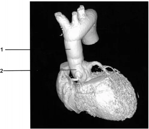
Electron beamed tomography images of a patient who

received a stentless valve as a whole root (Freestyle,

MEDTRONIC Inc, Minneapolis, MI, USA) for ascending

aortic aneurysm and aortic valve insuf®ciency 5 years after

the operation, with no sign of wall and lea¯et calci®cation

(Fig. 1).

European Journal of Cardio-thoracic Surgery 19 (2001) 929

1010-7940/01/$ - see front matter q 2001 Elsevier Science B.V. All rights reserved. PII: S1010-7940(01)00669-8

www.elsevier.com/locate/ejcts

Fig. 1. (1) Dacron graft used to extend the freestyle valve; (2) freestyle valve used as a total root.

* Corresponding author. Department of Cardiovascular Surgery, Florence Nightingale Hospital, Abide'i Hurriyet Cad. No:280, Sisli, Istanbul, Turkey. Fax: 190-21-2239-8791.

E-mail address: belh@turk.net (B. Akpõnar).

Şekil

Fig. 1. (1) Dacron graft used to extend the freestyle valve; (2) freestyle valve used as a total root.

Referanslar

Benzer Belgeler

Transesophageal long-axis (A) and short-axis echocardio- graphic views (B) showing the aneurysm of the left coronary cusp of the aortic valve resembling cystic mass (arrows) (C)

Transesophageal long-axis (A) and short-axis echocardio- graphic views (B) showing the aneurysm of the left coronary cusp of the aortic valve resembling cystic mass (arrows) (C)

Partial pericardial defect associated with ruptured aortic dissection of the ascending aorta: a rare feature presenting se- vere left hemothorax without cardiac

the subannular sutures; (ii) by attaching commissure holders to the aortic commissures, positioning of the device and aortic resuspension; (iii) using an

Bilateral coronary ostial patch angioplasty with autologous pericardium in Takayasu arteritis: a case requiring replacement of the aortic valve and ascending

Transesophageal echocardiography showed mobile vegetation of the bioprosthetic aortic valve, moderate to severe, eccentric aortic regurgitation, and a 14x34 mm aortic root

In this article, we present a successful valve-sparing aortic root replacement experience in a 19-year-old male patient of sinus valsalva aneurysm associated

Endovascular repair of a thoracoabdominal aortic aneurysm using ascending aorta as an access route in Leriche syndrome: a case report.. Leriche sendromunda torakoabdominal