Original Article / Özgün Araştırma
© Meandros Medical and Dental Journal, Published by Galenos Publishing House.
This is article distributed under the terms of the Creative Commons Attribution NonCommercial 4.0 International Licence (CC BY-NC 4.0).
Öz Abstract
Received/Geliş Ta rihi : 04.12.2018 Accepted/Ka bul Ta ri hi : 22.02.2019
ORCID ID: orcid.org/0000-0002-1916-8108
Ad dress for Cor res pon den ce/Ya zış ma Ad re si:
Sertaç Aksakallı MD,
İstanbul Aydın University Faculty of Dentistry, Department of Orthodontics, İstanbul, Turkey Phone : +90 533 354 86 85
E-mail : [email protected]
Keywords
Grape, expansion, rat, retention
Anah tar Ke li me ler
Üzüm, ekspansiyon, sıçan, retansiyon
Amaç: Medyan sutürün maksiller genişletilmesi ortodonti sıklıkla yapılan
bir uygulamadır. Retansiyon sonrasında bile genişlemiş sutürde geriye gidiş görülebilmektedir. Amacımız bu sutürdeki genişlemedeki kemikleşmeyi hızlandırmak ve kemikleşmeye etkisi olan üzüm çekirdeği ekstresi kullanarak retansiyon süresini kısaltmaktır.
Gereç ve Yöntemler: Yirmi adet rat iki gruba ayrıldı (n=20). Kontrol grubuna sadece
ekspansiyon yapıldı. Deney grubuna ise ekspansiyona ilaveten üzüm çekirdeği ekstresi verildi. Deney grubuna ekspansiyon sonrası orogastrik yöntemle sistemik olarak ekstreler verildi. Maksillaya springler uygulanıp aktiflendi. Beş gün sonra springler çıkartıldı ve köşeli tellerle retansiyon yapıldı. On iki gün pekiştirme yapıldı.
Bulgular: İki grup arasında yeni kemik alanı (p<0,05) ve yüzdeleri (p<0,05) açısından
anlamlı farklılıklar görüldü. İncelenen parametreler açısından, deney grubu daha iyi sonuçlar verdi.
Sonuç: Çene genişletmesinin erken dönemlerinde sistemik üzüm çekirdeği ekstresi
uygulanmasının midpalatal sutürde kemik oluşumunu artırdığı ve retansiyon
Objective: Widening the inter-maxillary suture is a preferred procedure in
orthodontics. However, relapse can occur in some patients after maxillary expansion therapy. This study aimed to increase bone formation in the inter-maxillary suture and decrease retention time with the help of grape seed extract (GS), which can stimulate bone tissue formation.
Materials and Methods: A total of twenty rats were separated into the following
two groups (n=20): the control group (group C) comprised rats that received only maxillary expansion therapy, and the other group (group GS) comprised rats that received maxillary expansion therapy along with GS. The grape seeds were systemically administered using an orogastric tube after maxillary expansion in group GS. Expansion springs were placed and activated to produce force. The springs were taken from the mouth 5 days later and exchanged with short rectangular wires for retention. This retention wire was placed for 12 days.
Results: Significant differences were found in the percentage of newly formed
bone (p<0.05) and bone area (p<0.05) between the two groups. Moreover, group GS had better bone formation than group C.
Conclusion: Systemic use of GS during the stages of the orthopaedic expansion of
the inter-premaxillary suture area increases newly formed bone and decreases the retention time.
Sertaç Aksakallı1, Şeref Ezirganlı2, Muhammet Birlik2, Hakkı Oğuz Kazancıoğlu2, Mehmet Şerif Aydın3
1İstanbul Aydın University Faculty of Dentistry, Department of Orthodontics, İstanbul, Turkey 2Private Practice
3İstanbul Medipol University, Regenerative and Restorative Medicine Research Center (REMER), İstanbul, Turkey
Üzüm Çekirdeği Özütünün İnter-premaksiller Sutürde
Kemikleşmeye Etkisi
Effect of Grape Seed Extract on Bone
Formation in The Expanded
Inter-premaxillary Suture
Introduction
Widening of upper jaw is a procedure in orthodontics for the theraphy of a small maxilla, posterior crossbite, or dental crowding (1). In rapid maxillary expansion (RME), the width of the posterior dentition increases first, followed by active bone formation in the expanded area (2). It is accepted that even after retention, there is a strong tendency to relapse in the expanded suture (3,4). Although the reasons for relapsing are not fully understood, some studies suggest that an insufficient retention period or changes in bone metabolism in the expanded suture might be responsible. After the active treatment, reorganization of the hard tissues in the suture begins and the ossification of the suture is complete in 60-90 days (3,4).Various experimental and clinical studies have focused on accelerating bone formation and consolidation in the expanded suture, in order to stabilize the maxilla and maxillary dentition and to shorten the retention time (5,6).
Several studies revealed that proanthocyanidine-type antioxidants prevent oxidative stress. In the dental literature, proanthocyanidines such as pine bark and grape seed extracts were reported to increase the low-bonding strength of composites after bleaching, which produces oxidation (7). Grape seed proanthocyanidine extract (GS) is derived from grape seeds during complex preservation and pharmaceutical processes (8). Proanthocyanidines are polyphenol extracts commonly found in vegetables, fruits, and flowers and have cytomodulating, antioxidant, antibacterial, antiviral, antiapoptotic, and anti-inflammatory properties. These compounds have a spectrum of pharmacological capabilities against oxidative stress, as well as a strong ability to scavenge oxygen free radicals (9,10). Recent studies revealed that GS could suppress bone destruction and promote bone formation in animal models (11). Ishikawa et al. (12) reported that GS had positive effect on mechanical properties associated with research animal mandibular condyle bone debility and some flavonoid functions increase osteoblast numbers and inhibit osteoclast activity.
Many studies in the field of orthodontics have investigated potential mechanisms for increasing bone formation during orthopedic expansion. For example, studies have shown that local application of
resveratrol during the early stages of inter-premaxillary suture expansion could stimulate bone formation and shorten the retention period (13). Similarly, Altan et al. (14) investigated the effect of propolis on the expanded suture and reported that systemic use of propolis could hasten new bone formation in rats. Therefore, the objectives of this study are to increase the osteoblastic processes in widened suture and accelerating bone formation can reduce the retention time by using GS.
Materials and Methods
Animals and Groups
Twenty 50- to 60-day-old male Sprague-dawley rats with a mean weight of 222.76±18.44 g were selected. The rats were placed in polycarbonate cages and subjected to a 12-h light-dark cycle at the constant temperature of 23 °C. The rats have been fed a standard pellet diet (Expanded pellets; Stepfield, Witham, Essex, UK) with tap water ad libitum. Permission to conduct the experiments was obtained from The Ethics Committee of Experimental Animals (approval no: 2013/107). The experiments were carried out in the Department of Experimental Animals, Research and Development Center in Bezmialem Vakıf University.
The research has been programmed as a parallel group design. In this programme, one group has been placed in the experimental protocol and the other has been placed in the control protocol. Power analysis was measured with G*Power ver.3.0.10 (Franz Faul, Universita ̈t Kiel, Germany) software. A size of 20 rats had greater than 90% power to detect significant differences including 0.40 effect size and a=0.05 (5). Rats were separated into two groups (control and experimental) of ten rats each with simple randomization.
Preparation of Grape Seed Extract
Grape seed extracts (Cactus Botanics, Long Beach, CA) have been placed under aseptic conditions and sterile volumetric flasks were used. A 25% GS was prepared using 3 g of grape seeds in 12 mL distilled water. The material was moderately shaken by magnetic mixer. It has been kept at room temperature. The solution was filtered under vacuum and the final concentrations were measured from the dry weights of the solutions as being 250 mg/mL. Specific dilutions
were made ready in the suitable culture medium. In the current research, we applied GS with dose of 100 mg/kg/d for Group GS.
Appliance Placement
The rats were anesthetized by intramuscular injection of 3 mg/kg xylazine hydrochloride (Rompuns, Bayer, Leverkusen, Germany) and 35 mg/kg ketamine hydrochloride (10% Ketasol®, Richter Pharma AG,
Wels, Austria). Helical springs prepared from 0.012-inch length of steel wire has been selected to widen the inter-premaxillary suture (Figure 1). The prepared springs has been put on a grid. Later they have been activated with pliers. The 30 gram force was calculated by using a gauge. To hold as retention, a groove on the distal sides of the maxillary incisor teeth have been performed. Next, 0.009-inch stainless-steel wire was used to keep in place the spring.
Twenty animals were randomly placed into two groups (n=10). The control group (group C) named as the maxillary expansion group. The maxillary expansion and GS group (group GS) is the other group. GS was given systemically with orogastric tubes when the expansion finished in the rate of 100 mg/kg/d.
The activated springs gave 30 g force and were not reactivated during the 5-day expansion peradverseiod. Five days later, the springs were taken from the mouth and short rectangular retaining wire
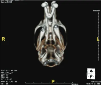
has been put. Tooth separation was maintained for 12 days. The consolidation phase started after an 5 days widening after distance of at least 1.5 mm was measured between maxillary incisors. It was confirmed by the literature that the distance of 1.5 mm was to be sufficient to induce the maximal rate of inter maxillary sutural widening (15). The inter-premaxillary suture was opened using helix springs and computed tomography revealed sufficient separation of the bones after the expansion period (Figure 2). The sutural width measurements were found to range between 338.32 and 390.68 μm. After period to consolodate of 15 days, the animals were euthanatized. 200 mg/kg of sodium pentothal (Pentothal; Abbot, North Chicago, Ill) were for this procedure. Surgically inter-premaxillary bone having the midpalatal suture cartilage was taken, then, for 24-48 hours, fixed in 10% formalin at room temperature.
The expansion of the inter-premaxillary suture was well tolerated. But, two rats were dispensed from the current research as a conclusion of spring problems. These animals were substituted with two another rats.
Histological Preparation
When fixation finished, the springs have been taken out. Demineralization has been performed in an aqueous 10% formic acid solution for specimens that were then dehydrated, later embedded in paraffin. To orient sections, the upper incisors accepted as the primary guide. A perpendicular cut performed
Figure 1. The expansion appliance in situ
Figure 2. Computed tomography of the inter-premaxillary suture after the expansion period
on the section as determined by two points, one was in the alveolar crest and the other one was 4 mm apical to the crest. The cut planes passed through the center of the gingival portion of the incisor crown. The paraffin blocks were cut into 5- μm-thin sections and made ready for hematoxylin - eosin staining prior to optical microscope examination. The bone histomorphometry measurements were centered on the inter-premaxillary suture, 175-250 mm (35th
-50th sections) under the surface of the ossified
palate facing the oral cavity, because surface bone formation was sometimes irregular and unsuitable for quantitative measurement.
The histologic and histomorphometric analyses were performed by the same histologist who was also blinded to the identity of samples. Histomorphometric analysis was performed centered around the inter-premaxillary suture and the sections under the surface of the ossified palate facing the oral cavity because bone formation on the surface was not regular and not suitable to make quantitative analysis. The presence of an inflammatory infiltrate, connective tissue, material resorption and bone regeneration were evaluated. Computer-assisted histomorphometric measurements were carried out using an automated image analysis system. The images of the histologic sections from all groups were examined using a fluorescent microscope (Nikon Eclipse i5, Tokyo, Japan), coupled with a video camera on a light microscope (Nikon, DS-Fi1c, Tokyo, Japan), and saved on a computer. Two flatways (straights) were defined on the sutura region. One of the flatway began at the incisors and the other was placed 2.5 mm from the first straight (Figure 3). Afterwards,
the formated new bone area (mm2) and percent
of the new bone formation were measured in the expanded suture area. For these measurements the NIS Elements version 4.0 image analysis system was used (Nikon Instruments Inc., Tokyo, Japan) with an original magnification of 40× on the fluorescent images (Figure 4).
Statistical Analysis
All variables and measurements were evaluated with the statistical package for social sciences, 15.0 (SPSS for Windows; SPSS Inc., Chicago, IL, USA). Quartiles were used on descriptive statistics (25th, 50th
- median - and 75th), minimum (Min) and maximum
(Max). The Mann-Whitney U test was used to evaluate differences between the two groups. The p value was set at <0.05.
Results
The expansion of the inter-premaxillary suture was well tolerated. No adverse effects such as inflammation, dehiscence, and mucosal trauma were observed in any of the rats. The mean body weight did not differ between the groups during the course of the experiment. Successfully the midpalatal suture was distracted after application of the active helical springs. New bone formation was compared between the groups and meaningful differences were found. The results revealed that new bone formation was significantly increased in the GS group over that observed in the control group (Figure 5 and 6).
Newly formed bone percentages (new bone area ratio to suture area) (p<0.05) and new bone area (p< 0.05) were significantly different between the two
Figure 3. Two straights were determined on the suture area. One of the straights was at the beginning of the incisors and the other was 2.5 mm from the first straight (g: Gingiva, s: Suture area, t: Tooth, *: New bone area)
Figure 4. The regenerated bones shown at the original magnification of 40× in the expanded suture area (A: Hematoxylin-eosin stain; B: The regenerated bone areas in the fluorescent image, hb: Host bone, rb: Regenerated bone, ct: Connective tissue, *: Capillary)
groups. For all investigated histologic parameters, better results about bone formation were found in Group GS (Table 1).
Discussion
Our objective was to study the effects of GS on new osteoblastic activity in the widened inter-premaxillary suture. This animal research is the first to study the effects of GS intake on expanded sutures.
For investigating RME, the rat model is one of the well-built model. Rats and rabbits are suitable for animal studies that focus on hard and sutural tissues (1). For our study, ethical considerations dictated that rats, as the smallest ideal animal model, should be used to test the new material on bone formation.
Weight loss, infection, and appliance problems such as failure were controlled in rats during the study. In a situation such as decrease in animal weight, the appearance of infection or appliance problem, the rats were taken out from research and replaced.
Histomorphometric method was selected to understand the effects of GS on the rate of new bone tissues during upper jaw widening procedure. A program for image analysis used to determine the histological changes objectively and revealed that total bone area amount correlated with newly formed bone amount. Technique named as bone histomorphometry is an acceptable technique that is usually chosen for the quantitative evaluation of bone formation (13,16). Similar studies also measured the area of new bone as an evaluation criterion. However, other parameters such as Feret’s diameter, bone
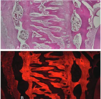
Figure 5. (A) Photomicrograph of a section in the expansion area of Group GS showing larger masses of new bone trabeculae. (HE 400× magnification) (B) Immunofluorescence analysis of tissues in Group GS.
GS: Grape seed proanthocyanidine extract
Figure 6. (A) Photomicrograph of a section in the expansion area of Group C showing abundant formation of bone trabeculae. (HE 400× magnification). (B) Immunofluorescence detection of tissues in group C.
Table 1. Results and statistical comparisons of biometric analysis for determination of expansion
Parameters Group n Min Max 25% Median 75% p value
Area investigated (μm2) C 10 101.23 149.59 106.39 130.98 140.5 0. 036
GS 10 125.52 176.79 134.04 155.39 168.73 -Newly formed bone (%) C 10 40.10 45.17 41.4 42.89 45.12 0.027
GS 10 43.90 54.90 44.62 47.72 52.66
-*Mean ± Standard deviation for number of osteoclasts were 6.61±0.85 in group GS, 3.43±0.40 in group C (p<0.05) n: sample size, Min: Minimum, Max: Maximum, C: Control, GS: Grape seed proanthocyanidine extract
perimeter, and osteoblast and osteoclast counts, were considered to be non-objective measurements. Particularly, the exact number of the osteoblasts and osteoclasts in the expanded area cannot be realistically determined without performing immunohistochemical staining. In addition, the amount of the new bone area is associated with osteoblast number. Therefore, we evaluated the new bone area and the percentage of newly formed bone in this study. However, investigators in other studies used different analytical methods. For example, da Silva et al. (17) evaluated osteogenic parameters and gene expression markers in cell culture experiments after treating the midpalatal suture expansion with low level laser theraphy. In addition, Rosa et al. (18) evaluated bone formation using Raman spectroscopy. Furthermore, Kobayashi et al. (19) determined alkaline phosphatase activity using histochemical staining.
Some investigators have investigated the space the upper incisors mesial side at the beginning and on the fifth day of the widening using calipers. It has been accepted that a 5-day widening time is sufficient to expand suture (16). Therefore, the space between the upper incisors was not used on the current research, although it has been verified there was space between the incisor teeth of all rats. Additionally, our histologist verified that the required widening was okay in the sutura palatina of all rats. Burstone and Schafer (20) (year) stated that sutural expansion of young rats over a period of 5 days resulted in a suture opening with an average length of 377±10 μm.In our study, the sutural width measurements ranged between 338.32 and 390.68 μm. The amount of expansion in our study was similar to previous investigations and was not significantly different between our two groups (p=0.58).
Numerous researches revealed a positive correlation: oxidative stress-bone metabolism. The free radical plurality which give harm biological mechanisms are oxygen-free radicals. They also were known as “reactive oxygen species” (ROS). Oxidative stress caused by ROS can have dangerous biological effects on bone through cell differentiation inhibition and in the marrow of the stromal cell line. ROS can directly promote osteoclast formation, and ROS or tumor necrosis factor can diminish osteoblast differentiations (21,22). Therefore, various host modulating agents, including antioxidants, have been
widely investigated for the capability ameliorate the oxidant-related problems of hard tissues and for the promotion of bone healing. The activity of GS has been attributed to the antioxidants flavan-3-ol or catechin that scavenge free radicals (23). Similarly, supplementation with extracts of grape seed proanthocyanidines more effectively reversed mandibular condyle bone debilities induced by a low-calcium diet compared to a standard diet or high-calcium diet alone (5). There are studies in the literature showing that GS increases bone formation/ strength but a study focusing on the effects of GS on an expanded inter-premaxillary suture have not been performed (11,12). In addition, treatment with antioxidants such as boron or propolis has been shown to enhance new bone formation.
GS is considered to be safe at low doses. On the current study, we preferred GS at a dose of 100 mg/ kg, similar to other studies (13,14), and found no adverse effects. Studies show that the body weight of rats given either a powdered diet or GS solution were not different from rats given a standard diet, but excessive GS can cause a reduction in body weight and reduce food intake (24). In this study, GS was used with orogastric feeding. Food consumption increase was detected, although it was not accompanied by an increase in body weight. In our study, over a 4-week experimental period, we did not observe significant differences in body weight between the groups.
In the first 14-day period of expanded suture healing in rats, osteoprogenitor cells proliferate and differentiate and ossification starts. GS can stimulate bone formation by osteoblasts and accelerate proliferation of osteoblasts (11,12). In our study, on the 10th day of retention, histologic and histomorphometric
evaluations revealed GS accelerated bone healing. GS may be useful for a wide range of applications such as treating osteoporosis, increasing bone formation and bone repair, and repairing bone defects.
Conclusion
Systemic administration of GS in rats during the beginning times of widened palatinal suture areas can raise bone formation. New researches are needed to evaluate GS effects in human being and to determine if GS must be used continuously or prophylactically until the end of retention.
Ethics
Ethics Committee Approval: Permission to conduct the experiments was obtained from Bezmialem University of The Ethics Committee of Experimental Vakıf Animals (approval no: 2013/107).
Peer-review: Externally peer-reviewed.
Authorship Contributions
Concept: S.A., Ş.E., Design: S.A., M.Ş.A., Data Collection or Processing: S.A., M.B., Analysis or Interpretation: H.O.K., M.Ş.A., Literature Review: S.A., Writing: S.A., S.E.
Conflict of Interest: No conflict of interest was declared by the authors.
Financial Disclosure: The authors declared that this study received no financial support.
References
1. Saito S, Shimizu N. Stimulatory effects of low-power laser irradiation on bone regeneration in midpalatal suture during expansion in the rat. Am J Orthod Dentofacial Orthop 1997; 111: 525-32.
2. Sawada M, Shimizu N. Stimulation of bone formation in the expanding mid-palatal suture by transforming growth factor-beta 1 in the rat. Eur J Orthod 1996; 18: 169-179.
3. Haas AJ. The treatment of maxillary deficiency by opening the midpalatal suture. Angle Orthod 1965; 35: 200-17.
4. Cleall JF, Bayne DI, Posen JM, Subtelny JD. Expansion of the midpalatal suture in the monkey. Angle Orthod 1965; 35: 23-35. 5. Uysal T, Ustdal A, Sonmez MF, Ozturk F. Stimulation of bone
formation by dietary boron in an orthopedically expanded suture in rabbits. Angle Orthod 2009; 79: 984-90.
6. Uysal T, Olmez H, Amasyali M, Karslioglu Y, Yoldas A, Gunhan O. Response of the expanded inter-premaxillary suture to intermittent compression. Early bone changes. Australian Orthodontic Journal 2010; 26: 49-55.
7. Aksakalli S, Ileri Z, Karacam N. Effect of pine bark extract on bond strength of brackets bonded to bleached human tooth enamel. Acta Odontol Scand 2013; 71: 1555-9.
8. Vidhya S, Srinivasulu S, Sujatha M, Mahalaxmi S. Effect of grape seed extract on the bond strength of bleached enamel. Oper Dent 2011; 36: 433-8.
9. Ozkan G, Ulusoy S, Orem A, Ersoz S, Alkanat M, Balaban Yucesan F, et al. Protective effect of the grape seed proanthocyanidin extract in a rat model of contrast-induced nephropathy. Kidney Blood Press Res 2012; 35: 445-53.
10. Ashtiyani SC, Najafi H, Firouzifar MR, Shafaat O. Grape seed extract for reduction of renal disturbances following reperfusion in rats. Iranian journal of kidney diseases 2013; 7: 28-35.
11. Yahara N, Tofani I, Maki K, Kojima K, Kojima Y, Kimura M. Mechanical assessment of effects of grape seed proanthocyanidins extract on tibial bone diaphysis in rats. J Musculoskelet Neuronal Interact 2005; 5: 162-9.
12. Ishikawa M, Maki K, Tofani I, Kimura K, Kimura M. Grape seed proanthocyanidins extract promotes bone formation in rat’s mandibular condyle. Eur J Oral Sci 2005; 113: 47-52.
13. Uysal T, Gorgulu S, Yagci A, Karslioglu Y, Gunhan O, Sagdic D. Effect of resveratrol on bone formation in the expanded inter-premaxillary suture: early bone changes. Orthod Craniofac Res 2011; 14: 80-7.
14. Altan BA, Kara IM, Nalcaci R, Ozan F, Erdogan SM, Ozkut MM, et al. Systemic propolis stimulates new bone formation at the expanded suture: a histomorphometric study. Angle Orthod 2013; 83: 286-91.
15. Takahashi O. [Histological investigations on the effect of interrupted expansion force applied to the midpalatal suture in the rat]. Nichidai Koku Kagaku 1990; 16: 212-36.
16. Uysal T, Amasyali M, Enhos S, Sonmez MF, Sagdic D. Effect of ED-71, a New Active Vitamin D Analog, on Bone Formation in an Orthopedically Expanded Suture in Rats. A Histomorphometric Study. Eur J Dent 2009; 3: 165-72.
17. da Silva AP, Petri AD, Crippa GE, Stuani AS, Stuani AS, Rosa AL, et al. Effect of low-level laser therapy after rapid maxillary expansion on proliferation and differentiation of osteoblastic cells. Lasers Med Sci 2012; 27: 777-83.
18. Rosa CB, Habib FA, de Araújo TM, Aragão JS, Gomes RS, Barbosa AF, et al. Effect of the laser and light-emitting diode (LED) phototherapy on midpalatal suture bone formation after rapid maxilla expansion: a Raman spectroscopy analysis Lasers Med Sci 2014; 29: 859-67.
19. Kobayashi ET, Hashimoto F, Kobayashi Y, Sakai E, Miyazaki Y, Kamiya T, et al. Force-induced rapid changes in cell fate at midpalatal suture cartilage of growing rats. J Dent Res 1999; 78: 1495-504.
20. Burstone CJ, Shafer WG. Sutural expansion by controlled mechanical stress in the rat. J Dent Res 1959; 38: 534-40. 21. Cadenas E. Biochemistry of oxygen toxicity. Annu Rev Biochem
1989; 58: 79-110.
22. Lean JM, Davies JT, Fuller K, Jagger CJ, Kirstein B, Partington GA, et al. A crucial role for thiol antioxidants in estrogen-deficiency bone loss. J Clin Invest 2003; 112: 915-23.
23. Pataki T, Bak I, Kovacs P, Bagchi D, Das DK, Tosaki A. Grape seed proanthocyanidins improved cardiac recovery during reperfusion after ischemia in isolated rat hearts. Am J Clin Nutr 2002; 75: 894-9.
24. Wren AF, Cleary M, Frantz C, Melton S, Norris L. 90-day oral toxicity study of a grape seed extract (IH636) in rats. J Agric Food Chem 2002; 50: 2180-92.