Nested Metamaterials for Wireless Strain Sensing
Rohat Melik, Student Member, IEEE, Emre Unal, Nihan Kosku Perkgoz, Brandon Santoni, Debra Kamstock,
Christian Puttlitz, and Hilmi Volkan Demir, Member, IEEE
Abstract—We designed, fabricated, and characterized metamaterial-based RF-microelectromechanical system (RF-MEMS) strain sensors that incorporate multiple split ring resonators (SRRs) in a compact nested architecture to measure strain telemetrically. We also showed biocompatibility of these strain sensors in an animal model. With these devices, our bioimplantable wireless metamaterial sensors are intended, to enable clinicians, to quantitatively evaluate the progression of long-bone fracture healing by monitoring the strain on the implantable fracture fixation hardware in real time. In operation, the transmission spectrum of the metamaterial sensor attached to the implantable fixture is changed when an external load is applied to the fixture, and from this change, the strain is recorded remotely. By employing telemetric characterizations, we reduced the operating frequency and enhanced the sensitivity of our novel nested SRR architecture compared to the conventional SRR structure. The nested SRR structure exhibited a higher sensitivity of 1.09 kHz/kgf operating at lower frequency compared to the classical SRR that demonstrated a sensitivity of 0.72 kHz/kgf. Using soft tissue medium, we achieved the best sensitivity level of 4.00 kHz/kgf with our nested SRR sensor. Ultimately, the laboratory characterization and in vivo biocompatibility studies support further development and characterization of a fracture healing system based on implantable nested SRR.
Index Terms—Biocompatibility, metamaterial, nested SRR, re-mote sensing, resonance frequency, sensitivity, split ring resonator (SRR), strain, telemetric.
I. INTRODUCTION
T
HE ABILITY to telemetrically measure strain is impor-tant in many aspects of daily life. But such a task brings about important challenges. In many sectors such as in civil en-gineering, measuring the strength of materials (e.g., concrete), remotely in real time, will help us to understand their transient Manuscript received June 5, 2009; revised June 29, 2009 and August 27, 2009. First published December 28, 2009; current version published April 7, 2010. This work was supported in part by the Turkish National Academy of Sciences Distinguished Young Scientist Award and European Science Foun-dation European Young Investigator Award Programs, and The Scientific and Technological Research Council of Turkey under Projects EEEAG 104E114, 106E020, 107E088, 107E297, 109E002, and 109E004. This work is also sup-ported by a grant from the United States National Institutes of Health (NIH) 5R01EB010035-02.R. Melik, E. Unal, and N. K. Perkgoz are with the Department of Electrical and Electronics Engineering, Nanotechnology Research Center, and Institute of Ma-terials Science and Nanotechnology, Bilkent University, 06800 Ankara, Turkey (e-mail: [email protected]; [email protected]; [email protected]). B. Santoni, D. Kamstock, and C. Puttlitz are with the Orthopedic Bioengineer-ing Research Laboratory, Department of Mechanical EngineerBioengineer-ing and School of Biomedical Engineering, Colorado State University, Fort Collins, CO 80523 USA (e-mail: [email protected]; [email protected]; [email protected]).
H. V. Demir is with the Department of Electrical and Electronics Engineer-ing, the Department of Physics, Nanotechnology Research Center, and Institute of Materials Science and Nanotechnology, Bilkent University, 06800 Ankara, Turkey (e-mail: [email protected]).
Color versions of one or more of the figures in this paper are available online at http://ieeexplore.ieee.org.
Digital Object Identifier 10.1109/JSTQE.2009.2033391
structural behavior better (e.g., before and after an earthquake). Real-time measurement of the flexural rigidity of aircraft com-ponents during service in avionics is also an important applica-tion of telemetric strain sensing. Another unrealized, yet critical, application area is human medicine.
One important clinical issue in which we are currently in-terested is objectively monitoring the healing processes of fractured long bones [1]. Orthopedic extremity injuries cur-rently present a large medical and financial burden to both the United States and worldwide communities, as can be seen in [2]. Severely comminuted-fracture patterns, commonly seen in high-energy fractures, are difficult to treat due to the inher-ent absence of mechanical support by the native osseous tissue. In these cases, the implanted hardware (intramedullary rods, bone plates, screws, etc.) must assume the total mechanical load in the early postoperative term, which frequently results in an aberrant course of healing and the onset of delayed union or nonunion. The most common treatment for these complications is additional surgery. These types of orthopedic injuries require prolonged time before patients return to full activity [3].
Approximately six million long bone fractures are reported per annum in the United States. Surprisingly, approximately 10% of these fractures do not heal properly. Though the ex-act mechanism through which the healing progression becomes impaired is poorly understood, many of these nonunions or pseudoarthroses result when there is a severe or communited condition that does not proceed through a stabilized (intramem-branous ossification) healing pathway [4]. Currently, clinicians may monitor healing visually by radiographs, and may examine the mechanical condition of the union through manually bend-ing the bone at the fracture [5]. However, the course of aberrant fracture healing is not easily diagnosed in the early time period when standard radiographic information of the fracture site is not capable of discriminating the healing pathway. The manual assessment of fracture healing is also subjective and, therefore, inadequate as a diagnostic tool in the early stages of healing, as shown in [6].
It has been shown in animal models that healing is criti-cally important in the early time period. Animal studies have demonstrated that the callus and bone assume an increasing proportion of the load as healing proceeds, reducing the load carried by the implanted hardware [1]. However, to date, many of the technologies that seek to exploit this bone-implant load-sharing phenomena have been considered too large in dimension or involve implantation of an associated power supply. Previous investigations have been successful in determining forces in the hip [7]–[9], spine [10]–[12], and femur [13], [14]. However, due to the relatively large size of the sensors and associated hard-ware (signal conditioning, modulation, etc.), most of the afore-mentioned telemetry systems have been implanted insidejoint 1077-260X/$26.00 © 2009 IEEE
replacement components or bulky internal fixators. The result is that these devices have produced data that have been use-ful in the understanding of bone-implant loading, but not been advantageous for large-scale implementation, as diagnostic and prognostic tools. Also, due to the complexity of the designs and requisite interconnectivity, manufacture of these systems could only be performed on a custom basis. The resulting expense could not justify their large-scale manufacture.
To overcome the aforementioned shortcomings, we have developed RF-microelectromechanical system (MEMS) strain sensors that take advantage of the recent advances in metamate-rials. To date, metamaterials have been extensively investigated and exploited for various applications [15]–[26]. In the previous literature, some of these applications include achieving negative refraction [18]–[21], obtaining plasmons using nanowires [22], making metamaterial antennas [23], focusing light [24], cloak-ing [25], and buildcloak-ing superlenses [26]. For metamaterials, sens-ing opens up a new direction where metametarials may provide unique benefits.
These split ring resonator (SRR)-based sensors are passively powered devices (with no implantable power source), which we implement to monitor the surface bending strains on implanted fracture fixation hardware. Another important feature of these wireless sensors is that they do not require an internal–external physical connection to sense and transmit in vivo biological data. Finally, the miniaturization of these metamaterial-based sensors allows for their use in various implant applications that otherwise would not be possible. As the SRR-based sensor dis-plays a characteristic resonance frequency under no load, by measuring the magnitude of operating frequency shift under ap-plied load, we are able to delineate the bending strain incurred in the fixation hardware and intend to relate this information to the progression of fracture healing.
For the sensing operation, four criteria are important and demonstrate the quality of the sensor. First, the sensor must have a low enough operating frequency (subgigahertz range) to avoid the background absorption of soft tissue. This poses a significant challenge as conventional fracture fixation devices have limited area to which the sensors can be affixed, and such reduced space tends to increase the operating frequency of the sensor. This, in turn, undesirably increases the background absorption of human body (soft tissue). At higher frequencies, the soft tissue limits the penetration depth of electromagnetic waves. Therefore, it is required to maintain a small layout of the sensor while decreasing its operating frequency as much as possible.
Second, the sensors must emit a strong and measurable read-out signal with sharp enough resonance behavior (high enough
Q-factor) to accurately track the shift in transmission spectra.
The third criterion pertains to the linearity of sensing, which is related to the quality factor of the sensor. A higher Q-factor leads to a higher SNR, which reduces errors in the measure-ment (e.g., from the network analyzer) and decreases the non-linearity error. The fourth criterion is sensor sensitivity. Since there are a limited number of data points in one frequency scan of the network analyzer, it is easier to resolve smaller shifts in the transmission spectra in response to the externally applied load when the sensitivity is higher. With the same
level of induced strain, higher sensitivity yields larger shifts in transmission.
Metamaterials provide the ability to make better wireless sen-sors compared to conventional RF structures (e.g., spiral coils) because of their advantages over conventional structures with respect to four criteria listed earlier. The benefits of SRRs in tele-metric sensing mainly stem from their unique structure, which features gaps (splits). Because of these gaps, SRRs exhibit lower resonance frequencies per unit area compared to spiral coils. This is critically important for bioimplant strain sensing within a limited space and in soft tissue environment. Furthermore, SRRs yield deeper and sharper dips, at resonance in their trans-mission spectra, compared to the spiral structures. The gaps of an SRR have much higher electric field density, which makes the resonance stronger and leads to a higher Q-factor, and, in turn, a higher SNR. This makes the SRR sensor more linear compared to the spiral coil sensor. As a final consequence of gap presence in the SRRs when an external load is applied, the change in the additional capacitance further shifts the operat-ing frequency, makoperat-ing the SRR more sensitive compared to the spiral structure. In our previous work as such, we used a single SRR structure in our strain sensor [27].
In this paper, we fabricate and characterize nested metamaterial-based strain sensors that incorporate multiple SRRs in a compact nested architecture on a single chip to sig-nificantly achieve enhanced sensitivity in telemetric sensing. This architecture introduced for implant sensing substantially features more gaps compared to the structure of a conventional SRR. This decreases the operating resonance frequency of the resulting nested SRR sensor compared to the classical SRR sen-sor. Moreover, when the external load is applied, the capacitance of our nested SRR sensor is changed to a greater degree than the classical SRR, resulting in larger shifts in the transmission spec-trum. This makes the nested SRR more sensitive than classical SRR in sensing.
Here we present the design, fabrication and characterization of our nested-metamaterial based RF-MEMS strain sensors, and compare the telemetric sensing operation of the classical SRR and nested SRR based sensors in telemetric sensing. Further, we characterize the site-specific biocompatibility and wound-healing response elicited against these sensors in a small animal model. Finally, we present the first proof-of concept telemetric demonstration of these nested SRR sensors using soft tissue
in vitro.
II. DESIGN ANDFABRICATION
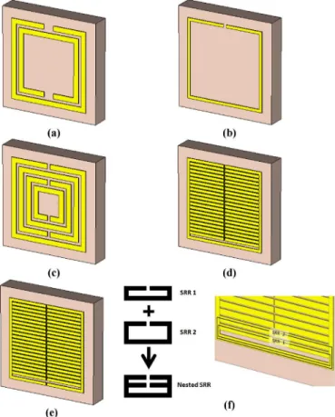
Fig. 1(a)–(c) shows schematics of three designs of conven-tional SRRs, with two gaps (in two turns) in Fig. 1(a) (classical SRR), with a single gap (in one turn) in Fig. 1(b), and with four gaps (in four turns) in Fig. 1(c), respectively. Here, increasing the number of turns decreases the operating resonance frequency because of the increased number of gaps, and thus, increased capacitance. However, the total possible reduction in the reso-nance frequency is limited by the space available on the chip, as each turn takes up a considerable amount of space. To make a compact sensor, we propose nesting of SRRs that share the same sides except where the gap is located, as depicted in Fig. 1(d)
Fig. 1. SRR with (a) two turns (classical SRR), (b) one turn, and (c) four turns. (d) Nested SRR architecture. (e) Comb-like structure. (f) Zoom-in view of nested SRR.
(nested SRR). With many more gaps available in the nested SRR, the resonance frequency is further reduced, consequently resulting in an increase in sensor sensitivity.
A key feature of the nested architecture is the connected bottom line of the RF structure, which confers continuity in our nested design. For comparison, Fig. 1(e) shows a pair of comb-like structures. These comb-comb-like structures yield undesirably high resonance frequencies (even higher than that of the classical SRR) due to the discontinuous bottom line. As can be seen in the zoom-in view of the nested SRR in Fig. 1(f), there are many SRRs nested together. These SRRs are added together with the same continuous bottom line, being the only difference with respect to Fig. 1(e).
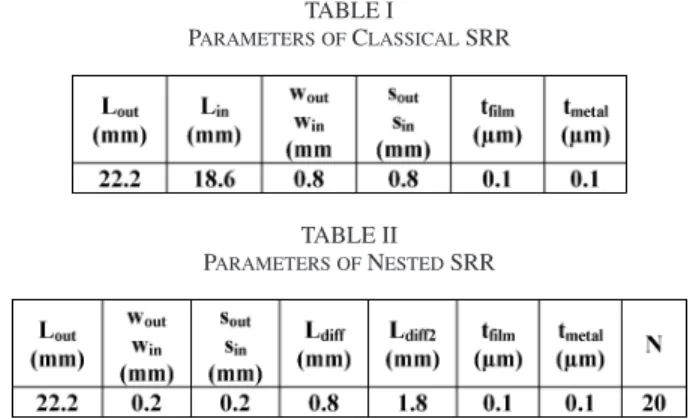
For the classical SRR, we denote the total length of the outer coil as Lout, total length of the inner coil as Lin, width of the outer coil as wout, width of the inner coil as win, spacing across the gap of the outer coil as sout, and spacing across the gap of the inner coil as sin. These parameters are depicted in Fig. 2(a). In the context of a design with fixed chip size (constant Lout), in-creasing Lindecreases the resonance frequency. But making Lin too much closer to Loutdecreases Q-factor due to the increased parasitic capacitance. Increasing Lin increases the capacitance between substrate and metal (Cdiel), decreasing the resonance frequency. Increasing win and wout increases the capacitance between gaps (Cgap) as well as Cdiel, which decreases the reso-nance frequency. Increasing sin and soutdecreases Cgap, thus, increasing the resonance frequency.
Fig. 2. Plain view pictures of (a) the classical SRR structure and (b) nested SRR structure.
The design parameters of the nested SRR are shown on a fabricated sample in Fig. 2(b). Ldiff is the distance between the combs, and Ldiff 2is the distance between the bottom line and the bottom comb teeth. The width of the top comb teeth is wout, and the width of the nested comb teeth is win. The spacing across the gap of top comb teeth is soutand that of the others is sin. Loutis the total length of the structure. For the design parameters, the same conditions, as in classical SRR, apply in general. Increas-ing winand woutdecreases the resonance frequency. Increasing
sin and soutincreases the resonance frequency. If the width is much greater than the spacing, then the parasitic capacitance dominates, so the Q-factor decreases. Therefore, we carefully choose the ratio of the width to the spacing.
The most important parameter for determining the resonance frequency of the nested SRR is the number of comb teeth (N ) in the design. As N increases, both Cgap and Cdiel increase, thus decreasing the resonance frequency. Another parameter of inter-est in the ninter-ested SRR is Ldiff. If we increase Ldiff, then we de-crease N . Subsequently, Cgapand Cdiel are decreased, thereby
TABLE I
PARAMETERS OFCLASSICALSRR
TABLE II PARAMETERS OFNESTEDSRR
increasing the resonance frequency. However, Ldiff should be greater than or equal to the width, otherwise the parasitic ca-pacitance dominates. Ldiff 2 is another important parameter. We decrease Ldiff 2 as much as possible to decrease the resonance frequency, since our goal is to maximize N . If Ldiff 2 is in-creased, then N is decreased. Therefore, Cgap and Cdiel are decreased and the resonance frequency is increased. We make
Ldiff 2 greater than or equal to 2Ldiff + win so that we do not ruin the sequence of the combs, and the parasitic capacitance does not dominate to decrease Q-factor.
The dielectric layer is also an important factor in our design. To have sufficient capacitance between the metal and the sub-strate, which serves as the distributed capacitance, we used a dielectric layer with a high dielectric constant [28]. On the other hand, to minimize the loss, a low-loss dielectric is required. Therefore, Si3N4 with a dielectric constant of eight and a loss tangent of 5× 10⁻⁴ was chosen as the dielectric film to satisfy these conditions. Its film thickness was set to 0.1 µm to confer maximum capacitance over the minimum area.
The metal type and substrate used in the design are also crit-ical, particularly for biocompatibility. We used Au as the metal layer and Si as the substrate. We also chose Si3N4as the dielec-tric layer because it is biocompatible [28]. For the fabrication process, we first deposited 0.1-µm-thick Si3N4 onto our sili-con substrate by plasma-enhanced chemical vapor deposition (PECVD). Then, by using standard lithography, metal evapo-ration, and lift-off, we deposited and patterned a 0.1-µm-thick Au film to make our sensors. The design parameters for the optimized classical SRR and the optimized nested SRR used in this paper are provided in Tables I and II.
III. EXPERIMENTALCHARACTERIZATION
In this section, we characterized differences in telemetric strain-sensing ability between the classical SRR- and nested SRR-based sensors. We then evaluate the biocompatibility of our sensors over a six-month time period in a small animal model. Finally, we demonstrated telemetric operation in soft tissue using our nested SRR sensor as a first proof-of-concept demonstration for implant applications.
A. Comparison of Classical and Nested SRR Strain Sensors
We experimentally characterized the metamaterial-based sen-sors using a custom-design mechanical testing setup. In this
pa-Fig. 3. (a) Mechanical apparatus and (b) coaxial probe antennas.
per, a uniaxial traction force was applied in a controlled manner to a polyamide beam rigidly coupled to a load cell and actuator, as shown in Fig. 3(a). Using this apparatus constructed at Bilkent University, loads were incrementally applied up to 300 kgf. The classic SRR and the nested SRR sensors were rigidly affixed to the polyamide beams with hard epoxy prior to testing.
Antennae made of coaxial probes with the same ground to decrease the noise during characterization were used to measure the change in RF spectrum of the loaded sensors during testing [see Fig. 3(b)]. The length of these probes was set to 2.5 cm, which was comparable to the size of our sensors. Because our sensors are small in size, i.e.,λo/30–λo/25, in comparison to
their operating wavelength, it was rather difficult to use stan-dard antennas with sizes comparable to our sensors to measure their transmission spectra. The distance between these probe an-tennas was set equal to Lout, and they were placed 0.5 cm away from the sensor, as shown in Fig. 3(b). In this configuration, the best signal is obtained with the probes parallel to the sen-sor. All these distance parameters were kept fixed throughout the calibration process and characterization process. For cali-bration purposes, the transmission of the polyamide beam was measured first with no sensor chip attached. Subsequently, the same measurement was repeated with the sensor attached under no load, and then, also following application of discrete tensile
loads. Transmission spectra referenced relative to the no-sensor condition were obtained as a function of the applied load.
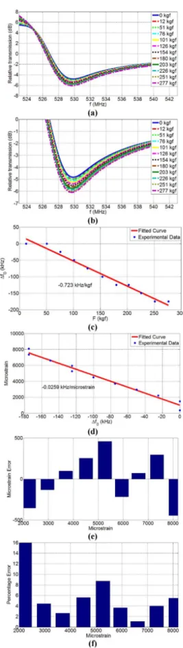
Relative transmission spectra (in decibel) are presented in Figs. 4 and 5 for the classical SRR and nested SRR sensors, re-spectively, with respect to the case of no sensor in semilog scale. Under no load, the operating frequency of the classic SRR was 529.8 MHz. At this point, the size of this classical SRR sensor corresponded toλo/25.5. With the applied load, the operating
frequency decreased, as seen in Fig. 4(a) and (b). Under the ap-plied tensile load, the gaps of SRR are decreased, hence, Cgapis increased. Also the dielectric area between substrate and metal layer is increased, thus Cdiel is increased [29]. Fig. 4(c) plots the operating frequency shift (with respect to the case of no load) as a function the applied load and indicates a sensitivity of 0.723 kHz/kgf for the classic SRR. Fig. 4(d) presents the induced strain (in microstrain) as a function of operating frequency shift. Here Young’s modulus of the cast polyamide beam was taken to be 2.37 GPa, which is separately verified using a wired strain gauge (Tokyo Sokki Kenkyujo Company, Ltd., Strain Gauges with a gauge factor of 2.1). This SRR sensor demonstrates a sensitivity of 0.0259 kHz/microstrain. In Fig. 4(e), we observed that this sensor had less than 500-microstrain nonlinearity er-rors, which corresponded to 16% nonlinearity error, as shown in Fig. 4(f).
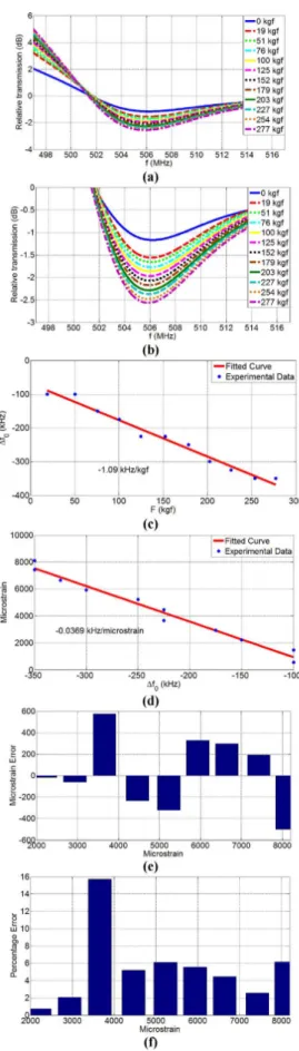
Transmission spectra of our novel SRR structure (nested SRR) for different levels of the applied load are illustrated in Fig. 5(a) and with zoom-in view in Fig. 5(b). Here the operating frequency was 506.2 MHz under no load, which was lower than that of the classical SRR, and a function of an increased number of in the nested SRR. The size of this nested SRR corresponded toλo/26.7. Fig. 5(c) plots the change in the operating frequency
with respect to the case of no load as a function of the applied load and indicates a sensitivity of 1.09 kHz/kgf. The sensitivity of the nested SRR was significantly increased compared to the classical SRR as a result of the multiple gaps used in the nested SRR. Fig. 5(d) plots the strain versus frequency shift demon-strating a sensitivity of 0.0369 kHz/microstrain. In Fig. 5(e), the nested SRR sensor had less than 600-microstrain nonlin-earity errors, corresponding to less than 16% nonlinnonlin-earity error in Fig. 5(f). The nonlinearity errors of the nested SRR sensors are nearly the same as those of the classical SRR. However, the nested SRR sensor exhibited reduced operating frequency and increased sensitivity.
B. Biocompatibility Experiments of our Sensors
New Zealand white rabbits (Harlan, Inc., Indianapolis, IN) were chosen as an appropriate animal model to investigate the sit-specific biocompatibility of our MEMS sensors due to their size and ability to house multiple sensor chips. A total of four rabbits each implanted with four sensors and two control ma-terial implants (6 implants/rabbit; 16 sensors and 8 controls to-tal) were used to investigate the biocompatibility in accordance with ASTM Standards F981-04 and F763-04. Animals were hu-manely euthanized six months post-operatively at which time critical gross pathology and microscopic evaluation of the im-plant sites for an imim-plant-associated tissue reaction was pursued.
Fig. 4. Experimental characterization of the classical SRR sensor under ten-sion. (a) Relative transmission spectra. (b) Zoom-in view of the transmission shift. (c) Frequency shift (∆fo) versus applied load (F ). (d) Induced strain
(mi-crostrain) versus frequency shift (∆fo). (e) Nonlinearity error (in microstrain).
Fig. 5. Experimental characterization of the nested SRR sensor under ten-sion. (a) Relative transmission spectra. (b) Zoom-in view of the transmission shift. (c) Frequency shift (∆fo) versus applied load (F ). (d) Induced strain
(mi-crostrain) versus frequency shift (∆fo). (e) Nonlinearity error (in microstrain).
(f) Nonlinearity error percentage.
This study was approved by the Institutional Animal Care and Use Committee (IACUC #07–057A-01) at Colorado State Uni-versity. New Zealand white rabbits were placed on a constant temperature-heating pad (32–37◦C), and premedicated and se-dated with intramuscular acepromazine (1 mg/kg) and anes-thetized by inhalation of 4% isoflurane delivered after intuba-tion with a cuffed 3 mm endotracheal tube (Harvard apparatus dual phase control respiratory pump-canine, Harvard Appara-tus Company, South Natic, MA). Respiration was accomplished with a tidal volume of 15 mL/kg and frequency of 20–30/min. The peak airway pressure was 20 cm H2O. Anesthesia was maintained by 1.5% isoflurane.
Prior to surgery, all implant sensors weresterilized terminally via two cycles of autoclave for 25 min/10 min dry at 121◦C. The surgical sites were denuded of all hair on both sides of the spinal column. The skin was swabbed lightly with diluted alcohol and dried prior to sample implantation. Each rabbit received six (n = 6) sterilized samples (four test materials and two controls) each implanted in individual sites of the lumbar paravertebral musculature. Two sensor chips (5 mm× 5 mm) and one con-trol specimen (aluminum oxide Al2O3; ASTM F603-00) were placed parallel to and on either side of the spinal column ap-proximately 2.5 to 5 cm from midline and 2.5 cm apart from each other, resulting in a total of three implants per side.
Animals were euthanized six months postoperatively by in-travenous injection of sodium pentobarbital (150 mg/kg). This method is consistent with the recommendations of the Panel on Euthanasia of the American Veterinary Medical Associa-tion. The tissue surrounding and overlying each implant, both for the sensor material and the Al2O3 control material, was macroscopically evaluated for evidence of internal and external lesions in accordance with a semiquantitative scoring system developed in our laboratory. Each implant was then removed with an intact envelope (∼4 mm) of surrounding tissue and fixed for 24 hours in 10% neutral buffered formalin. After fix-ation, each implant was removed from the tissue envelope, and the tissue specimens were routinely processed, embedded in paraffin, sectioned at 5 µm, and stained with Hematoxylin and Eosin (H&E) for semiquantitative evaluation of the cellular and tissue response to the sensor and control materials. Microscopic evaluation was performed by a single board certified pathologist who was blinded to the treatment groups so as to avoid observer bias.
The animal surgeries were uneventful and vital signs were normal. During convalescence, there were no complications re-sulting from the surgical procedure, no evidence of postopera-tive infection, and no mortality in the six-month survival period. Gross examination of tissue adjacent to these sensor materials did not reveal any visible signs of adverse reactions manifested as external or internal lesions to the test materials. No infection or inflammation was grossly noted in the musculature surround-ing implanted materials.
Microscopic examination of the H&E stained slides con-firmed that there was no adverse tissue reaction to the sensor materials either immediately adjacent to or peripheral to the implantation site, as shown in Fig. 6. Examination of histolog-ical slides confirmed the absence of abnormal macrophage or
Fig. 6. (a) 2× and (b) 4× images of the H&E stained tissue adjacent to the implanted sensor. After six months in vivo, no evidence of inflammation or adverse tissue response was documented adjacent to any of the implanted sensors demonstrating adequate biocompatibility.
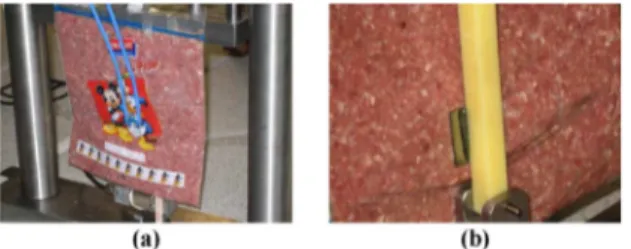
Fig. 7. In vitro characterization with soft tissue in (a) front view showing
antennas and (b) back view showing the sensor.
lymphocytic cellular activity. As a result, inflammation scores for the sensor and Al2O3control materials were 0± 0 and 0 ± 0 (mean± standard deviation), respectively. Minimal fibrosis was noted, surrounding both the control and sensor materials (1.0± 0.5 and 0.94± 0.24, respectively), and the general toxicity score for the test and control materials was zero.
C. Soft Tissue Experiments
After showing enhanced functional performance with the nested SRR sensor relative to the classic SRR design and demon-strating biocompatibility of our sensors, we investigated the fidelity with which we could detect the shift in transmission spectra in a scenario that more closely resembles the intended application of the device: one in which soft tissue separates the sensor and the antennae, as would be the case while monitor-ing bendmonitor-ing strains on internal hardware in human patients [see Fig. 7(a) and (b)]. Fig. 8(a) and (b) depicts the transmission spec-tra of the nested SRR with the soft tissue parameterized with respect to the applied load. In the soft tissue experiment, the operating frequency of the nested SRR was 474.2 MHz under no load, which was lower than that measured in free space (see Fig. 5). This is an exact consequence of the soft tissue, as it has a very high dielectric constant of 56.445 around 500 MHz [30], thereby decreasing the resonance frequency of the device. The sensitivity of the sensor [see Fig. 8(c)] under these test condi-tions was also increased to 4.00 kHz/kgf because the soft tissue better focuses on electromagnetic waves compared to the free space because of its high dielectric constant at low frequencies. Consequently, the mechanical deformation under load affects the operating frequency more strongly, which leads to better sensitivity in the soft tissue.
Using the soft tissue medium is advantageous for our sensing application since the operating frequency is lowered. It should be noted that the space between the antennae and soft tissue should
Fig. 8. Experimental characterization of the nested SRR sensor using soft tissue under tension. (a) Relative transmission spectra. (b) Zoom-in view of the transmission shift. (c) Frequency shift (∆fo) versus applied load (F ).
(d) Induced strain (microstrain) versus frequency shift (∆fo). (e) Nonlinearity
be kept at an absolute minimum. If there is sufficient free space between them, then the antenna signal decreases dramatically. Also, in Fig. 8(d), we show the strain versus operating frequency shift and obtain 0.17 kHz/microstrain sensitivity. In Fig. 8(e), the nested SRR sensor had a maximum of 1500-microstrain nonlinearity errors, corresponding to 35% nonlinearity error, as shown in Fig. 8(f), in the soft tissue, which is larger than in the free space. We are currently working on understanding the nonlinearity error sources.
IV. CONCLUSION
In conclusion, we designed, fabricated, and characterized a novel nested SRR for implant strain sensing. We demonstrated that the nested SRR sensor outperforms the classical SRR sen-sor with regard to operating frequency and sensitivity, which was a direct result of the increased number of gaps in the nested architecture. The unloaded operating frequency of nested SRR (506.2 MHz) was decreased relative to the classical SRR (529.8 MHz) in the free-space experiments. Further, the sen-sitivity of the nested SRR (1.09 kHz/kgf) was increased with respect to the classical SRR (0.72 kHz/kgf) in the free space.
We also demonstrated biocompatibility of our metamaterial sensors by implanting them into New Zealand white rabbits and observing no evidence of inflammation or adverse tissue re-sponse over a period of six months. As a first proof-of-concept demonstration using soft tissue in a situation that approximates the clinical condition, we demonstrated that the unloaded op-erating frequency and sensitivity of the nested sensor were 474.2 MHz and 4.00 kHz/kgf, respectively. These findings were a direct consequence of the interposed soft tissue, which exhibits a very high dielectric constant and effectively served to better focus on the incident electromagnetic waves. The results pre-sented herein support the continued development and character-ization of a fracture healing system based on these implantable metamaterial sensors with nested architecture.
ACKNOWLEDGMENT
We would like to thank to K. B. Alici for his help in making our external antenna.
REFERENCES
[1] K. Stoffel, K. Klaue, and S. M. Perren, “Functional load of plates in fracture fixation in vivo and its correlate in bone healing,” Injury, vol. 31, pp. 37–50, 2000.
[2] J. D. Heckman and J. Sarasohn-Kahn, “The economics of treating tibia fractures. The cost of delayed unions,” Bull. Hosp. Joint Dis., vol. 56, pp. 63–72, 1997.
[3] E. W. Lambert, R. B. Simpson, A. Marzouk, and D. V. Unger, “Or-thopaedic injuries among survivors of USS COLE attack,” J. Orthop.
Trauma, vol. 17, pp. 436–441, 2003.
[4] M. S. Gaston and A. H. Simpson, “Inhibition of fracture healing,” J. Bone
Joint Surg. Br., vol. 89, pp. 1553–1560, 2007.
[5] A. H. Simpson, T. N. Gardner, M. Evans, and J. Kenwright, “Stiffness, strength, and healing assessment in different bone fractures—A simple mathematical model,” Injury, vol. 31, pp. 777–781, 2000.
[6] J. Webb, G. Herling, T. Gardner, J. Kenwright, and A. H. Simpson, “Man-ual assessment of fracture stiffness,” Injury, vol. 27, pp. 319–320, 1996. [7] G. M. Kotzar, D. T. Davy, J. Berilla, and V. M. Goldberg, “Torsional
loads in the early postoperative period following total hip replacement,”
J. Orthop. Res., vol. 13, pp. 945–955, 1995.
[8] G. M. Kotzar, D. T. Davy, V. M. Goldberg, K. G. Heiple, J. Berilla, Jr., K. G. Heiple, R. H. Brown, and A. H. Burstein, “Telemeterized in vivo
hip joint force data: A report on two patients after total hip surgery,” J.
Orthop. Res., vol. 9, pp. 621–633, 1991.
[9] D. T. Davy, G. M. Kotzar, R. H. Brown, K. G. Heiple, V. M. Goldberg, K. G. Heiple, J. Berilla, and A. H. Burstein, “Telemetric force measure-ments across the hip after total arthroplasty,” J. Bone Joint Surg. Am., vol. 70, pp. 45–50, 1998.
[10] A. Rohlmann, F. Graichen, R. Kayser, A. Bender, and G. Bergmann, “Loads on a telemeterized vertebral body replacement measured in two patients,” Spine, vol. 33, pp. 1170–1179, 2008.
[11] A. Rohlmann, F. Graichen, and G. Bergmann, “Loads on an internal spinal fixation device during physical therapy,” Phys. Ther., vol. 82, pp. 44–52, 2002.
[12] A. Rohlmann, F. Graichen, U. Weber, and G. Bergmann, “2000 Volvo Award winner in biomechanical studies: Monitoring in vivo implant loads with a telemeterized internal spinal fixation device,” Spine, vol. 25,
pp. 2981–2986, 2000.
[13] S. J. Taylor and P. S. Walker, “Forces and moments telemetered from two distal femoral replacements during various activities,” J. Biomech., vol. 34, pp. 839–848, 2001.
[14] S. J. Taylor, P. S. Walker, J. S. Perry, S. R. Cannon, and R. Woledge, “The forces in the distal femur and the knee during walking and other activities measured by telemetry,” J. Arthroplasty, vol. 13, pp. 428–437, 1998. [15] W. E. Kock, “Metal-lens antennas,” Proc. IEEE, vol. 34, pp. 828–836,
1946.
[16] V. G. Veselago, “The electrodynamics of substances with simultaneously negative values of epsilon and mu,” Usp. Fiz. Nauk, vol. 92, pp. 517–526, 1967.
[17] R. M. Walser, Introduction to Complex Mediums for Electromagnetics
and Optics. Bellingham, WA: SPIE, 2003.
[18] D. R. Smith, “The reality of negative refraction,” Phys. World, vol. 16, pp. 23–24, 2003.
[19] D. R. Smith, J. B. Pendry, and M. C. K. Wiltshire, “Metamaterials and negative refractive index,” Science, vol. 305, pp. 788–792, 2004. [20] V. M. Shalaev, W. Cai, U. K. Chettiar, H.-K. Yuan, A. K. Sarychev,
V. P. Drachev, and A. V. Kildishev, “Negative index of refraction in optical metamaterials,” Opt. Lett., vol. 30, pp. 3356–3358, 2005.
[21] V. M. Shalaev, “Optical negative-index metamaterials,” Nature Photon., vol. 1, pp. 41–48, 2007.
[22] V. A. Podolskiy, A. K. Sarychev, and V. M. Shalaev, “Plasmon modes in metal nanowires and left-handed materials,” J. Nonlinear Opt. Phys.
Mater., vol. 11, pp. 65–74, 2002.
[23] K. B. Alici and E. Ozbay, “Electrically small split ring resonator antennas,”
J. Appl. Phys., vol. 101, pp. 083104-1–083104-4, 2007.
[24] J. B. Pendry and S. A. Ramakrishna, “Focusing light using negative re-fraction,” J. Phys. [Condensed Matter], vol. 15, pp. 6345–6364, 2003. [25] D. Schurig, J. J. Mock, B. J. Justice, S. A. Cummer, J. B. Pendry, A. F.
Starr, and D. R. Smith, “Metamaterial electromagnetic cloak at microwave frequencies,” Science, vol. 314, pp. 977–980, 2006.
[26] D. R. Smith, “How to build a superlens,” Science, vol. 308, pp. 502–503, 2005.
[27] R. Melik, E. Unal, C. Puttlitz, and H. V. Demir, “Wireless metamaterial RF-MEMS strain sensors,” Appl. Phys. Lett., vol. 95, pp. 011106-1– 011106-3, 2009.
[28] R. Melik, N. K. Perkgoz, E. Unal, Z. Dilli, and H. V. Demir, “Design and realization of a fully on-chip high-Q resonator working at 15 GHz on silicon,” IEEE Trans. Electron Devices, vol. 55, no. 12, pp. 3459–3466, Dec. 2008.
[29] R. Melik, N. K. Perkgoz, E. Unal, C. Puttlitz, and H. V. Demir, “Bio-implantable passive on-chip strain sensors for the assessment of bone fractures,” J. Micromech. Microeng., vol. 18, no. 11, pp. 115017-1– 115017-9, 2008.
[30] [Online]. Available: http://niremf.ifac.cnr.it/tissprop/htmlclie/htmlclie.htm
Rohat Melik (S’07) was born on Jan. 29, 1982 in Diyarbakir, Turkey. He graduated from Sanliurfa Anatolian High School and from Ankara Science High School, Ankara. He received the B.Sc. degree from the Department of Electrical and Electronics Engineering and Department of Economics (Minor in International Economics) from Middle East Technical University, Ankara, in 2004. He is currently working toward the Ph.D. degree in electrical and electronics engineering with Nanotechnology Research Center and Institute of Materials Science and Nanotechnology, Bilkent University, Ankara under the supervision of Prof. H. V. Demir.
His research interests include bioimplant RF-MEMS devices and sensors, including the design, fabrication, and characterization of these components at Bilkent University.
Emre Unal received the B.Sc. degree in electrical and electronics engineering from Hacettepe University, Ankara, Turkey, in 2005.
He is currently a full-time Research Engineer with Nanotechnology Research Center and Institute of Materials Science and Nanotechnology, Bilkent Univer-sity, Ankara, under the supervision of Prof. H. V. Demir. His research interests include the development of microwave and optoelectronic devices at Bilkent University.
Nihan Kosku Perkgoz received the B.Sc. degree in electrical and electron-ics engineering from Middle East Technical University, Ankara, Turkey, in 1999, and the M.S. and Ph.D. degrees in semiconductor electronics and inte-grated science from Hiroshima University, Hiroshima, Japan, in 2003 and 2005, respectively.
In April 2007, she was a Postdoctoral Research Fellow and Projects Coordi-nator in the Demir Devices and Sensors Research Group, Nanotechnology Re-search Centre, and Institute of Materials Science and Nanotechnology, Bilkent University, Ankara. Her current research interests include RF and optoelectronic devices.
Brandon Santoni received the B.S. degree in microbiology and biochemistry from Texas Tech University, Lubbock, in 1998, and the M.S. and Ph.D. degrees in mechanical engineering from Colorado State University, Fort Collins, in 2003 and 2006, respectively.
He is currently a Research Scientist in the Orthopedic Bioengineering Research Laboratory, Department of Mechanical Engineering and School of Biomedical Engineering, Colorado State University. His research inter-ests include bone biology, regeneration, and healing, as well as orthopedic biomechanics.
Debra A. Kamstock received the D.V.M. degree from the University of Florida, Gainesville and the Ph.D. degree in pathology from Colorado State University, Fort Collins.
In 2007, she was a Diplomate of the American College of Veterinary Patholo-gists, where she specialized in anatomic pathology. She is currently an Assistant Professor with the Orthopedic Bioengineering Research Laboratory, Depart-ment of Mechanical Engineering and School of Biomedical, Colorado State University . Her research interests include oncological pathology, with em-phasis in carcinogenesis, tumor angiogenesis, tumor immunology, and cancer vaccines.
Christian Puttlitz received the B.S. degree in materials science and mechan-ics from Michigan State University, East Lansing, in 1992, the M.S. degree in bioengineering from Clemson University, Clemson, SC, in 1993, and the Ph.D. degree in biomedical engineering from the University of Iowa, Iowa City, in 1999.
From 1999 to 2001, he was a Postdoctoral Research Fellow in orthopedic biomechanics with the University of California, San Francisco (UCSF). In May 2001, he was an Assistant Professor in the Department of Orthopedic Surgery at the UCSF and Director of the Orthopedic Biomechanics Laboratory at the San Francisco General Hospital, San Francisco, CA. In August 2004, he was a Fac-ulty Member with the Department of Mechanical Engineering, Colorado State University (CSU), Fort Collins, where he is currently an Associate Professor and Head of the Orthopedic Bioengineering Research Laboratory. His current research interests include experimental and computational investigations in or-thopaedic biomechanics as it relates to normal function, disease states, and treatments.
Hilmi Volkan Demir (S’97–M’04) received the B.Sc. degree in electrical and electronics engineering from Bilkent University, Ankara, Turkey, in 1998, and the M.S. and Ph.D. degrees in electrical engineering from Stanford University, Stanford, CA, in 2000 and 2004, respectively.
Since 2004, he has been a Faculty Member with Bilkent University, with joint appointments at the Department of Electrical and Electronics Engineering and the Department of Physics. He is the Principle Investigator of the Devices and Sensors Laboratory and the Associate Director of Nanotechnology Research Center, Bilkent University. He also received the Docent title (Associate Profes-sor) from Turkish Council of Higher Education, in 2007. His current research interests include the development of innovative optoelectronic devices and novel bioimplant RF devices.
Dr. Demir received the European Union Marie Curie Fellowship in 2005, the Turkish National Academy of Sciences Distinguished Young Scientist Award in 2006, and the European Science Foundation European Young Investigator Award in 2007.