IMMUNOMODULATORY EFFECTS OF TLR LIGANDS AND
POLYSACCHARIDE COMBINATIONS: STRATEGIES TO
AUGMENT INNATE IMMUNE RESPONSE
A THESIS SUBMITTED TO
THE DEPARTMENT OF MOLECULAR BIOLOGY AND GENETICS AND THE INSTITUTE OF ENGINEERING AND SCIENCE OF
BILKENT UNIVERSITY
IN PARTIAL FULFILLMENT OF THE REQUIREMENTS FOR THE DEGREE OF MASTER OF SCIENCE
BY
GİZEM TİNCER SEPTEMBER 2007
I certify that I have read this thesis and that in my opinion it is fully adequate, in scope and in quality, as a thesis for the degree of Master of Science.
Assist. Prof. Dr. İhsan Gürsel
I certify that I have read this thesis and that in my opinion it is fully adequate, in scope and in quality, as a thesis for the degree of Master of Science.
Assist. Prof. Dr. Cengiz Yakıcıer
I certify that I have read this thesis and that in my opinion it is fully adequate, in scope and in quality, as a thesis for the degree of Master of Science.
Assist. Prof. Dr. İsmail Şimşek
Approved for the Institute of Engineering and Science
Director of Institute of Engineering and Science
Prof. Dr. Mehmet Baray
ABSTRACT
IMMUNOMODULATORY EFFECTS OF TLR LIGANDS AND
POLYSACCHARIDE COMBINATIONS: STRATEGIES TO
AUGMENT INNATE IMMUNE RESPONSE
Gizem Tincer
M.Sc. in Molecular Biology and Genetics Supervisor: Assist. Prof. Dr. İhsan Gürsel
September 2007, 90 Pages
Microbial infection initiates multiple TLR ligand mediated signaling cascade on innate immune cells. While some TLRs trigger a Th1 biased immune activation, others may lead to a Th2 dominant immune response. Extracellular (TLR1, 2, 4, 5, 6, 10, and 11) vs endosome-associated TLRs (TLR3, 7/8, and 9) display differential immune activation and cytokine milieu. Understanding contrasting and synergistic behaviors of these TLR subclasses when mixed together may lead to more potent formulations for immunotherapy. Delivery and retaining the stability of nucleic acid based labile TLR ligands to the site of immunologically relevant cells is also a challenge.
In the first part of the thesis, optimum TLR combinations with differential immune effects will be brought into light. Next, immunomodulatory effect of a natural polysaccharide (PS) will be characterized. Finally the ability of a PS carrier to form complex with ligands of nucleic acid sensing TLRs and its potential as a controlled delivery vehicle to stimulate the immune cells will be documented.
In brief, our results suggest that different PS types extracted from various mushroom sources are immunostimulatory and are targeted to TLR2/6 for delivery of other relevant stimulants. Moreover, certain TLR ligand combinations can be harnessed to induce more robust immune activation compared to their stand alone counterparts.
This knowledge will pave the way for establishing an effective PS based carrier of DNA/RNA ligands thus, more effective immunotherapeutic strategies for treating infectious and other local or systemic diseases be possible.
Keywords: TLR, polysaccharide, cooperation, innate immunity, vaccine, immunotherapy
ÖZET
TOLL-BENZERİ ULAK VE POLİSAKKARİT
BİRLEŞİMLERİNİN BAĞIŞIKLIK DÜZENLEYİCİ ETKİLERİ:
DOĞAL BAĞIŞIKLIK SİSTEMİNİ SAĞLAMLAŞTIRMAK İÇİN
STRATEJİLER.
Gizem Tincer
Moleküler Biyoloji ve Genetik Yüksek Lisans Tez Yöneticisi: Doç. Dr. İhsan Gürsel
Eylül 2007, 90 Sayfa
Mikrobiyal enfeksiyonlar birçok TLR ulağının, doğal bağışıklık hücrelerini yönlendirmiş olduğu sinyal yolaklarını başlatır. Bazı TLR’lar Th1 eğilimli bağışıklık cevabını harekete geçirirken, diğerleri Th2’nin baskın olduğu bağışıklık yanıtına neden olabilirler. Hücre yüzeyi (TLR1, 2, 4, 5, 6, 10, ve 11) ve endozomlara bağlı TLR’lar (TLR3, 7/8, ve 9) farklı bağışıklık tepkileri ve sitokin salımları gerçekleştirebilir. Bu TLR alt sınıflarını birbirleriyle beraber karıştırıp kullanarak, sinerjistik ve karşıtık etkilerini anlayarak daha etkin immün tedavi formülasyonları elde edilebilir. Bazı kararsız TLR ulaklarını bağışıklıkla ilgili hücrelere iletilene kadar kararlı tutup, salacak taşıyıcıları tasarlamak da başa çıkılması gereken bir sorundur.
Tezin ilk kısmında, bağışıklık hücreleri üzerine en çarpıcı fark yaratan TLR bileşenleri ortaya çıkarılıp etkin dozları tayin edilecektir. Sonra, mantar kökenli polisakkaridlerin (PS) doğal bağışıklığı uyarıcı özellikleri karakterize edilecektir. Son olarak da en etkin PS taşıyıcısıyla nükleik asit ulaklarının kompleksleri oluşturulup kontollu salım aracı olarak bağışıklık hücrelerini uyarma şiddeti dökümanlanacaktır.
Özetle, bulgularımız farklı mantarlardan saflaştırılan değişik PS tiplerinin bağışıklığı etkinleştirici ve diğer ilgili uyarıcı ulakların salımı için TLR2/6 almacına hedeflenerek uyardığını göstermektedir. Ayrıca, değişik ulak karışımları, TLR ulaklarının tek tek kendi başlarına yaptıkları immün etkiye göre, birleşimlerin bu etkiyi oldukça çok arttırdığı da saptanmıştır.
Bu bilgiler, çeşitli bulaşıcı ya da lokal ve sistematik hastalıkların tedavisi için, PS temelli etkin DNA/RNA ulaklarını taşıyabilen daha güçlü etki gösterebilen immünterapi yaklaşımlarının yolunu açacaktır.
Anahtar kelimeler: TLR, polisakkarit, karışım, doğal bağışıklık, aşı, immün tedavisi
To my parents Sevtap, Teoman and my sister Ezgi who have
encouraged me and have made this possible with their
constant help and guidance both during my time as a M. Sc.
candidate and M. Sc. studies.
ACKNOWLEDGEMENTS
I would like to express my deepest appreciation to my advisor, Assist. Prof. Dr. İhsan Gürsel, for the opportunity he extended to me in agreeing to be my supervisor and allowing me to become a part of his “fresh” laboratory. I would additionally like to thank him for his invaluable guidance, support, teaching and patience during my studies.
I would like to thank to my laboratory mates, Fuat, Hande, Rashad and one of the nicest senior student, Seda for their help, precious friendship and support in the lab and in my experiments.
I would also like to thank to The Scientific and Technological Research Council of Turkey (TÜBİTAK) for their financial support throughout my experiments.
Thanks to Prof. Dr. Oktay Erbatur and his group, from Çukurova University, for kindly providing the polysaccharide extracts for my studies.
Moreover, I would like to thank to my dearest friends Zeynep, Çiğdem, Esen, Koray and Rasim for their support and for always being there whenever I needed.
I wish to thank to, Bâlâ, Sevgi, Elif and Tolga for their friendship and being there with all knowledge whenever I had dilemma with my studies.
My sincere thanks go to MBG Family for their guidance, companionship and assistance.
I would like to thank to Ezgi Karasözen for boosting my morale in times of difficulty. Without my family, none of the exceptional things in my life would have been
possible, and I would like to express my love and gratitude for their everlasting support in life.
Finally, I would like to thank to Yasin who always trusted and supported me in every moment.
TABLE OF CONTENTS
SIGNATURE PAGE………..ii
ABSTRACT... iii
ACKNOWLEDGEMENTS... vi
TABLE OF CONTENTS... vii
LIST OF TABLES... x
ABBREVIATIONS ... xiii
1. INTRODUCTION ... 1
1.1 The Immune System ...3
1.1.1 Induction of Immune System upon Exposure to Pathogens ...4
1.2. Innate Immune System ...5
1.2.1. Pathogen Recognition Receptors ...6
1.2.2. Toll-like Receptors...7
1.2.1.1 TLRs in Innate and Adaptive Immunity ...7
1.2.1.2. The TLR Family Members ...8
1.2.2.2.1. TLR 2, TLR1 and TLR6 ...9 1.2.2.2.2. TLR3 ...10 1.2.2.2.3. TLR4 ...10 1.2.2.2.4. TLR5 ...11 1.2.2.2.5. TLR7 and TLR8...11 1.2.2.2.6. TLR9 ...12 1.2.2.3. TLR Signaling Pathways ...13 1.2.2.3.1. MyD88-Dependent Pathway...14
1.2.2.3.2. MyD88-Independent/TRIF Dependent Pathway ...15
1.2.2.4 TLR Cooperation ...16
1.2.2.5. Delivery of TLR Ligands...19
1.2.2.6. Therapeutic Implications of TLRs ...20
2. AIM OF STUDY ... 23
3. MATERIALS AND METHODS... 24
3.1. MATERIALS...24
3.1.1. Polysaccharides...25
3.1.2. Standard Solutions, Buffers, Media ...25 vii
3.2. METHODS ...25
3.2.1. The Maintenance of the Animals...25
3.2.2. Cell Culture...25
3.2.2.1. Spleen Cell Preparation...25
3.2.2.2. Cell Lines ...26
3.2.2.2.1. RAW 264.7 ...26
3.2.2.2.2. HEK 293 hTLR2/6...26
3.2.2.3. Cell Number Detection with Thoma Cell Counter ...26
3.2.2.4. Cell Distribution...27
3.2.3. Stimulation with Ligands and Polysaccharides ...27
3.2.4. Delivery of TLR Ligands with Polysaccharides...28
3.2.5. Enzyme Linked-Immunosorbent Assay (ELISA)...28
Cytokine ELISA...28
3.2.6. NO2 Detection...29
Griess Assay...29
3.2.7. Transfection of Cells...29
3.2.8. Luciferase Assay...29
3.2.9. Determination of the Gene Expression ...30
3.2.9.1. Total RNA Isolation from the Cells...30
3.2.9.2. cDNA Synthesis and PCR ...31
3.2.9.2.1. cDNA Synthesis...31
3.2.9.2.1.2. PCR ...31
3.2.9.2.1.2.1. Primers ...31
3.2.9.2.1.2.2. Semi-Quantitative RT-PCR ...33
3.2.9.2.1.2.3. Agarose Gel Electrophoresis...33
3.2.10. Atomic Force Microscopy (AFM) ...34
3.2.11. Statistical Analysis...34
4. RESULTS ... 35
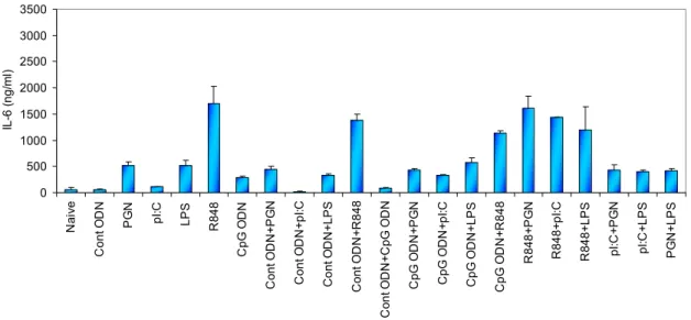
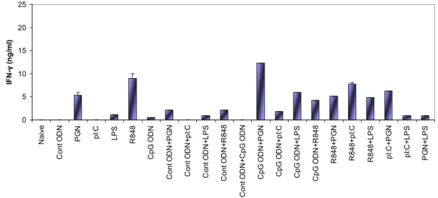
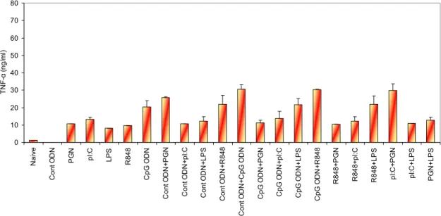
4.1 Studies to Establish the Benefit of Using More Than One TLR Ligand Combinations in Immunotherapy ...35
4.1.1 Determining the Most Adequate Combination of TLR Ligands ...35
4.1.1.1 Cytokine ELISA...36
4.1.1.2. mRNA Expression Profile of TLRs...43
4.2 Immunomodulatory Effects of Polysaccharides Extracted from Ganoderma
lucidum and Shiitake Mushrooms ...48
4.2.1 Effect of Polysaccharides on Cytokine Secretion and Nitric Oxide Production by Macrophages ...49
4.2.1.1 Preliminary Study of Dose and Time Dependent Cytokine ELISA and NO Assay Polysaccharides ...49
4.2.1.2 Dose-dependent TNF-α and NO secretion Profiles of Polysaccharide Extracts ...53
4.2.1.3 Effect of Incubation Time on Ctokine and NO Production by Polysaccharide Extracts ...55
4.2.2 Effect of Polysaccharides on TLR and Cytokine, Chemokine Gene Expression...56
4.2.2.1 Polysaccharide Stimulation Alters mRNA Expression Profile of TLRs ...56
4.2.2.2 Expression Profile of Cytokines and Chemokines upon PS Treatment ...58
4.2.3 Analysis of PS mediated NF-κB activation...59
4.3 Delivery of TLR Ligands with Polysaccharides...61
4.3.1. Cytokine ELISA and NO Assay ...61
4.3.2. Stimulation of Spleen Cells with PS4 and TLR ligands ...67
4.3.3. TLR Expression Pattern of Spleen Cells ...70
4.3.4. Cytokine and Chemokine Expression Pattern of Spleen Cells ...70
4.3.5. Particle Size Determination of PS4-complexed TLR ligand ...71
5. DISCUSSION ... 73 6. FUTURE STUDIES... 78 7. REFERENCES ... 80 9. APPENDICES ... 88 9.1 Appendix A...88 9.2 Appendix B ...88 ix
LIST OF TABLES
Table 1.1 Ligands specificity, immunological fate and cellular specificity of the Toll-like receptors...2 Table 1.2 Types of ligands that pathogens expressed, for multiple TLRs...16 Table 1.3 Potential role of TLRs as therapeutic targets in disease………...21 Table 3.1 The sequences, the sizes and the sources of the primers used ...31–32 Table 3.2 PCR reaction composition………33 Table 3.3 PCR running conditions………...33 Table 4.1 The list of TLR ligands which were used for the stimulation of spleen cells...36 Table 4.2 Upregulated and downregulated cytokine profiles compared to single TLR ligand induction level………...42 Table 4.3 Upregulated and downregulated mRNA message, based on band intensities of single TLR ligand...47 Table 4.4 The fold induction table of TLR ligand combinations…..…...………48
LIST OF FIGURES
Figure 1.1 TLR family members recognize specific patterns of microbial
components……….9 Figure 1.2 MyD88-dependent and MyD88-independent TLR signaling
pathways………...14 Figure 4.1 IL-6 production from spleen cells stimulated with various TLR ligand combinations (optimum doses)... 37 Figure 4.2 IL-6 production from spleen cells stimulated with various TLR ligand combinations (suboptimum doses)...38 Figure 4.3 IFN-γ production from spleen cells stimulated with various TLR ligand combinations (optimum doses)...38 Figure 4.4 IFN-γ production from spleen cells with various TLR ligand combinations (suboptimum doses)...39 Figure 4.5 TNF-α production from spleen cells stimulated with various TLR ligand combinations (optimum doses)...40 Figure 4.6 TNF-α production from spleen cells stimulated with various TLR ligand combinations (suboptimum doses)...40 Figure 4.7 IL-4 production from spleen cells stimulated with various TLR ligand combinations (optimum doses)...41 Figure 4.8 IL-4 production from spleen cells stimulated with various TLR ligand combinations (suboptimum doses)...41 Figure 4.9 Agarose gel picture of the RT-PCR products of the tlr genes………...43 Figure 4.10 Fold induction graphs of tlr genes………..………44–46 Figure 4.11 Cytokine production of IL-6 for 24h and 42h with PSs…….……….50–51 Figure 4.12 Cytokine production of TNF-α for 24h and 42h with PSs………...51–52 Figure 4.13 NO production for 24h and 42h with PSs………...…52–53 Figure 4.14 TNF-α and NO production by different PS extracts at different doses……...54 Figure 4.15 Time dependent TNF-α and NO production by different PS extracts...55–56 Figure 4.16 Alteration of TLR mRNA expression mediated by polysaccharides…....57
Figure 4.17 Time dependent relative expression change of TLR gene message
mediated by polysaccharide stimulation………..58 Figure 4.18 Cytokine and chemokine mRNA expression from polysaccharides…….59 Figure 4.19 Relative luciferase activity of different PS extracts………..60 Figure 4.20 IL-6 ELISA of RAW 264.7 cells when stimulated with PS-4 complexed TLR ligands………....62–64 Figure 4.21 NO production by RAW 264.7 cells when stimulated with PS-4
complexed TLR ligands………..………...65–67 Figure 4.22 IL-6 ELISA of spleen cells when stimulated with PS-4 complexed TLR ligands.………..……….68–69 Figure 4.23 TLR mRNA expression of PS4 complexed pI:C or R848………....70 Figure 4.24 RT-PCR analysis for various chemokines and cytokines…..…………...71 Figure 4.25 AFM images of PS-4 unsonicated/sonicated, pI:C, PS-4 + pI:C……...72
ABBREVIATIONS
AFM Atomic Force Microscopy
APC Antigen presenting cell
AVA Anthrax vaccine adsorbed
bp Base pairs
BCG Bacille Calmette Guerin of Mycobacterium bovis
BCR B-cell receptor
CD Cluster of differentiation
cDNA Complementary Deoxyribonucleic Acid
CFA Complete Freund’s adjuvant
CMV Cytomegalovirus
CpG Unmethylated cytosine-phosphate-guaniosine motifs
CXCL CXC-chemokine ligand
DC Dendritic cell
DMEM Dulbecco's Modified Eagle's Medium
DNA Deoxyribonucleic acid
dsRNA Double-stranded RNA
ELISA Enzyme Linked-Immunosorbent Assay
ER Endoplasmic reticulum
FBS Fetal Bovine Serum
HBV Hepatitis-B Virus
HEK Human embryonic kidney
HIV Human Immunodeficiency Virus
HPV Human papillomavirus
Ig Immunoglobulin
IκK Inhibitor kappa B kinase
IL Interleukin
iNOS Inducible Nitric Oxide Synthase
IFN Interferon
IRAK IL-1 receptor-associated kinase
IRF3 Interferon-regulatory factor 3
LBP LPS-binding protein
LPS Lipopolysaccharide
LRR Leucine-rich repeats
LTA Lipotheicoic Acid
MALP Mycoplasmal lipopeptide
MAP Mitogen-activated protein
MCP Monocyte Chemoattractant Protein
MDP Muramyl dipeptide
MHC Major Histocompatibility Complex
MIP Macrophage Inflammatory Protein
moDC Myeloid dendritic cells
MSR Macrophage scavenger receptor
MyD-88 Myeloid Differentiation Primary Response gene 88
NF-κB Nuclear factor-kappa B
NK Natural killer
NLR Nucleotide-binding oligomerization domain like
proteins or receptors
NO Nitric oxide
NOD Nucleotide-binding oligomerization domain
ODN Oligodeoxynucleotide
OVA Ovalbumin
PA Protective-antigen
PAMP Pathogen associated molecular patterns
PBS Phosphate buffered saline
PCR Polymerase chain reaction
pDC Plasmacytoid dendritic cells
PGN Peptidoglycan
pI:C Polyriboinosinic polyribocytidylic acid
PLG Polylactide-co-glycolide
PNPP Para-nitrophenyl pyro phosphate
PRR Pattern recognition receptors
PS Polysaccharide
RANTES Regulated upon activation, normal T-cell
expressed, and secreted
RIP Receptor-interacting protein
RNA Ribonucleic acid
RPMI Roswell Park Memorial Institute
RSV Respiratory Syncytial Virus
RT Reverse transcriptase
SA-AKP Streptavidin Alkaline-phosphatase
SLE Systemic Lupus Erythematosus
SPG β-(1 Æ3)-D-glucan schizophyllan
SSCL Sterically stabilized cationic liposomes
ssRNA Single-stranded RNA
STF Soluble tuberculosis factor
TCR T-cell receptor
TH T-helper
TIR Toll/IL-1 receptor
TIRAP Toll/IL1 receptor-associated protein
TLR Toll-like Receptor
TNF Tumor Necrosis Factor
TRAF TNF-associated factor
TRAM TRIF-related adaptor molecules
TRIF TIR domain containing adaptor inducing IFN-β
UV Ultraviolet
1. INTRODUCTION
All kinds of vertebrates from birth to death face debilitating and potentially life-threatening infectious agents varying from viruses to fungi. Survival of the infected organism depends on its ability to recognize infectious pathogens and to respond with an appropriate defense system. “Immunity” which is derived from the Latin word immunis, meaning exemption from military service, tax payments or other public services in Roman Empire, refers protection from disease especially infectious disease almost for the last 300 years.
A reaction to foreign substances “antigens”, including bacteria, viruses, as well as to macromolecules such as proteins or polysaccharides, alerts the immune system and the physiological response begins. This response is divided into two subcategories known as innate and adaptive immunity. Innate immunity (also called natural and native) is the first line of host defense against pathogens and consists of cellular and biochemical defense mechanisms that are in place even before infection. Physical and chemical barriers such as epithelia, antimicrobial peptides, lysozymes, phagocytic cells; neutrophils, macrophages, natural killer (NK) cells, complement blood proteins, other types of proteins called cytokines which coordinate and regulate many of the mechanisms of the cells, are the main components that build up the innate immune system (Janeway, 2004).Despite innate immunity, adaptive immunity – also called as specific or acquired immunity – is mediated by clonally expanded T and B lymphocytes and characterized by specifity and memory.
The innate immune system recognizes microorganisms via germline-encoded pattern recognition receptors (PRR). Among the members of PRRs that recognize pathogen-associated molecular patterns (PAMPs) are Toll-like receptors, which are evolutionarily conserved from C. elegans to highly organized mammals (Janeway 2002). There are at least 13 members of the TLR family characterized to date (Akira, 2005).
In humans ten functional Toll-like receptors (TLR) can be subdivided according to their subcellular localization. TLR1, 2, 4, 5, 6 and 10 are expressed on
the cell surface, however TLR3, 7, 8 and 9 are expressed in intracellular compartments, principally in endosomes and the endoplasmic reticulum (ER) (Kanzler, 2007) (Table 1.1). They can occur as dimers, for instance TLR1 and TLR2 heterodimerizes as they recognize bacterial triacylated lipopeptides, TLR2 and TLR6 heterodimerization results in the recognition of diacylated bacterial lipopeptides (O’Neill, 2007).
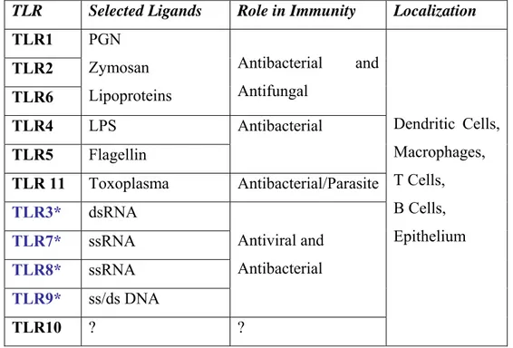
Table 1.1. Ligands specifity, immunological fate and cellular specifity of the Toll-like receptors.
TLR Selected Ligands Role in Immunity Localization
TLR1 TLR2 TLR6 PGN Zymosan Lipoproteins Antibacterial and Antifungal TLR4 LPS TLR5 Flagellin Antibacterial TLR 11 Toxoplasma Antibacterial/Parasite TLR3* dsRNA TLR7* ssRNA TLR8* ssRNA TLR9* ss/ds DNA Antiviral and Antibacterial TLR10 ? ? Dendritic Cells, Macrophages, T Cells, B Cells, Epithelium
*Nucleic acid sensing TLRs are endosome associated, others are expressed at the cell surface.
In addition to hetero-homo dimerization, TLRs also affect the immune system as by acting synergistically. TLR2 and TLR4 synergistically act on macrophages synergize to induce production of inflammatory cytokines (Sato, 2000). Costimulation with TLR4 and TLR2 or TLR9 induces synergistic release of interferon-gamma (IFN-γ) and tumor necrosis factor- alpha (TNF-α) (Equils, 2003). TLR3 and TLR9 promotes enhanced antitumor and cytokine activity (Whitmore, 2004). Some of the combinations such as TLR3 and TLR9 were also searched if they could be used as vaccine adjuvants, combined with cationic liposomes (Zaks, 2006).
TLR4, TLR5, TLR7/8 and TLR9 agonists have potential to therapeutic vaccination for cancer and chronic viral infections, including human-immuno deficiency virus (HIV) and hepatitis-B virus (HBV) (Kanzler, 2007). Conjugating of HIV Gag protein (HIV vaccine candidate) to TLR7/8 agonist (Yarovinsky, 2006) and AVA (Anthrax Vaccine Adsorbed, the licensed anthrax vaccine for human use) to TLR9 ligand CpG substantially enhances the immune response (Klinman, 2006). Not only single TLR ligand but also the combination of more than one TLR ligand are and could be used as vaccine adjuvants with a liposomal delivery systems, mixed with licensed vaccines. The variety of TLR ligands may contribute significantly to inflammation, and appropriate agonists may represent a new class of therapeutic agents for diseases including; Hepatitis, Influenza, cancer and Human papillomavirus (HPV).
Mushrooms, Ganoderma lucidum and Shiitake have been investigated for their medicinal benefits, most notably their anti-tumor properties in laboratory mice. These studies have also identified the polysaccharide lentinan, a (1-3) β-D-glucan, as the active compound responsible for the anti-tumor effects (Kim, 1999). Balachandran et. al. (2006) showed Spriluna (microalgae rich in protein) polysaccharides showed TLR-2 dependent immune activation through monocytes. However this is one of the unique example that reveals the relationship between non-bacterial derived polysaccharides and TLR, so relationship polysaccharide and TLR-mediated immune response should be established for usage of polysaccharides as immuno-therapeutic or immuno-carrier agent.
1.1 The Immune System
Immunity meaning “the ability of an organism to resist infection” can be mainly divided into two subcategories such as; innate and adaptive immunity. Adaptive immune system can provide specific recognition of foreign antigens, immunological memory of infection and pathogen-specific adaptor proteins, but this type of immune response is also responsible for allergy, autoimmunity and the rejection of tissue grafts (Janeway, 2002). The defining characteristics of adaptive immunity are unique specificity for distinct molecules and ability to remember and respond more vigorously to repeated exposures to the same microbe. Therefore,
contributions of adaptive immune systems to pathogen elimination and vaccine design have been extensively studied (Medzhitov, 1997). Innate immunity was formerly thought to be a nonspecific immune response characterized by engulfment and digestion of microorganisms and foreign substances by phagocytic cells such as macrophages and lymphocytes (Akira, 2001). However, recent studies of host defense against microbial pathogens, have demonstrated that the type of effector response generated is strongly dependent on the strength of innate immune response initiated (Medzhitov, 1997). Indeed, the innate immunity help to shape the final adaptive immunity therefore, they act as hand in hand. The importance of innate immunity was fueled by the discovery of pathogen-associated molecular patterns (PAMP) which are recognized by the pattern-recognition receptors (PRR). The role, PRRs play in the elimination of pathogen and activity as adjuvant has reverted the interest in the importance of the initially ignored field of innate immunity.
1.1.1 Induction of Immune System upon Exposure to Pathogens
Defense against infections by both adaptive and innate immune system cells start with the recognition of a pathogen through binding of a PAMP to a PRR through either by antigen-presenting cells (APC) such as macrophages, NK cells, dendritic cells (DC), B-cells found at the site of infection like skin and mucosal epithelia that expresses PRRs and produce antimicrobial chemicals. DCs which were distributed throughout the body, encounters pathogen at different sides of the body such as the mucosal surfaces or the skin, phagocytose then process and present the major histocompatibility complex (MHC) I or MHC II complexed antigenic epitopes to T and B cells. Moreover, activated DCs produce cytokines and chemokines that will be act on pathogen and also alert and recruit other immune cells to the site of insult/infection (Lee, 2007). Activated T and B cells expressing T cell receptor (TCR) and B cell receptor (BCR) will migrate to the infected site upon chemokine and cytokine production (Luster, 2002). These cells rapidly differentiate into effector cells whose main function is to control ongoing infection. Therefore, the innate immune system can instruct the adaptive immunity about the nature and location of pathogenic infection.
1.2. Innate Immune System
The innate immune system detects the presence of and the nature of infection, provides the first line of host protection, and controls the initiation and determination of the effector class of the adaptive immune response (Medzhitov, 2001). The components of innate immunity recognize the structures that are shared by various classes of microbes and are not present in host cells (self/non-self differentiation). The innate immune system is composed of epithelia, which provide barriers to infection, cells in the circulation and in the tissues, and several plasma proteins, such as members of the complement system. The other component of the innate immune system is phagocytes, (i.e. neutrophils, monocytes/macrophages, DCs and B cells). Both types of immune cells recognize microbes in blood and extravascular tissues by surface receptors that are specific to microbial products such as Toll-like receptors. The recognition of pathogens by these phagocytic cells leads to engulfment of the microbes and activation of the phagocytes to kill the ingested agent (Bancroft, 1994). NK cells are a class of lymphocytes that respond to Interleukin-12 (which is produced by macrophages) (IL-12), kills microbe infected cells and produces Interferon-γ (IFN-γ) that activates the other components of immune system. In addition to IL-12, NK cells’ effector functions are induced by a range of cytokines including IL-15, IL-18 and type I IFNs (produced by DCs). NK cells have a crucial role in anti-viral immunity, by recognizing and eliminating Cytomegalovirus (CMV), Hepatitis C and HIV infected cells (Hammerman, 2005). In response to microbes, macrophages and other cells secrete proteins called cytokines and chemokines. These chemoattractants can mediate many of the cellular reactions of innate immune cells. The main cytokines/chemokines appear during the onset of innate immune activation are; TNF-α, IL-1α/β, IP-10, macrophage inflammatory protein-1α (MIP-1α), MIP-3TNF-α, monocyte chemoattractant protein (MCP) and Regulated upon activation, normal T-cell expressed, and secreted (RANTES). These mediators can induce fever, apoptosis, neutrophil activation, recruitment of T and B cells and induction of inflammation as well as regulating the trafficking of immune effector cells to the site of infection.
Other indispensable cytokines such as; IL-12, (which directs T-helper 1 (TH1)
differentiation), Type I IFNs (IFN-α and IFN-β; important for anti-viral response), IL-6 (stimulates and promotes B cell proliferation), IL-15 and IL-18 (helps NK and T cell proliferation) and IL-10 (that is known to induce inhibitory/stimulatory effect on other immune cells) are involved in the orchestral activation/regulation of innate immunity.
1.2.1. Pathogen Recognition Receptors
Pathogen recognition receptors are able to discriminate self from non-self. They evolved to recognize special non-self pathogen-associated signature structures not present on the host (Medzhitov, 1997). They can be expressed on the cell surface, in intracellular compartments (i.e. endosomal organelles or ER) or secreted into bloodstream and tissue fluids, such as opsonins. Opsonins (enhancement of the process of phagocytosis) include: mannan-binding lectin, C-reactive protein and serum amyloid proteins are the secreted molecules produced by the liver (Fraser, 1998). PRR functions include opsonization, activation of proinflammatory signaling pathways, induction of apoptosis and phagocytosis. Several pattern recognition receptors are expressed in the cytosol where they detect these intracellular pathogens and induce responses that block their replication. The protein kinase (PKR) activates, nuclear factor-kappa B (NF-κB) and mitogen-activated protein (MAP) kinase signaling pathways upon binding to dsRNA, which leads to the induction of the antiviral type-I IFN genes (Clemens, 1997). PKR also inhibits viral spread by inducing apoptosis in infected cells (Williams, 1999).
Another group of proteins likely involved in intracellular pattern recognition is the family of Nucleotide-binding oligomerization domain protein-like receptors (NLR). The full range of ligands recognized by NOD proteins is currently unknown, but both NOD1 and NOD2 are reported to activate NF-κB in response to LPS, presumably through binding to their leucine-rich repeats (LRR) regions. Besides their common ligand, NOD1 recognizes a molecule called meso-DAP, that is a peptidoglycan constituent of only Gram negative bacteria and NOD2 proteins
recognize intracellular muramyl dipeptide (MDP), which is a peptidoglycan constituent of both Gram positive and Gram negative bacteria (Inohara, 2001).
1.2.2. Toll-like Receptors
The best characterized PRRs, Toll-like receptors were identified in mammals as a family of type I transmembrane receptors, that are homologous to the Drosophila Toll receptor (Medzhitov, 1997). TLRs are a group of evolutionarily conserved proteins belonging to the IL-1R superfamily, characterized by an extracellular LRR and an intracellular Toll/IL-1 receptor like (TIR) domain. TIR domain of Toll proteins is a conserved protein-protein interaction module, which is also found in a number of transmembrane and cytoplasmic proteins in animals and plants have a role in host defence (Medzhitov, 2001). Ten TLRs are identified to date in mammals. They differ from each other in ligand specificities, expression patterns, and presumably in the target genes they can induce.
1.2.1.1 TLRs in Innate and Adaptive Immunity
TLRs in the innate immune system serve an essential role not only in recognition of pathogen, but also in directing the course and type of innate immune response generated following exposure to foreign antigen (Takeda, 2003). TLRs have been demonstrated to have a wide array of functions including initiation of proinflammatory responses and antiviral responses, up-regulation of costimulatory molecules on antigen presenting cells (APC), release of chemokines to induce migration of responder cells to the site of infection, and induction cross-priming of T cells by DCs (Takeda, 2005). TLRs are responsible for the adjuvant activity that is required to initiate immune responses both in natural infection and in vaccine responses (Lien, 2003). TLRs have emerged as essential not only in innate immune responses but also in shaping adaptive immune responses to pathogen. The signals for activation of adaptive immunity are mostly provided by DCs. TLR-mediated recognition of pathogens by DCs induces the expression of costimulatory molecules such as CD80/CD86 (which provides a costimulatory signal necessary for T cell activation and survival) and production of inflammatory cytokines such as IL-12
(Akira, 2001). DCs subsets can induce TH1 and TH2 responses. Activation of TLR9 in
DCs induces production of IL-12, thereby changing the Th cell differentiation toward TH1 type (Sousa, 2001). LPS stimulates TLR4 signaling pathway and DCs to support
TH1 and TH2 cell differentiation (Kaisho, 2002). In addition to that some
pathogen-derived adjuvants such as Complete Freund’s Adjuvant (CFA), Bacille Calmette Guerin of Mycobacterium bovis (BCG) are recognized by TLRs; TLR9 and TLR2, TLR4 respectively, which may explain the involvement of TLRs in adaptive immunity (Akira, 2003).
1.2.1.2. The TLR Family Members
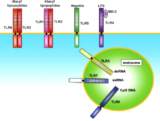
Mammalian TLRs comprise of a large family consisting of at least 13 members. TLRs play important roles in recognizing specific microbial components derived from pathogens including bacteria, fungi, protozoa and viruses. Human TLR4 was the first characterized mammalian Toll (Poltorak, 1998). It is expressed in a variety of cell types, most predominantly in the cells of the immune system, including macrophages and DCs (Medzhitov, 1997). TLRs can be subcategorized according to their localization in the cells. TLR1, 2, 4, 5, 6 and 10 which are seemed to specialized in the recognition of mainly bacterial products; are located on the plasma membrane, whereas TLR3, 7, 8 and 9 that are specialized in viral and intracellular bacteria detection and nucleic acids, are located in the intracellular endosomal and/or ER compartments (Iwasaki, 2004 and Latz 2004) (Figure 1.1).
Figure 1.1. The TLR family members recognize specific patterns of microbial components. TLR1 and TLR6 cooperate with TLR2 to discriminate differences between triacyl and diacyl lipopeptides, respectively. TLR4 is the receptor for bacterial LPS. TLR9 is essential in CpG DNA recognition. TLR3 is implicated in the recognition of viral dsRNA, whereas TLR7 and TLR8 recognizes viral-derived ssRNA. TLR5’s ligand is bacterial flagellin. (Adopted from Takeda, 2005, Nature).
1.2.2.2.1. TLR 2, TLR1 and TLR6
TLR2 responds to various microbial products, including lipoproteins, Gram-positive bacterial PGN and LTA, lipoarabinomannan from mycobacteria, glycosylphosphatidylinositol anchors from a protozoan Trypanosoma cruzi, a phenol-soluble modulin from Staphylococcus epidermis, Zymosan from fungi (Takeda, 2005). One of the aspects proposed for the wide spectrum recognition of microbial components TLR2 recognizes, is that TLR2 forms heterophilic dimers with other TLRs such as TLR1 and TLR6, both of which are structurally related to TLR2. The studies done with the TLR6 or TLR1 deficient mice showed no inflammatory response to mycoplasma-derived triacyl and diacyl lipopeptides respectively. This proves that TLR1 and TLR6 functionally associate with TLR2 and discriminate between diacyl or triacyl lipopeptides. In addition to that TLR2 has been shown to
functionally collaborate with distinct types of receptors such as dectin-1, a lectin family receptor for the fungal cell wall component β-glucan. The bacterial components which were mentioned above, act on immune system from monocytes and macrophages to produce inflammatory cytokines such as TNF-α and IL-6 (Sato, 2000). Gil et. al. (2006) reported that TLR2 triggers TNF-α and MIP-2 secretion from macrophages through the MyD88 signaling pathway with yeast C. albicans. In 2003, it has been found that PGN, could also be delivered to the cytosol for NOD1 recognition from extracellular sites or from phagocytosed bacteria (Chamaillard, 2003). Therefore we can suggest that for the recognition of PGN, TLR and NLR could act together.
1.2.2.2.2. TLR3
The discovery of double-stranded (ds) RNA as the ligand for endosomal located TLR3 helped recognize that TLRs may have a key role in the host defense against viruses by enhancing NF-κB and interferon (IFN)-regulatory factor 3 (IRF3) pathways (Alexopoulou, 2001). dsRNA is produced by most viruses during their replication and induces the synthesis of type I interferons (IFNα/β), which exert anti-viral and immunostimulatory activities. NK cells are the major players in the antianti-viral immune response and express TLR3 and are activated directly in response to synthetic dsRNA, polyriboinosinic polyribocytidylic acid (poly I:C) (Schimdt, 2004). Also myeloid DCs mainly produce IL-12 and IFN-β on TLR3 stimulation (Ito, 2002).
1.2.2.2.3. TLR4
As mentioned above TLR4 is the first identified mammalian Toll. This extracellular TLR is expressed in variety of cell types, most predominantly in macrophages and DCs (Medzhitov, 1997). TLR4 functions as the signal-transduction for signal-transducing receptor for lipopolysaccharide (LPS) which is a major component of the outer membrane of Gram-negative bacteria (Hoshino, 1999). Recognition of LPS by TLR4 is complex and requires several accessory molecules. LPS is first bound to a serum protein, LPS-binding protein (LBP), which functions by transferring LPS monomers to CD14 (Wright, 1999). CD14 is a high-affinity LPS
receptor that can either be secreted into serum, or expressed as a glycophosphoinositol (GPI)-linked protein on the surface of macrophages. Another component of the LPS receptor complex is MD-2 (Shimazu, 1999). Although its precise function is not known, MD-2 is required for LPS recognition (Schromm, 2001). In addition to LPS, TLR4 is involved in the recognition and is considered to be an accessory protein other ligands, including LTA, and a heat-sensitive cell-associated factor derived from Mycobacterium tuberculosis (Li, 2001). Interestingly, TLR4 and CD14 were also shown to trigger a response to the fusion (F) protein of respiratory syncytial virus (RSV). Since it is not clear yet whether the F protein of RSV represents an example of a viral PAMP, an alternative possibility is that the RSV evolved the ability to stimulate TLR4 for its own benefit (Kurt-Jones, 2000).
1.2.2.2.4. TLR5
TLR5 recognizes flagellin, the protein subunits that make up bacterial flagella. TLR5 is expressed on the basolateral side of the intestinal epithelium, where it can sense flagellin from pathogenic bacteria, such as Salmonella. Flagellin induces lung epithelial cells to induce inflammatory cytokine production (Hawn, 2003).
1.2.2.2.5. TLR7 and TLR8
Both of these TLRs are structurally highly conserved proteins, and recognize the same ligand in some cases. Although both TLRs are expressed in mice, mouse TLR8 appears to be nonfunctional (Akira, 2006). It has been revealed that murine and human TLR7 (but not murine TLR8) recognizes synthetic compounds, imidazoquinolines (R848), which are clinically used for treatment of genital warts associated with viral infection (Hemmi, 2002). Murine TLR7 and human TLR8 recognize guanosine or uridine-rich single-stranded RNA (ssRNA) from viruses such as HIV, vesicular stomatitis virus and influenza virus. ssRNA is abundant in host, but usually host-derived ssRNA is not detected by TLR7 or TLR8. This might be due to the fact that TLR7 and TLR8 are expressed in the endosome, and host-derived ssRNA is not delivered to the endosome (Lund, 2004).
1.2.2.2.6. TLR9
One of the most popular TLR, TLR9 is the receptor for unmethylated bacterial genomic DNA which are primarily expressed on B cells, NK cells and DCs to proliferate, mature and secrete various cytokines (IL-12, IFN-γ, IL-6), chemokines or immunoglobulins (Ig) (Krieg, 2000). A single nucleotide substitution or methylation of a cytosine residue within the CpG motif completely abrogates the immunostimulatory property of bacterial DNA (Krieg, 1995). Because bacteria lack cytosine methylation, and most CpG is methylated in the mammalian genome, CpG motifs might signal the presence of microbial infection. There are at least two types of synthetic CpG DNA, termed A or D-type CpG DNA and B or K-type CpG DNA (Klinman, 2004). B/K-type CpG DNA is made up of phosphorothioate backbone and possesses >1 CpG motifs on a single backbone, and is a potent inducer of inflammatory cytokines such as IL-12, IL-6 and TNF-α, B cell proliferation and IgM secretion. A/D-type CpG DNA is structurally different from B/K CpG DNA, which are phosphodiester/phosphorothioate mixed backbone, and G-runs at 3’–5’ ends, and a single CpG motifs has a greater ability to induce IFN-α production from pDCs, but inability to induce B-cells (Gursel, 2006 and Verthelyi, 2001). TLR9 has been shown to be essential for the recognition of both types of CpG DNA (Hemmi, 2003). In addition to bacterial CpG DNA, TLR9 has been shown to recognize viral-derived CpG DNA in pDC such as Mouse cytomegalovirus MCMV (Krug, 2004). While TLR9 is essential for CpG mediated effect the mechanism of the observed dichotomy between K and D type CpG-ODN on human cells was elusive. Recently, Gursel et. al. revealed that pDC but not B cells expresses a co-receptor known as CXCL16 and IFN-α induction by pDC trigerred by D-type ODN is significantly dependent on the CXCL16 expression. In addition to bacterial and viral CpG DNA, TLR9 is presumably involved in pathogenesis of autoimmune disorders. The immunoglobulin-G2a (IgG2a) is bound and internalized by the B cell receptor, and the chromatin,
including hypomethylated CpG motifs, is then able to engage TLR9, thereby inducing rheumatoid factor. Chloroquine is clinically used for treatment of autoimmune diseases such rheumatoid arthritis and systemic lupus erythematosus (SLE) (Boule, 2004). Since chloroquine blocks TLR9-dependent signaling (Hacker, 1998), it act as an anti-inflammatory agent by inhibiting TLR9-dependent immune response. More
than a dozen of human clinical trails have been initiated utilizing TLR9 agonists. It seems likely that the targeted activation of TLR9 using CpG ODN will enhance the treatment of cancer and infectious diseases, as well as showing new hopes for reducing the harmful inflammatory responses such as, asthma and other allergic diseases (Krieg, 2006).
1.2.2.3. TLR Signaling Pathways
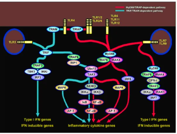
Activation of TLRs by PAMPs leads to induction of various genes that involved in host defense, including inflammatory cytokines, chemokines, MHC and co-stimulatory molecules. Mammalian TLRs also induce multiple effector molecules such as inducible nitric oxide synthase (iNOS) and antimicrobial peptides, which can directly eliminate microbial pathogens. Although both TLRs and IL-1Rs rely on TIR domains to activate NF-κB and MAP kinases and can induce some of the same target genes, a growing body of evidence points to several differences in signaling pathways activated by individual TLRs (Thoma-Uszynski, 2001). Besides, activation of specific TLRs lead to slightly different patterns of gene expression profiles. For example, activation of TLR3 and TLR4 signaling pathways results in induction of type I IFNs, (Doyle, 2002) but activation of TLR2- and TLR5-mediated pathways does not (Hoshino, 2002). In addition to TLR3 and TLR4, TLR7, TLR8 and TLR9 signaling pathways also lead to induction of type I IFNs but in a different manner (Ito, 2002). Although myeloid differentiation primary response gene (88) (MyD 88) is common in all TLR pathways. It has been revealed that there are dependent and MyD88-independent/TRIF dependent signaling (Figure 1.2).
Figure 1.2. The MyD88-dependent and MyD88-independent TLR signaling pathways (Adopted from Akira, 2005).
1.2.2.3.1. MyD88-Dependent Pathway
The role of Toll-mediated recognition in the control of MyD88 protein was studied using MyD88-deficient mice. A MyD88-dependent pathway is analogous to signaling pathways through the IL-1 receptors. MyD88, including a C-terminal TIR domain and an N-terminal death domain, joins with the TIR domain of TLRs. After stimulation, MyD88 recruits IL-1 receptor-associated kinase-4 (IRAK-4) to TLRs by the interaction of the death domains of both molecules, and facilitates IRAK-4-mediated phosphorylation of IRAK-1. Activated IRAK-1 then associates with TRAF6, leading to the activation of two distinct signaling pathways. One pathway leads to activation of AP-1 transcription factors through activation of MAP kinases. Another pathway activates the TAK1/TAB complex, which enhances activity of the Inhibitor kappa B kinase (IκK) complex. Once activated, the IκK complex induces phosphorylation and subsequent degradation of IκB, which leads to nuclear
translocation of transcription factor NF-κB (Takeda, 2004 and Klinman, 2004). MyD88-deficient mice do not show production of inflammatory cytokines such as TNF-α and IL-12p40 in response to all TLR ligands (Takeuchi, 2000 and Klinman, 2004). This once again proves that MyD88 is essential for inflammatory cytokine production through all TLRs. MyD88-deficient macrophages, show impaired inflammatory cytokine production in response to TLR4 and TLR2 ligands in contrast to TLR3, TLR5, TLR7 and TLR9 ligands (Yamamoto, 2002).
1.2.2.3.2. MyD88-Independent/TRIF Dependent Pathway
TLR4 ligand-induced production of inflammatory cytokines is not observed in MyD88-knock-out macrophages; on the other hand, delayed NF-κB expression is observed. This shows that although TLR4 signaling depends on MyD88-dependent pathways, a MyD88-independent component exists in TLR4 signaling. TLR4-induced activation of IRF-3 leads to production of IFN-β. IFN-β in turn activates Stat1 and induces several IFN-inducible genes, like TLR3 (Yoneyama, 1998 and Alexopoulou, 2001). TRIF-deficient mice generated by gene targeting showed impaired expression of IFN-β- and IFN-inducible genes in response to TLR3 and TLR4 ligands (Yamamoto, 2002). Studies with the other TRIF-related adaptor molecules (TRAM)/TICAM-2 showed that TRAM is involved in TLR4-mediated, but not TLR3-mediated, activation of IRF-3 and induction of IFN-β and IFN-inducible genes (Yamamoto, 2003), so TRAM is essential for the TLR4-mediated MyD88-independent/TRIF-dependent pathway. Key molecules that mediate IRF-3 activation have been revealed to be non-canonical IκKs, Tank binding kinase-1 (TBK1) and IκKi/IκKe (Fitzgerald, 2003). It has been recently reported that, complete MyD88 and TRIF expression is required for the effective cooperation, resulting in the induction of IL-12, IL-6, and IL-23 but not of TNF-α and IP-10 upon MyD88- and TRIF-dependent TLR stimulation. Downstream of MyD88, TRIF and IRF5 were identified as an essential transcription factor for the synergism of IL-6, IL-12, and IL-23 gene expression (Ouyang, 2007). Since TRAF6 is critically involved in TLR mediated NF-κB activation, and TRAF6 associates the N terminal portion of TRIF (Gohda, 2004) and the association of C-terminal portion of TRIF with Receptor-interacting protein-1 (RIP1) (Meylan, 2004) leads to NF-κB activation.
1.2.2.4 TLR Cooperation
Because TLR-family members can collectively recognize biomolecules such as lipid, carbohydrates, peptides and even nucleic-acids that are broadly expressed by different groups of microbes, (Table 1.2) recently scientists are trying to understand the synergistic/antagonistic relationships between TLRs.
Table 1.2. Types of ligands that pathogens expressed, for multiple TLRs (Adopted from; Trinchieri, 2007).
The early reports for TLR cooperation are shown that, there is a synergism between TLR2 and TLR4. Sato et. al. (2000) and Beutler et. al. (2001), showed that a TLR2 agonist: Mycoplasmal lipopeptide (MALP-2) or MDP and TLR4 ligand: LPS
synergistically act on peritoneal macrophages and induce production of inflammatory cytokines such as; TNF-α and IL-6. However, the induction of cross-tolerance between the two receptors and the use of MDP as a putative TLR2 ligand, which has recently been formally identified as a ligand for NOD2 (Girardin, 2003). In one of these studies complicates the interpretation of these early reports. A subsequent study showed that stimulation of mouse macrophages with both polyI:C and CpG DNA induced more-than-additive levels of TNF, IL-6 and IL-12 p40 which confirmed that cooperation between certain TLRs does exist (Whitmore, 2004). Equils et. al. in 2003, in addition to their early reports that shows, TLR4 mediates LPS induction of HIV-Long terminal repeat (LTR) trans-activation through IL-1R signaling molecules and NF-κB activation, TLR2 ligand; soluble tuberculosis factor (STF) with the combination of TLR9 ligand plays a central role in HIV-LTR transactivation. Also costimulation with TLR4 and TLR2 or TLR9 elevates synergistic release of Th1 cytokines, IFN-γ and TNF-α in HIV-1 transgenic mouse spleen cells. In human and mouse DCs, TLR3 and TLR4 potently acted in synergy with TLR7, TLR8 and TLR9 in the induction of a selected set of genes. Synergic TLR stimulation increased production of IL-12 and IL- 23 from DCs However, the expression of a few genes, were also downregulated in a synergistic manner by the combined TLR stimulation (Napolitani, 2005). Cytokine production can also be negatively regulated by simultaneous signalling through certain TLRs. Especially, the production of IL-10 after TLR2 stimulation was shown to block the expression of IL-12 p35 and CXC-chemokine ligand 10 (CXCL10; also known as IP10) by human DCs in response to either TLR3 or TLR4 ligands (Re, 2003). Stimulation of mouse or human DCs with the TLR7 and TLR8 ligand R848 and either polyI:C or the TLR4 ligand LPS results in higher amounts of IL-12 p70 than the amounts induced by the individual TLR ligands (Roelofs, 2005). However, when a TLR2 ligand was combined with any other TLR ligand the synergy for IL-12 p70 production was low of absent. Only a low-level synergy for IL-12 p40 (induced by MyD88 pathway), TNF-α and IL-6 production was observed when a TLR2 ligand was combined with a TLR3, TLR4 or TLR9 (Bekeredjian-Ding, 2006). In addition to these works, CpG (TLR9) and LPS (TLR4) can cooperate in a functional manner. The synergistic effect on cytokine production from DCs was restricted to IL-12p40 and IL-12p70, but not IL-6, TNF-α or IL-10, and required a time window of about 4h pretreatment with CpG before LPS (Theiner,
2007). An example of the synergism between TLRs is that, pretreatment of mouse macrophages with the TLR9 and TLR7 ligands results in substantial decrease in the secretion of IL-6 and TNF-α in response to B. antracis infection of macrophages (Sabet, 2006). This result indicates that combination of TLR ligands could be used as vaccine adjuvants for the treatment of bacterial diseases.
In addition to the cooperation between different ligands of TLRs, synergistic induction of cytokine production has also been observed for DCs or macrophages activated by a TLR ligand combined with ligands for other PRRs. Especially NOD1 and NOD2 can synergize with many TLR ligands, including TLR2 ligands, for the induction of TNF and IL-12 p40 production (Tada, 2005). Because of the degradation of bacterial PGNs into different compounds that can activate NOD proteins, the synergy between TLR- and NLR family receptors can boost the response not only to a single pathogen but also to a single component of a pathogen (Girarin, 2005). Besides, a TLR5 ligand flagellin could induce the immune system via both TLR and NLRs (Franchi, 2006).
To evaluate the TLR cooperation in vivo, various double knock-out mice were studied. Tlr2-/- or Tlr9-/- mice at high doses of aerosol-challenge of M. tuberculosis are
clearly susceptible to infection than are their wild-type counterparts (Bafica, 2005). This indicates that under high levels of infectious stress, the function of each TLR involved in pathogen recognition becomes more crucial for the control of microbial growth, an explanation with possible TLR cooperation in host defence. Tlr2 and Tlr4 double-knockout mice are more susceptible to infection than either of the Tlr2–/– or Tlr4–/– single knock-out parental strains (Weiss, 2004 and Reiling, 2002). During T. cruzi and M. tuberculosis infection, as judged by the bacterial load, the Tlr2–/–Tlr9–/– double-knockout mice were clearly less susceptible to infection than Myd88–/– mice, therefore indicating that other MyD88-dependent signaling events, in addition to TLR2 and TLR9 signaling, are involved in host resistance to these pathogens (Trinchieri, 2007).
The idea that more than one TLR–ligand interactions are required for the induction of effective host resistance to pathogens has important implications for the design of superior vaccines and immunotherapy against infectious diseases. Individual
TLR7, TLR8 and TLR9 agonists have already been used successfully as adjuvants to improve CD4+ and CD8+ T-cell responses, to candidate microbial vaccine antigens. These agonists seem to be particularly effective when they are covalently conjugated to the immunogens (Krieg, 2006).
Because of the results as mentioned above we aimed to study the cooperation of TLR ligands in dose-specific manner for the induction of immune response in a synergistic way. The determination of appropriate TLR ligands combination will guide and teach us how to include of more than one TLR ligand into vaccine formulation that induces stronger immune response, thus avoid combinations that will lower the overall immune response.
1.2.2.5. Delivery of TLR Ligands
Starting from 1990’s the use of liposomes as carriers of peptide, protein, and DNA vaccines requires simple, easy-to-scale-up technology capable of high-yield vaccine entrapment. Liposomes are vesicles consisting of one or more concentric bilayers alternating with aqueous compartments. They are usually made up of phospholipids or other amphiphiles such as nonionic surfactants (Gregoriadis, 1999). Gursel et. al. showed a technique that has been developed for the entrapment of live microbial vaccines into giant liposomes under conditions which retain their viability in 1995. They indicated that these kinds of liposomes (containing microbial vaccines and other soluble antigens or cytokines if required) could be used as carriers of vaccines in cases. Even though more stable backbone is used to synthesize CpG-ODN (a phosphorothioate modified form) when used in vivo still eliminated rapidly from the circulation due to the adsorption onto serum proteins and degradation by serum nucleases (Barry, 1999), prolonging the bioavaliabilty and duration of CpG ODN by liposomal capsulation can improve their therapeutic efficiency. Pioneering studies by Gursel et. al. (2001) revealed that, sterically stabilized cationic liposomes (SSCL) contain positively charged and hydrophilic elements can efficiently encapsulate CpG ODN and significantly enhance DNA uptake by cells of the immune system. The immunostimulatory activity of SSCL-encapsulated ODN significantly exceeded that of free ODN in vitro and in vivo. In particular, coencapsulation of CpG ODN with a model Ag ovalbumin (OVA) increased Ag-specific IFN-γ production (10-fold) and
IFN-γ-dependent IgG2a anti-OVA antibody production (40-fold), consistent with the
preferential induction of a TH1-biased immune response. In addition to that in 2005,
Xie et. al. confirmed that co-administrating CpG ODN polylactide-co-glycolide (PLG; another cationic microparticle that improves the uptake and processing of immune adjuvants) with the licensed anthrax vaccine, “AVA”, resulting in a more rapid and stronger anti-protective antigen (PA; the core of anthrax vaccine) antibody response; IgG, than immunization with AVA alone in vivo. Not only CpG ODN but also pI:C a TLR3 ligand has been co-administrated with cationic liposomes and thereby elevated the type I IFN, IFN-α production and have a unique effective on CD8+ T cell
responses in vivo (Zaks, 2006). Besides synthetically produced liposomes, the CpG delivery could be achieved by natural carriers, such as a β-(1Æ3)-D-glucan schizophyllan (SPG) polysaccharide from a fungus called Schizophyllan commune. SPG when modified with other peptides and cholesterol, and when the phosphorothioate CpG ODN complex made then exposed to macrophages, dramatic enhancement in the secretion of cytokines; like IL-6 and IL-12 secretion is observed (Mizu, 2004).
Since some polysaccharides could be used as carrier molecules of CpG DNA. We postulated that polysaccharides from different types of edible mushrooms could reproduce the same effect thus, could serve as a novel delivery agent for nucleic acid ligands such as: CpG ODN, R848, pI:C. The enhanced in vivo action would implicate that these nucleic acids were protected from agile biological milieu targeted naturally to the cells of the immune system via TLR2/6 or TLR1/2 systems.
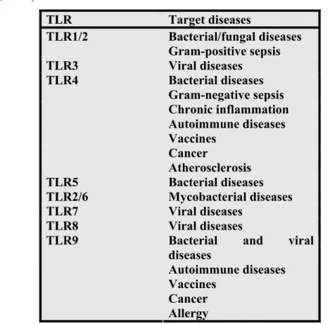
1.2.2.6. Therapeutic Implications of TLRs
The discovery of TLRs has opened up a whole new range of therapeutic possibilities, for infectious, autoimmune diseases, sepsis and cancer. (Table 1.3) Preclinical studies suggest that TLR3, TLR4, TLR7/8 and TLR9 agonists have potential to enhance therapeutic vaccination for cancer and chronic viral infections, HIV and HBV (O’Neill, 2003). Advantage of therapeutic applications of TLR ligands compared to current vaccines are; TLR agonists enhances CD8+ T-cell (kill cells that are infected with viruses) responses to protein antigens and overcoming tolerance to
self-antigens, probably necessary for generating responses to tumor-associated antigens (Hodi, 2006).
Table 1.3. Potential role of TLRs as therapeutic targets in disease. (Adopted from O’Neill, 2003). TLR Target diseases TLR1/2 Bacterial/fungal diseases Gram-positive sepsis TLR3 Viral diseases TLR4 Bacterial diseases Gram-negative sepsis Chronic inflammation Autoimmune diseases Vaccines Cancer Atherosclerosis TLR5 Bacterial diseases TLR2/6 Mycobacterial diseases TLR7 Viral diseases TLR8 Viral diseases
TLR9 Bacterial and viral
diseases
Autoimmune diseases Vaccines
Cancer Allergy
Since TLR3 is a key initiator of anti-viral host defense, stimulating TLR3 would be predicted to have an anti-viral adjuvant effects, in the other hand blocking TLR3 might be useful in limiting viral virulence. Targeting of TLR3 with Ampligen (with a synthetic mismatched dsRNA,) for HIV is currently in phase II trials (Hemispherx). Other viral TLR7/8 ligands, imiquimod and resiquimod have been studied in cutaneous disorders, like basal cell carcinoma and moles that are caused by HPV and shown that they induce cytokine production and elevate cutaneous immune responses (Licenced by 3M Pharma company).
The probable key driver of TNF during sepsis, TLR4 have been targeted mainly in allergic diseases. Blocking of TLR4 is of use in the prevention of over-exuberant immune response induced in sepsis and autoimmune diseases such as Familial Mediterranean Fever and uveitis. Indeed TLR 4 antagonists have been in
phase III clinical trials for the treatment of severe sepsis (Takeda Pharmaceutical and Eisai).
TLR9 ligands; CpG ODNs has been in clinical trials for treatment of cancers, like melanoma, breast cancer, leukemia, non-small-cell lung, renal and colorectal cancers (Krieg, 2007). Conjugation of hepatitis B virus plus CpG ODN developed 15-fold higher anti-hepatitis B antibody titers than did animals immunized with vaccine alone (Klinman, 2004). Consequently Hepsilav, a candidate HBV vaccine is in phase III trials (Dynavax Technologies). Also VaxImmune (CpG B class ODN) for anthrax and influenza antigens with CpG ODN for influenza are in clinical phase studies. In monkey models, CpG ODN are capable of inhibiting airway hyper responsiveness, eosinophilia and even features of airway remodeling (Fanucchi, 2004). Therefore, four different CpG DNAs are targeted to suppress asthma (Dynavax Technologies, Coley Pharmaceuticals, Idera Pharmaceuticals).
TLR4 and TLR9 agonists are being developed for the cure of allergic rhinitis because of their ability to induce strong Th1 responses (Racila, 2005)
Many companies with preclinical antagonists to the intracellular TLRs 7, 8 and 9 have shown efficiency in models of SLE (Dynavax Technologies, Coley Pharmaceuticals and Idera Pharmaceuticals).
In conclusion, variety of TLR agonists or antagonists for the most severe diseases are undergoing preclinical and various stages of clinical trials.
Since more than one TLR ligand could induce more powerful immune activation, we propose that introduction of two or more TLR ligands in vaccination could provide more efficient, robust and long-lasting immune-response.
2. AIM OF STUDY
Studying the immunobiology of the Toll-like receptors is one of the most popular fields in basic and applied immunology in recent years. There is no doubt that TLR therapeutics soon will be in the clinics. The TLR ligand(s) are strong candidate drugs as an immunotherapeutic agent against diseases like cancer, allergy, and infectious diseases (including viral, parasitic and bacterial infections) as well as vaccine adjuvant or immunoprotective agent where there is no available vaccine. Recently, accumulating data indicate that two or more TLR ligands can cooperate to mount a suitable immune response in order to contain a pathogenic insult. We proposed that including more than one TLR ligands in vaccine formulations would provide an added benefit and help to establish a more efficient and stable immune response.
Because of this, the first part of this study was dedicated to reveal the antagonistic/synergistic cooperation of several candidate TLR ligands. These were the most promising, and potent extracellular and endosomal-associated TLR ligands members planned for the clinical trials (i.e. TLR2/6, TLR3, TLR4, TLR7/8 and TLR9).
The second part of the study is dedicated to establish a novel natural delivery depot system especially designed for the targeted delivery of candidate nucleic acid based TLR ligands (because of their lability in biological milieu as well as low level accumulation within relevant immune cells). For this, we have selected candidate polysaccharides extracted from different mushrooms and first characterized their stand alone immunostimulatory potential and then prepared complexes with the candidate RNA and DNA ligands and tested their synergistic effect on spleen cells.
3. MATERIALS AND METHODS
3.1. MATERIALS
All cell culture media components were from Hyclone (USA) unless otherwise stated. Cytokine pairs and recombinant proteins for ELISA were from Endogen (USA) unless otherwise mentioned. TLR ligands; for stimulation assays were as follows and supplied from several vendors: peptidoglycan (PGN) (isolated from B.subtilis; Fluka, Switzerland), pI:C (Amersham, UK), lipopolysaccharide (LPS) (isolated from E.coli; Sigma, USA), Zymosan (isolated from S.cerevisiae; Invivogen, USA), phosphorothioate backbone modified synthetic CpG ODN 1555 (15mer)
(GCTAGACGTTAGCGT), CpG ODN 2006 (24mer) ('TCGTCGTTTTGTCGTTTTGTCGTT or CpG ODN K3 (20mer) (ATCGACTCTCGAGCGTTCTC) as control ODN; and 1612 ODN (GCTAGATGTTAGCGT) (Alpha DNA, Canada) K3 Flip ODN (ATGCACTCTGCAGGCTTCTC), R848 or gardiquimod (Invivogen, USA). Four different polysaccharide extracts were kind gift from Prof. Dr. Oktay Erbatur (Cukurova University, Chem Dept., Adana, Turkey). They were isolated from Shiitake and the different strains (Balcali and Alata) of Ganoderma lucidum, under subcritical water conditions at 100°C (AROsm, BROsm) and 150°C (AR150, BR150). For RNA isolation and for cDNA synthesis, obtained from TRIdity G (AppliChem, Germany) and DyNAmoTM cDNA Syntesis kit (Finnzymes, Finland) respectively and were used according to the manufacturer’s protocol. HEK 293 and RAW cells were transfected with Fugene6 (Roche, Germany) or Lyovec (Invivogen, USA) and Luciferase acitivity was detected with using Promega kit. Several plasmids were expanded in house and purified using Endotoxin free plasmid isolation kit from Qiagen (Germany).
3.1.1. Polysaccharides
For the coding of four different polysaccharide extracts, see appendix A
3.1.2. Standard Solutions, Buffers, Media See appendix B
3.2. METHODS
3.2.1. The Maintenance of the Animals
Adult male or female BALB/C mice (8-12 weeks old) were used for the experiments. The animals were kept in the animal holding facility of the Department of Molecular Biology and Genetics at Bilkent University under controlled ambient conditions (22o C ±2) regulated with 12 hour light and 12 hour dark cycles. They were provided with unlimited access of food and water. Our experimental procedures have been approved by the animal ethical committee of Bilkent University (Bil-AEC).
3.2.2. Cell Culture
3.2.2.1. Spleen Cell Preparation
Spleens were removed from the BALB/C female mice after cervical dislocation. Single cell suspensions were obtained by smashing of spleens with the back of the sterile syringes by circular movements suspended in the 2% FBS supplemented regular RPMI media. The cells were washed 2-3 times at 1500 rpm for 10 mins. The cell pellet was gently dislodged with fresh media, the tissue debris was removed and finally the splenocyte suspension was counted and adjusted to 2-4x10⁶/ml unless otherwise stated.
3.2.2.2. Cell Lines
3.2.2.2.1. RAW 264.7
Macrophage / monocyte like RAW 264.7 (Mus musculus) cells (ATCC) were cultured with RPMI 1640 plus 5% regular FBS. Adherent RAW 264.7 cells were passaged in every 3-4 days when they reached >90% confluency with fresh media, following washing (2% FBS containing RPMI).
3.2.2.2.2. HEK 293 hTLR2/6
Adherent HEK cells, that stably expresses hTLR2/6 genes (Invivogen) were sustained in a High-glucose DMEM media with 5% or 10% regular FBS (Hyclone) supplemented with 10µg/ml Blasticidin S (Invivogen). Cells were passaged by scraping in every 3-4 days intervals as they get over 90% confluency.
3.2.2.3. Cell Number Detection with Thoma Cell Counter
After the spleen cells, RAW cells, HEK 293 hTLR2/6 were pooled, washed and precipitated, they were suspended in 10 ml of 5% regular RPMI-1640 media. Cells were diluted 10 fold and micropipetted on a hemocytometer.
The number of cells in the chamber was determined by counting under the light microscope from these gridlines as indicated with red areas:
The cell number was calculated according to the following formula: __Cell number__ 106 = Total cell number in 10 ml media
4
3.2.2.4. Cell Distribution
For NO Assay and Cytokine ELISA; 2-4x10⁶/ml cells were distributed into 96 well plates with a final volume of 200µl or 250µl media per well. After 6 to 42 hours stimulation, supernatants were collected from the plates and stored at -20°C. Supernatants were layered on the 96 well plates with or without dilution for two assay as previously mentioned.
Spleen cells (6-8 x 10⁶) were splitted into 6 well plates or 15 ml falcons with a final concentration 2-3 ml, for RNA isolation after stimulation with TLR ligands for 2 and 4 hours.
For the transfection assay 2-4x10⁶/ml HEK 293 hTLR2/6 cells were added to 24 well plates, in a 500 µl/ well media final volume.
3.2.3. Stimulation with Ligands and Polysaccharides
Spleen cells were stimulated with the combination of two TLR ligands in various doses (i) PGN; 5 µg/ml, (ii) pI:C; 20 µg/ml, (iii) LPS; 1 µg/ml, (iv) R848; 1 µg/ml and (v) CpG ODN 1555, including control either K3 Flip ODN or 1612 ODN; 1 µM. These ligands were diluted in 5x, 25x, 125x, 625x doses and used for stimulations.
For the stimulation assays including polysaccharides, RAW and HEK 293 hTLR2/6 cells were incubated with PS-1, PS-2, PS-3 and PS-4 for concentration ranges between 20 µg/ml to 0,002 µg/ml. PGN; 20 µg/ml- 0,002 µg/ml or Zymosan 20 µg/ml- 0,02 µg/ml and LPS 5 µg/ml- 0,5 µg/ml including control ODN; 3 µg/ml- 0,3 µg/ml.
Cells were incubated with 5% oligo FBS RPMI-1640 or 5% oligo FBS DMEM when stimulated with ODNs and cultured with 5% regular RPMI-1640 or 5% regular FBS supplemented DMEM as they stimulated with other TLR Ligands and polysaccharides.