ORIGINAL ARTICLE
IJPHY
ABSTRACT
Background: Friedreich’s ataxia is a progressive degenerative disorder caused by deficiency of the frataxin protein. Expanded GAA repeats in intron 1 of the FXN gene lead to its heterochromatinization and transcriptional silencing. Strategies being trialed to treat Friedreich’s ataxia include drugs that improve mitochondrial function and reduce oxidative injury. It has been shown that mesenchymal stem cell (MSC)-derived factors can restore cellular homeostasis and function to frataxin deficient cells.
Case Summary: Here, we report three FRDA cases treated with four consecutive allogeneic transplantations of umbilical cord-derived MSCs with 30 days interval, upon per patient regulatory approvals for advanced cellular therapy.
Outcome Measures: The cases were followed up after the treatment in means of the therapeutic effect of the cellular treatment by attenuating the neurological findings and gene expression parameters.
Conclusions: Closely followed promising safety and efficacy outcomes demonstrated that the MSC treatment for FRDA might positively affect the clinical results caused by the defect in this genetic-based disease.
Keywords: Friedreich’s ataxia, Hereditary Ataxia, Cellular Treatment, Regenerative Medicine, Stem Cell. Received 28th December 2020, accepted 07th March 2021, published 09th March 2021
DOI: 10.15621/ijphy/2021/v8i1/903
CORRESPONDING AUTHOR
Int J Physiother. Vol 8(1), 31-35, February (2021) ISSN (P): 2349-5987, ISSN (O): 2348-8336
Safety and Efficacy of The Stem Cell Transplantation in
Friedreich’s Ataxia: A Report of Three Cases
¹Riza Azeri, MD*2,3Duygu Koyuncu Irmak, PhD 2,3Eda Sun, MSc
2,3,4,5Erdal Karaöz, PhD
*2Duygu Koyuncu Irmak, PhD
Istinye University, School of Medicine, Department of Histology & Embryology, Istanbul, Turkey.
Topkapi Campus, Maltepe Mah., Teyyareci Sami Sk., No.3, Zeytinburnu, Istanbul, 34010, Turkey. E-mail: [email protected]
¹Istinye University, School of Medicine, Liv
Hospital/Ulus, Department of Physical Therapy and Rehabilitation, Istanbul, Turkey.
*2Istinye University, School of Medicine, Department of Histology & Embryology, Istanbul, Turkey. Email: [email protected]
³Istinye University, Stem Cell, and Tissue Engineering R&D Center, Istanbul, Turkey.
⁴Istinye University, 3D Bioprinting Design & Prototyping R&D Center, Istanbul, Turkey. ⁵Liv Hospital/Ulus, Center for Stem Cell and
Regenerative Therapies (LivMedCell), Istanbul, Turkey.
This article is licensed under a Creative Commons Attribution-Non Commercial 4.0 International License.
INTRODUCTION
Friedreich ataxia (FRDA) is a progressive neurodegenerative disorder characterized by clinical manifestations of progressive gait, limb ataxia, dysarthria, dysphagia, oculomotor dysfunction, loss of deep tendon reflexes, pyramidal tract signs, and scoliosis sometimes accompanied by cardiomyopathy, diabetes mellitus, loss in vision and hearing (orphanet.org). In etiology, the mitochondrial protein frataxin’s reduced expression (FXN) is an accountable factor. Besides, more than 96% of patients were shown to bear homozygous expansions of GAA trinucleotide repeating in intron 1 of the FXN gene [1]. Due to the Frataxin insufficiency, iron-sulfur cluster protein deficits appear, oxidative stress increases, mitochondrial iron accumulates, and ultimately cell death occurs, with primary pathology sites being the large sensory neurons of the dorsal root ganglia and the dentate nucleus of the cerebellum [1].
A wide range of therapeutic strategies, including antioxidants, iron chelators, frataxin-increasing compounds, and histone deacetylase inhibitors, are the subject of several clinical trials [2]. Despite these efforts, there is currently no effective treatment for FRDA yet [3]. The cellular treatment has been under investigation in neurodegenerative disease modeling. A potential cellular therapy path with the background knowledge of the regeneration occurring in the nervous system causes clinical symptoms improvement [4]. The therapeutic potential, mesenchymal stem cells (MSCs), is mediated by their innate talent to migrate toward damaged tissues [5]. Engrafted cells secrete bioactive mediators, such as growth factors, cytokines, and extracellular vesicles that exert immunosuppressive, anti-apoptotic, antifibrotic, angiogenic, and anti-inflammatory effects [6]. Here, we report three cases of FRDA who were successfully responded to this intervention of cellular therapy.
Patient Information:
Three patients admitted to the Liv Hospital in Istanbul fulfilled the FRDA diagnostic criteria according to the Functional Staging for Ataxia [7], were evaluated.
Physical Examination:
Demographic data and the disease stages identified by the physical examination before the treatment are summarised in Table 1.
Diagnostic and Assessment:
The diagnosis was confirmed by the Polymerase Chain Reaction (PCR) analysis of the FRDA gene expression. Upon obtaining approval from the Turkish Competent Authority with numbers of authorization letters 09.06.2017-56733164/203-E.1692, 27.10.2017-56733164/203-E.3236, and 27.10.2017-56733164/203-E.3237 respectively, patients and their legally authorized representatives were informed about this regenerative cellular therapy, and they provided written consents by the regulatory requirements.
Age Gender FRDA Stage at Hospital Admission
Case 1 10 Female Capable of doing some daily workStage 3.0 Case 2 39 Male Not capable of walking without assistance, Stage 4.0
limited efforts of working Case 3 22 Male Not capable of walk or work, confined to Stage 6.0
bed.
Table1: Demographic data and the FRDA stages of the cases at the time of hospital admission. (The Scale used for
the staging is Functional Staging for Ataxia 7).
Patients were treated and followed up on the neurological examination and neurophysiological findings’ efficacy and safety parameters, along with the adverse and severe adverse events. Besides, following the treatment cures completion, FRDA gene expression was analyzed by RT-PCR for all cases.
Interventions:
Stem cell production and quality control
Transplanted MSCs were isolated, expanded, and characterized based on our previous protocols [8]. MSCs to be transplanted were isolated, expanded, and analyzed in Good Manufacturing Practices (GMP)-certified laboratory conditions.
Cellular Therapy Protocol
Each case received four consecutive UC-MSCs ‘treatment cures’ 30 days. Each treatment cure consisted of two infusions, at 12 hours interval, which was ‘Infusion-1’ (1× 10⁶ cells/kg body weight in 3 ml normal saline, infused intrathecally (it), in 80 minutes) and ‘Infusion-2’ (1x10⁶/ kg in body weight suspended in 30 ml normal saline, diluted in 200 ml saline and infused intravenously (iv) in 80 minutes) (Figure 1). To facilitate the spreading of the UC-MSCs, 2 ml of saline was injected after the infusion of the UC-MSCs.
Figure 1: The details of the UC-MSCs treatment cures are illustrated. (UC-MSCs: Umbilical Cord Derived Mesenchymal Stem Cells, it: Intrathecal, iv: Intravenous) Follow up and Outcomes:
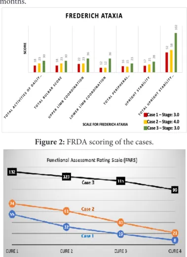
The efficacy and safety of the allogeneic UC-MSC treatment in FRDA cases were evaluated by the clinical and analytical measures of Functional Assessment Rating Scale (FARS), Muscle Strength Scale (MAS), which was evaluated before
each cure application, and side effects follow up which were evaluated before each cure application. RT-PCR analysis for FRDA gene expression was evaluated one week after the fourth cure application.
Case 1: According to the FARS scale of 159 points, the pre-treatment score measured as 55 decreased to 8 as measured after the 4th treatment cure. The spasticity was present at the first evaluation. At the end of all treatment cures, the spasticity decreased; the case became able to do some day-to-day work, continued attending school and daily-life activities with various self-care skills. Her physical therapy and rehabilitation, a clinical psychologist, and speech physiotherapy were still ongoing at 3 months after all the cures were completed.
In pre-treatment MAS, muscle strength was detected as 3 while the patient was raised to 4 in post-treatment. Some movement restrictions were reduced, and the patient can move more independently. Self-care skills and individual independence have also increased. Considering all patient treatment processes, neurological and neurophysiological follow-ups, significant progress has been detected in terms of functional stability, posture, quality of life, and coordination. In the initial stage, while it was able to remain stable for a short time accompanied by a walking disorder, it was found that it could walk 700 m unsupported and balanced in its final assessment. Case coordination was much better in active participation during physical therapy and reached a very positive point in independent participation in daily life.
Case 2: According to the FARS scale of 159 points, the pre-treatment was measured as 74, and post-treatment was measured as 21. The muscle strength was determined as 3 (right) / 3 + (left) as a result of the “Muscle Strength Assessment,” while it increased to 4 in the post-treatment term. Some movement restrictions were reduced, and the patient became more independent. While the patient could not walk independently before the MSC cures, however after the cures were completed, he has been able to walk on his own without shaking, and he has been able to speak more clearly. After his treatment cures were completed, his physical therapy and rehabilitation continued; at three months was continuing his exercises and continued to work.
At the end of the treatment period, we recorded the significant improvement in functional stability, posture and quality of daily life, and neurological findings, especially in balance coordination. Specifically, in comparison with the pre-and the post-treatment evaluations patient showed the effectiveness of the treatments applied by coming to a stage in which he can stand up without tremors in the sitting position, walk without support in the flat area, can remain stable while losing his balance even with a slight jolt, and can provide a voluntary extension in his knees.
Case 3: The patient was diagnosed with stage 6 for FRDA. According to the FARS scale of 159 points, the pre-treatment was measured as 132, and post-pre-treatment was measured as 99. MAS was dropped from 5 to 3+ after the treatment. Spasticity was very high in pre-treatment evaluation; as the cures were completed, the spasticity
decreased, and the muscles relaxed in the post-treatment term. The muscle strength, which was raised from 2-(right) / 2- (left) to as 3 (right) / 3+ (left) as a result of the Muscle Strength Assessment. Some movement restrictions were reduced, and the patient became more independent. While the patient cannot walk independently, he has been able to walk on his own without shaking at the end of the applications, and he has been able to speak more clearly. All after his treatment, his physical therapy, and rehabilitation continue, he continues his exercises.
Neurological and neurophysiological evaluations showed that this case improved motor functions, thus reaching a better daily quality of life, coordination, and cognitive aspects. This case was not completely independent for 3 months.
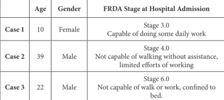
Figure 2: FRDA scoring of the cases.
Figure 3: The Functional Assessment Rating Scale (FARS) evaluation results of all cases measured at each treatment
cure.
Figure 4: Muscle Strength Scale evaluation of all cases right (R) and left (L)(*16)
The FRDA scoring is shown in Figure 2. FARS and MAS evaluations are illustrated in Figures 3 and 4.
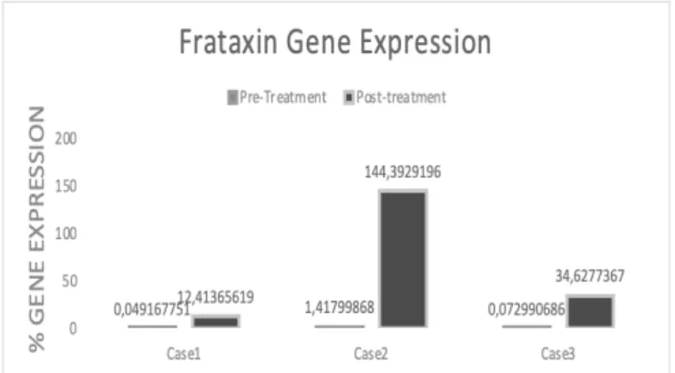
By RT-PCR, we measured changes in the amount of FXN after treatment; we found a 12,4%, 144,4%, 34,6% increase in gene expression level in cae1, 2, and 3, respectively (Figure 5).
Figure 5: Frataxin gene expression alteration obtained by RT-PCR.
The physicians on-site consistently monitored the cases throughout the treatment to track the adverse and serious adverse events. The adverse events observed in the cases were a slight ache at the MSC injection site, followed by fever and mild headache. None of these were related to the cellular treatment, but no severe adverse events were seen.
DISCUSSION
The decreased FARS scores indicate that the limb ataxia, daily activities, and limb coordination symptoms improved for all three cases we treated. A few clinical studies showed that Spinocerebellar Ataxia patients had improved ataxia after 1-year follow-up of MSC transplantation [9]. Miranda et al. also emphasized that repetitive MSC transplants in ataxia treatment sustained motor behavior and neuropathology alleviation improvement as they protected the cell pool [10]. One of our patients can walk and work without any trouble. The other case continues to school and her life with various self-care skills. As Tajiri et al. showed in MSC transplanted patients, differentiation into peripheral neurons and cardiomyocytes with their proliferative and pluripotency properties [11], MSCs constitute the leading cause of FA’s death, myocardial fibrosis, cardiac failure, and neurodegeneration [12]. This suggests MSC could be effective in slowing down these pathologies with the combination of and clinical findings. The transplantation route of MSCs is one of the important factors determining the effectiveness of the treatment. Our study design was based on two different routes as intrathecal and intravenous, because of the anti-inflammatory effects of intravenous injections [13]; by the intrathecal injection, a local regeneration as the lost tissue caused by the disease triggers differentiation and triggers the formation of new myofibrils can be provided and a wider systemic effect of intra-arterial [14].
MSCs are not only able to cytokine and paracrine secretions but also release their genomic material. The cellular fusions helped compensate for the amount of dystrophin in a DMD clinical trial; we again showed upregulation of FXN expression in FA patients [8]. We have shown the FXN
gene expression increase in all cases; this gene prevents the generation of toxic hydroxyl radicals in mitochondria and plays a vital role in neurogenesis [15]. Both in terms of the potential of preventing muscle degeneration and differentiation into the host cell, it is essential to consider the repeated MSCs injections in such genetic-based muscle diseases.
CONCLUSION
Closely followed promising safety and efficacy outcomes demonstrated that the MSC treatment for FRDA might positively affect the clinical results caused by the defect in this genetic-based disease. Our results have suggested that MSC treatment may have a substantial potential for the treatment of FRDA, thus improving life quality.
REFERENCES
[1] Koeppen AH. Friedreich’s ataxia: Pathology, pathogenesis, and molecular genetics. J Neurol Sci. 2011;303(1-2):1-12.
[2] Schulz JB, Boesch S, Bürk K, et al. Diagnosis and treatment of Friedreich ataxia: A European perspective. Nat Rev Neurol. 2009;5(4):222-234.
[3] Koeppen AH. Neuropathology of the inherited ataxias. In: The Cerebellum and Its Disorders. ; 2010:387-406. [4] Bürk K, Schulz SR, Schulz JB. Monitoring progression
in Friedreich ataxia (FRDA): The use of clinical scales. J Neurochem. 2013;126(SUPPL.1):118-124.
[5] Hmadcha A, Martin-Montalvo A, Gauthier BR, Soria B, Capilla-Gonzalez V. Therapeutic Potential of Mesenchymal Stem Cells for Cancer Therapy. Front Bioeng Biotechnol. 2020;8.
[6] J. Braga Osorio Gomes Salgado A, L. Goncalves Reis R, Jorge Carvalho Sousa N, et al. Adipose Tissue Derived Stem Cells Secretome: Soluble Factors and Their Roles in Regenerative Medicine. Curr Stem Cell Res Ther. 2010;5(2):103-110.
[7] Subramony SH, May W, Lynch D, et al. Measuring Friedreich ataxia: Interrater reliability of a neurologic rating scale. Neurology. 2005;64(7):1261-1262.
[8] Dai A, Baspinar O, Yesilyurt A, et al. Efficacy of stem cell therapy in ambulatory and nonambulatory children with Duchenne muscular dystrophy – Phase I–II. Degener Neurol Neuromuscul Dis. 2018;8:63-77.
[9] Tsai YA, Liu RS, Lirng JF, et al. treatment of spinocerebellar ataxia with mesenchymal stem cells: A phase I/IIa clinical study. Cell Transplant. 2017;26(3):503-512.
[10] Oliveira Miranda C, Marcelo A, Silva TP, et al. Repeated Mesenchymal Stromal Cell Treatment Sustainably Alleviates Machado-Joseph Disease. Mol Ther. 2018;26(9):2131-2151.
[11] Tajiri N, Staples M, Kaneko Y, Kim SU, Zesiewicz TA, Borlongan C V. Autologous stem cell transplant with gene therapy for Friedreich ataxia. Med Hypotheses. 2014;83(3):296-298.
[12] Cook A, Giunti P. Friedreich’s ataxia: Clinical features, pathogenesis and management. Br Med Bull. 2017;124(1):19-30.
JE. Human Umbilical Cord Perivascular Cells and Human Bone Marrow Mesenchymal Stromal Cells Transplanted Intramuscularly Respond to a Distant Source of Inflammation. Stem Cells Dev Published online. 2018:scd.2017.0248.
[14] Das AK, Abdullah BJJ Bin, Dhillon SS, Vijanari A, Anoop CH, Gupta PK. Intra-arterial allogeneic mesenchymal stem cells for critical limb ischemia are safe and efficacious: Report of a phase i study. World J Surg. 2013;37(4):915-922.
[15] Pandolfo M. Frataxin deficiency and mitochondrial dysfunction. Mitochondrion 2002;2(1-2):87-93. [16] Aids to the Examination of the Peripheral Nervous
System LONDON: HER MAJESTY’S STATIONERY OFFICE.; 1976.
Conflicts of interest:
The authors certify no conflict of interest with any financial organization regarding the material discussed in the manuscript.