Atlı et al. (2020) Comm. J. Biol. 4(2): 96-99.
DOI: 10.31594/commagene.754697 e-ISSN 2602-456X
Research Article / Araştırma Makalesi
*Corresponding author: [email protected]
Effects of Acute Sodium Nitrite and Sodium Nitrate on the Development
of Drosophila melanogaster
Emel ATLI1*, Hadi ESHRAGHI2, Alper ORHAN2
1Nevsehir Haci Bektaş Veli University, Faculty of Education, Department of Mathematics and Science Education, 50300, NEVŞEHIR, TURKEY 2Hacettepe University, Faculty of Science, Department of Biology, 06800, ANKARA,
ORCID ID: Emel ATLI: https://orcid.org/0000-0002-0220-546X; Hadi ESHRAGHI: https://orcid.org/0000-0002-4641-1767; Alper ORHAN:
https://orcid.org/0000-0003-1732-6368
Received: 18.06.2020 Accepted: 18.09.2020 Published online: .21.09.2020 Issue published: 31.12.2020 Abstract: Sodium nitrite (NaNO2) and sodium nitrate (NaNO3) are used as food preservatives. In addition to teratogenic,
mutagenic, and carcinogenic effects, these products are also thought to have endocrine disrupting effects. This study aimed to investigate the effects of sodium nitrite (NaNO2) and sodium nitrate (NaNO3) on the development of Drosophila melanogaster.
Third instar larvae of Canton S strain (wild type) were treated to 25 mM, 50 mM, 75 mM NaNO2, and NaNO3 for 6 hours. The
pupation and maturation percentages and times were determined. There was no significant difference in the percentage of pupation and maturation compared to the control group (p>0.05). However, 50 and 75 mM NaNO2 exposures caused
developmental delay in both the mean pupation and the mean maturation times (p<0.05). Additionally, it was found that mean maturation time was delayed with 50 and 75 mM NaNO3 exposures (p<0.05).
Keywords: Food additives, Drosophila, pupation, maturation, developmental time delay.
Akut Sodyum Nitrat ve Sodyum Nitrit Uygulamasının Drosophila melanogaster’in Gelişimi Üzerine Etkileri
Öz: Sodyum nitrit (NaNO2) ve sodyum nitrat (NaNO3) gıda koruyucusu olarak kullanılmaktadır. Teratojenik, mutagenic ve
kanserojen etkilere ek olarak, bu ürünlerin endokrin bozucu etkileri de vardır. Bu çalışmada, sodyum nitrit (NaNO2) ve
sodyum nitratın (NaNO3) Drosophila melanogaster gelişimi üzerindeki etkileri araştırılmıştır. Yabanıl tip Canton S soyunun
üçüncü evre larvalarına, 6 saat boyunca 25 mM, 50 mM, 75 mM NaNO2 ve NaNO3 uygulanmıştır. Pupalaşma ve erginleşme
yüzdeleri ve süreleri belirlenmiştir. Pupalaşma ve erginleşme yüzdelerinde kontrol grubuna göre istatistiksel olarak anlamlı bir fark saptanmamıştır (p>0.05). Ancak, 50 ve 75 mM NaNO2 uygulamaları hem ortalama pupalaşma hem de ortalama
erginleşme sürelerinde gelişimsel gecikmeye neden olmuştur (p<0.05). Ayrıca ortalama erginleşme süresinin 50 ve 75 mM NaNO3 uygulaması ile ertelendiği saptanmıştır (p<0.05).
Anahtar kelimeler: Gıda katkı maddeleri, Drosophila, pupalaşma, erginleşme, gelişim zamanı gecikmesi.
1. Introduction
Natural or synthetic food additives are substances added to almost all foods. Coloring agents, preservatives, flavors, emulsifiers, and stabilizers are the most well-known food additives (Helal, Zahkok, Soliman, Al-Kassas, & Wahed, 2008). Sodium and potassium salts of nitrate and nitrite are widely used in meat products as they contribute to the development of the characteristic color and flavor, control lipid oxidation and antimicrobial effect on pathogenic microorganisms, especially Clostridium botulinum that produces a deadly neurotoxin causing food poisoning (Özdestan & Üren 2010; Turp & Sucu 2016; Basak, Chatterjee, Ghosh, & Chakravarty, 2017). Contrary to the beneficial applications of these salts, excessive use in the human diet can cause health-damaging effects (Ikemoto, Teraguchi, & Kobayashi, 2002; Ansari, Ali, & Mahmood, 2015). It is also reported that nitrate and nitrite cause developmental defects (Griffis-Kyle, 2007; Hannas, Das, Li, & LeBlanc, 2010; Simmons, Karimi, Talwar, & Simmons, 2012).
The most commonly used food additives, sodium nitrite and sodium nitrate, have been tested for different biological organisms (Sarıkaya & Cakır, 2005; Sarıkaya, Cakır, & Solak, 2006; Helal, et al., 2008; Özdestan & Üren
2010; Ansari et al., 2015; Basak et al., 2017). Suzuki, Iijima, Scobie, and McColl (2005) found out that dietary nitrates are metabolized to nitrite after absorption from the intestine. In the acidic content of the stomach, it was determined that these nitrites react with amines and amides to form nitroso compounds. It was determined that nitrites and nitrosamines increased the amount of ROS (reactive oxygen species) in human hepatoma cells. Increased ROS levels are known to induce oxidative damage. In other studies, nitrite, nitrate, and nitroso-compounds have been reported to be associated with developmental disorders (Brender et al., 2011; Simmons et al., 2012). These studies have shown that nitrite and nitrates may have effects on development and directed us to work with these chemicals.
In the present study, the effects of sodium nitrite
(NaNO2) and sodium nitrate (NaNO3) on the development
of Drosophila melanogaster were examined considering the changes in the developmental stages. D. melanogaster is a dipteran insect that is often preferred in biological studies due to its short life, well-defined genetics, easy cultivation in the laboratory environment, and its genetic similarity to mammals. Thus, this organism is used for the detection of mutagenic, morphological, and developmental effects of different chemical agents. In addition, D. melanogaster is a
Atlı et al. (2020) Comm. J. Biol. 4(2): 96–99.
97
very useful model organism for developmental biology and toxicology studies (Basak et al., 2017). For these reasons, it is thought that the results of the experiments with Drosophila can be adapted to the effects of food additives in humans.2. Material and Methods
2.1. Strain, media and environmental conditions
Fly strains used in this study were the wild-type Canton S strain of D. melanogaster. The flies were reared on a standard cornmeal Drosophila medium. They were kept in a
Drosophila culture room at 25 ± 1°C, with 50-60% relative
humidity and in 12 hours light, 12 hours dark cycle. Virgin female and male flies of the same age were mated in culture bottles. After 8 hours, the flies were removed from the bottles and the third instar larvae were collected 72 ± 4 hours later.
2.2. Chemicals and treatment procedure
The test solutions were prepared by using solid
compounds of sodium nitrite (NaNO2) and sodium nitrate
(NaNO3). Sodium nitrite (CAS No: 7632-00-0, 99.5%
purity) was obtained from Carlo Erba (Italy). Sodium nitrate (CAS No. 7631-99-4, 99% purity) was supplied from
Sigma (USA). The amounts of NaNO2 and NaNO3 to be
tested were weighed and dissolved in 1 liter of 5% sucrose (Merck; Darmstadt, Germany) solution to obtain required concentrations (25 mM, 50 mM and 75 mM).
In this study, Canton S third instar larvae of D.
melanogaster were treated to 25 mM, 50 mM, and 75 mM
NaNO2 and NaNO3 for six hours acute exposure. During
the nitrite and nitrate exposures, larvae were placed in glass tubes (2.5´7.5 cm) containing filter papers that had absorbed 100 µL stock solutions. In this way, larvae were exposed to these chemicals by nutrition as well as by absorption from the skin. While choosing the dose, the results of previous studies were investigated (Sarıkaya & Cakır, 2005; Sarıkaya et al., 2006). Moreover, the effects of
NaNO2 and NaNO3 on the survival of 72-hour larvae of D.
melanogaster were investigated in the dissertation studies
conducted by Özcan (2011). Therefore, the most appropriate doses were selected based on the available literature findings and the results of previous experiments. Doses of 25 mM, 50 mM, and 75 mM were selected to investigate the effects of these chemicals on the development of D. melanogaster.
2.3. Developmental stages observations
NaNO2 and NaNO3 exposed and non-exposed (control
groups) larvae were placed in glass tubes (2.5´7.5 cm) that contained a standard D. medium. Every tube was contained ten larvae. The development of all experimental groups was followed at 4-hour intervals. The numbers and transition times of individuals passing from larvae to pupae and from pupae to adults were recorded.
2.4. Statistical analysis
The statistical analyses were applied by using the Statistical Package for the Social Sciences (SPSS) 15 program. Analysis of variance (ANOVA) test was used to determine the significance of the pupation and maturation percentages. The comparison of the pupation and maturation times was done by a two-variable t-test. The significance level was p<0.05 for all analyzes.
3. Results
3.1. Effect of the sodium nitrite (NaNO2) and sodium
nitrate (NaNO3) on the transition percentages from larvae
to pupae
Pupae developed from NaNO2 and NaNO3 exposed and
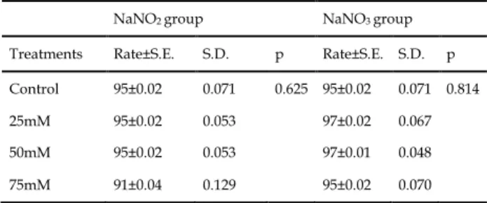
control larvae were counted separately and their pupation percentages were detected. The statistical (one-way analysis of variance) analyses showed that nitrite and nitrate applications did not cause any statistical difference in pupation percentages (p>0.05) (Table 1).
Table 1. The changes of pupation percentages depending on NaNO2 and NaNO3 exposures
NaNO2 group NaNO3 group Treatments Rate±S.E. S.D. p Rate±S.E. S.D. p Control 95±0.02 0.071 0.625 95±0.02 0.071 0.814
25mM 95±0.02 0.053 97±0.02 0.067
50mM 95±0.02 0.053 97±0.01 0.048
75mM 91±0.04 0.129 95±0.02 0.070
S.E.: Standard error, S.D.: Standard deviation, p>0.05
3.2. Effects of the NaNO2 and NaNO3 on the mean
pupation time
In this study, the mean pupation time for NaNO2 and
NaNO3 at different concentrations was also determined.
The mean pupation times in the experimental and control groups were compared with a two-variable t-test and the results are given in Table 2. In the control group, the mean pupation time was 52.17 hours. However, the mean pupation time increased to 52.90 and 54.41 hours in the 50
mM and 75 mM NaNO2 treatment groups, respectively.
These increases were statistically significant (p<0.05). It
can be said that the application of 50 and 75 mM NaNO2
exposures cause a developmental delay by extending the transition period from larvae to pupae. In contrast to these findings, no statistically significant change in mean
pupation time was observed in NaNO3 exposure groups
compared to the control (p>0.05) (Table 2).
Table 2. Mean pupation times of control and NaNO2 and NaNO3
exposure groups
Treatment Mean Pupation Time (hour)
NaNO2 group NaNO3 group
Control 52.17 52.17
25 mM 52.05 51.87
50 mM 52.90* 52.83
75 mM 54.41* 52.85
*: Indicates significant delay in pupation time compared to control (p<0.05)
3.3. Effect of the sodium nitrite (NaNO2) and sodium
nitrate (NaNO3) on the transition percentages from pupae
to adult
The transition rates of NaNO2 and NaNO3 exposure
groups and control group from pupae to adults were determined and then compared by using one-way ANOVA. The results of the variance analysis showed that nitrite and nitrate applications did not cause statistically
Atlı et al. (2020) Comm. J. Biol. 4(2): 96–99.
98
significant differences in maturation percentages compared to the control group (p>0.05) (Table 3).Table 3. The changes of maturation percentages depending on NaNO2 and NaNO3 exposures
NaNO2 group NaNO3 group
Treatments Rate±S.E. S.D. p Rate±S.E. S.D. p Control 86.9±0.04 1.117 0.467 86.9±0.04 0.071 0.796 25mM 90.5±0.03 0.078 88.9±0.03 0.099 50mM 91.5±0.03 0.083 88.7±0.04 0.125 75mM 93.2±0.01 0.078 91.8±0.03 0.095
S.E.: Standard error, S.D.: Standard deviation, p>0.05
3.4. Effects of the NaNO2 and NaNO3 on the mean
maturation time
The times to transition from pupae to adults were also followed at 4-hour intervals and the mean maturation times were determined for all groups. The mean maturation times in the experimental and control groups were compared with a two-variable t-test and the results are given in Table 4. The mean maturation time of the control group was found to be 70.92 hours. However, there were statistically significant increases in the mean maturation times of 50 mM and 75 mM exposure groups (p<0.05) but it was determined that hour changes in 25 mM nitrite and nitrate treatment groups were not statistically significant (p>0.05) (Table 4).
Table 4. Mean maturation times of control and NaNO2 and NaNO3
exposure groups
Treatment Mean Maturation Time (hour) NaNO2 group NaNO3 group
Control 70.92 70.92
25 mM 71.93 71.18
50 mM 73.65* 72.82*
75 mM 76.24* 73.66*
*: Indicates significant delay in maturation time compared to control (p<0.05)
4. Discussion
We chose sodium nitrite (NaNO2) and sodium nitrate
(NaNO3) for our study. They are commonly used in meat
as a food coloring and flavoring agent. Nitrites and nitrates also prevent the growth of Clostridium botulinum and the formation of its toxins (Sarıkaya & Cakır, 2005). In our experiments, it was observed that sodium nitrite and sodium nitrate applications did not affect the pupation and maturation percentages but the mean pupation time
was delayed in 50 mM and 75 mM NaNO2 exposure
groups (p<0.05). Besides, the maturation time was
prolonged in 50 mM, 75 mM NaNO2 and NaNO3 exposure
groups (p<0.05).
Basak et al. (2017) investigated the developmental effects of sodium and potassium salts of nitrite and nitrate by using Drosophila melanogaster. The concentration of 40
mM, NaNO2 and KNO2 stopped Drosophila development
completely. In 20 and 30 mM NaNO2 and KNO2 exposure
groups, the developmental delay was observed. However,
NaNO3 and KNO3 exposures did not lead to any
significant changes compared to control in the
developmental outcomes of Drosophila. In another study,
Sarıkaya et al. (2006) investigated the effect of NaNO2,
NaNO3, KNO2 and KNO3 on the life span of D.
melanogaster. 75mM doses of these substances were found
to be statistically significant reduction in the life span compared to the control group. Also, Sarıkaya et al. (2006) found that the effect of nitrite was higher than nitrate.
Cheng and Chen (2002) conducted a study with the
Asian tiger shrimp Panaeus monodon and applied NaNO2
and NaNO3 to these organisms. They investigated the
accumulation of nitrite and nitrate in the tissues. Cheng and Chen (2002) found out that nitrite and nitrate have similar toxicological mechanisms but nitrite accumulates in the tissues more than nitrate. In addition, it is stated that organisms convert nitrate to nitrite and toxicity occurs in
this way. In the present study, NaNO2 was effective on
both pupation and maturation times whereas NaNO3 only
delayed maturation time. This may be due to the fact that nitrate given by feeding is converted to nitrite in D.
melanogaster's tissues and shows its effect in this way.
Therefore, the effect of nitrate on development may have emerged at the time of maturation.
Siikavuopio and Sæther (2006) investigated the effects of nitrite concentration on growth in juvenile cod,
Gadus morhua. Significantly reduced growth was found in
fish exposed to all treatment levels during later periods. Similarly, Balangoda, Deepananda, and Wegiriya (2018) examined the potentially toxic effects of environmentally relevant nitrate concentrations on the development, growth, and mortality of early life stages of common hour-glass tree frog, Polypedates cruciger. No behavioral changes or abnormalities due to nitrate exposure were reported in the control and nitrate-induced group. However, it was found out that nitrate exposures significantly increased (p<0.05) the growth of the tadpoles up to 25 days old.
Guillette and Edwards (2005) examined the effect of NOx. It was stated that these compounds may be disrupting the reproductive physiology of wildlife. They suggested that the toxicity of NOx may be due to binding to heme groups of steroidogenic enzymes and inhibiting hormone synthesis. Panesar and Chan (2000) proposed that NOx converts to nitric oxide which then binds to heme groups of steroidogenic enzymes in cells and suppresses their activities. This conversion is thought to be one of the main causes of endocrine disruption.
Ecdysteroids are steroid hormones that have essential roles in the development of D. melanogaster (Atlı & Ünlü, 2012). Hannas et al. (2010) reported that nitric oxide acts by lowering ecdysteroid levels. They hypothesized that nitrite and nitrate are converted to nitric oxide in Drosophila cells and disrupt normal endocrine activities. In the same study, it was stated that nitrite was converted to nitric oxide more effectively than the nitrate and its cellular uptake was higher. In addition, it was
proposed that NO2 was more toxicologically significant
than was NO3 due to its reproductive and developmental
toxicity at low exposure levels. In our study, it was found
that NaNO2 changed both pupation and maturation time
but NaNO3 only affected maturation time. It is a
conclusion to support the results of Hannas et al. (2010). In
our study, NaNO2 disrupts the development more quickly
Atlı et al. (2020) Comm. J. Biol. 4(2): 96–99.
99
References
Ansari, F.A., Ali, S.N., & Mahmood, R. (2015). Sodium nitrite induced oxidative stress causes membrane damage, protein oxidation, lipid peroxidation and alters major metabolic pathways in human erythrocytes. Toxicology in Vitro, 29, 1878-1886. https://doi.org/10.1016/j.tiv.2015.07.022
Atlı, E., & Ünlü, H. (2012). Developmental and Reproductive Effects of Bisphenol A (Bpa) in Drosophila melanogaster. Hacettepe Journal of Biology and Chemistry, 40(1), 61-68.
Balangoda, A., Deepananda, K.H.M.A., & Wegiriya, H.C.E. (2018). Effects of environmental contamination and acute toxicity of n-nitrate on early life stages of endemic arboreal frog, Polypedatesm cruciger (Blyth, 1852). Bulletin of Environmental Contamination and Toxicology, 100, 195–201. https://doi.org/10.1007/s00128-017-2261-9
Basak, A.K., Chatterjee, T., Ghosh, S.K., & Chakravarty, A. (2017). Impacts of dietary exposure to sodium or potassium salts of nitrate and nitrite on the development of Drosophila melanogaster. Interdisciplinary Toxicology, 10(2), 70-78. https://doi.org/10.1515/intox-2017-0012 Brender, J.D., Werler, M.M., Kelley, K.E., Vuong, A.M., Shinde, M.U.,
Zheng, Q., … Canfield, M.A. (2011). Nitrosatable drug exposure during early pregnancy and neural tube defects in offspring: National Birth Defects Prevention Study. American Journal of Epidemiology, 174(11), 1286–1295.
Cheng, S.Y., & Chen, J.C. (2002). Accumulations of nitrite and nitrate in the tissues of Penaeus monodon exposed to a combined environment of elevated nitrite and nitrate. Archives of Environmental Contamination and Toxicology, 43, 64–74. https://doi.org/10.1007/s00244-001-0056-8 Griffis-Kyle, K.L. (2007). Sublethal effects of nitrite on eastern tiger
salamander (Ambystoma tigrinum tigrinum) and wood frog (Rana sylvatica) embryos and larvae: implications for field populations. Aquatic Ecology, 41, 119-127. https://doi.org/10.1007/s10452-006-9047-1
Guillette, L.J., & Edwards, T.M. (2005). Is nitrate an ecologically relevant endocrine disruptor in vertebrates? Integrative and Comparative Biology, 45, 19-27. https://doi.org/10.1093/icb/45.1.19
Hannas, B.R., Das, P.C., Li, H., & LeBlanc, G.A. (2010). Intracellular conversion of environmental nitrate and nitrite to nitric oxide with resulting developmental toxicity to the crustacean Daphnia magna. Plos One, 5(8), 1-8. https://doi.org/10.1371/journal.pone.0012453 Helal, E., Zahkok, S., Soliman, G.Z.A., Al-Kassas, M., & Wahed, H. A.
(2008). Biochemical studies on the effect of sodium nitrite and/or glutathione treatment on male rats. The Egyptian Journal of Hospital Medicine, 30, 25-38.
Ikemoto, Y., Teraguchi, M., & Kobayashi, Y. (2002). Plasma levels of nitrate in congenital heart disease: Comparison with healthy children. Pediatric Cardiology, 23(2), 132-136. https://doi.org/10.1007/s00246-001-0036-9 Özcan, P.Ö. (2011). Propolisin antimutajenik etkilerinin Drosophila
melanogaster'de araştırılması (MSc Thesis), Hacettepe University, Ankara, Turkey.
Özdestan, Ö., & Üren, A. (2010). Nitrate and nitrite in foods. Academic Food Journal, 8(6), 35-43.
Panesar, N.S., & Chan, K.W. (2000). Decreased steroid hormone synthesis from inorganic nitrite and nitrate: Studies in vitro and in vivo. Toxicology and Applied Pharmacology, 169, 222-230. https://doi.org/10.1006/taap.2000.9079
Sarıkaya, R., & Cakır, S. (2005). Genotoxicity testing of four food preservatives and their combinations in the Drosophila wing spot test. Environmental Toxicology and Pharmacology, 20, 424-430. https://doi.org/10.1016/j.etap.2005.05.002
Sarıkaya, R., Cakır, S., & Solak, K. (2006). Gıdalardaki koruyucu maddelerin Drosophila melanogaster’de (mwhxflr) ömür uzunluğuna etkisi. Kastamonu Eğitim Dergisi, 14(1), 173-184.
Siikavuopio, S.I., & Sæther, B. (2006). Effects of chronic nitrite exposure on growth in juvenile Atlantic cod, Gadus morhua. Aquaculture, 255, 351– 356. https://doi.org/10.1016/j.aquaculture.2005.11.058
Simmons, A.E., Karimi, I., Talwar, M., & Simmons, T.W. (2012). Effects of nitrite on development of embryos and early larval stages of the zebrafish (Danio rerio). Zebrafish, 9, 200–206. https://doi.org/10.1089/zeb.2012.0746
Suzuki, H., Iijima, K., Scobie, G., & McColl, K.E.L. (2005). Nitrate and nitrosative chemistry within Barrett’s oesophagus during acid reflux. Gut, 54, 1527-1535. https://doi.org/10.1136/gut.2005.066043 Turp, Y.G., & Sucu, C. (2016). Et Ürünlerinde Nitrat ve Nitrit Kullanımına
Potansiyel Alternatif Yöntemler. Celal Bayar Üniversitesi Fen Bilimleri Dergisi, 12(2), 231-242.