Effect of lisinopril on oxidative stress in brain tissues of
rats with L-Name induced hypertension
[L-Name ile hipertansiyon oluşturulan sıçanların beyin dokularındaki oksidatif
stres üzerine lisinoprilin etkisi]
Research Article [Araştırma Makalesi]
Türk Biyokimya Dergisi [Turkish Journal of Biochemistry–Turk J Biochem] 2013; 38 (2) ; 163–168
Yayın tarihi 30 Haziran, 2013 © TurkJBiochem.com [Published online 30 June, 2013]
TÜ R K BİY OKİMYA DERNEĞİ DER G İSİ TÜ R K BİY OKİMYA DERNEĞİ DER G İSİ 1976 TÜ R K BİY OKİMYA DERNEĞİ DER G İSİ TÜ R K BİY OKİMY A DERNEĞ İ D ERG İS İ 1976 ORJİNAL 1. ÖRNEK 2. ÖRNEK Serkan Kirbas1, Suleyman Kutluhan2, Aynur Kirbas3, Recep Sutcu4, Ahmet Kocak5, Ertugrul Uzar6
Recep Tayyip Erdogan University Faculty of Medicine, Deparments of 1Neurology, 3Biochemistry, Rize; 2Suleyman Demirel University Faculty of Medicine, Deparment of Neurology, Isparta;
4 Katip Celebi University, Faculty of Medicine, Deparment of Biochemistry, Izmir; 5 Dumlupınar University, Faculty of Medicine, Deparment of Histology and Embriology, Kütahya; 6 Dicle University, Faculty of Medicine, Deparment of Neurology, Diyarbakir,
Turkey
Yazışma Adresi [Correspondence Address] Yrd.Doç.Dr.Serkan Kirbas
Department of Neurology, Faculty of Medicine, Re-cep Tayyip Erdogan University, 53100, Rize, Turkey Phone. +90 464 212 30 09
Fax. +90 464 217 03 67 E-mail. [email protected]
Registered: 29 July 2012; Accepted: 28 February 2013 [Kayıt Tarihi: 29 Temmuz 2012; Kabul Tarihi: 28 Şubat 2013]
ABSTRACT
Objective: Arterial hypertension is often associated with pathologies related with oxidative stress. Angiotensin converting enzyme (ACE) inhibitors have been used as a safe and effective treatment of hypertension and coronary heart disease. However, the significance of ACE inhibitor usage in hypertension-induced cerebrovascular and neurodegenerative diseases is still unknown. In this study, we aimed to investigate the effects of lisinopril, an ACE inhibitor, on oxidative stress and antioxidant enzyme activities in brain tissues of rats with L-NAME (Nω-Nitro-L-Arginine Methyl Ester hydrochloride) induced hypertension. Methods: Thirty-two Sprague-Dawley rats were divided into four groups: Control, L-NAME, L-NAME plus lisinopril, and only lisinopril. Hypertension was induced by oral administration of the L-NAME (75 mg/kg/day) in drinking water for 6 weeks. Rats were treated with Lisinopril (10 mg/kg/day) for six weeks. Systolic blood pressures were measured at the first, third and sixth weeks by using tail cuff method. Malondialdehyde (MDA), Superoxide dismutase (SOD), Catalase (CAT) and Glutathione peroxidase (GSH-Px) activity were measured from the brain tissue. Nitric oxide (NO) levels were measured from plasma. Results: Our results showed that L-NAME leads to an increase in systolic blood pressure of animals. The antihypertensive effect of lisinopril was observed. MDA level was significantly increased, and antioxidant enzymes activities were decreased in L-NAME given group (p<0.05). However, there was no statistically significant differences between the lisinopril given and other groups according to antioxidant enzymes activities (p>0.05).
Conclusion: In our study, hypertension led to oxidative damage in brain tissues. Although lisinopril prevents the hypertension induced oxidative damage, direct antioxidant effect was not observed. Further studies are needed in order to gain certainty effect of lisinopril in brain tissue. Key Words: L-NAME (Nω-Nitro-L-Arginine Methyl Ester hydrochloride), hypertension, lisinopril, oxidative stress, antioxidant enzymes
Conflict of Interest: There is no conflict of interest among the authors who contributed to the present study.
ÖZET
Amaç: Arteriyel hipertansiyon sıklıkla oksidatif stresle bağlantılı patolojilerle ilişkilidir. Anjiotensin Dönüştürücü Enzim (ADE) inhibitörleri hipertansiyon ve koroner kalp hastalıkları tedavisinde etkili ve güvenilir bir şekilde kullanılmaktadır. Ancak hipertansiyonla ilişkili serebrovasküler ve nörodejeneratif hastalıklardaki ADE inhibitörleri kullanımının önemi henüz netlik kazanmamıştır. Bu çalışmada, bir ADE inhibitörü olan lisinoprilin L-NAME (Nε-nitro-L-Arjinin Metil Ester hidroklorid) ile hipertansiyon oluşturulan sıçanların beyin dokularındaki oksidatif stres ve antioksidan enzim aktiviteleri üzerine olan etkisini araştırmayı amaçladık.
Yöntem: 32 adet Sprague-Dawley cinsi sıçan 4 gruba bölündü: Kontrol, L-Name, L-Name ve lisinopril, sadece lisinopril. Hipertansiyon, sıçanların içme sularına 75mg/kg L-Name katılıp, ağız yoluyla 6 hafta verilerek oluşturuldu. Sıçanlar 6 hafta süreyle 10mg/kg dozunda lisinopril ile tedavi edildi. Tail cuff metoduyla birinci, üçüncü ve altıncı haftalarda sistolik kan basınçları ölçüldü. Malondialdehid (MDA), süperoksit dismutaz (SOD), katalaz (CAT) ve glutatyon peroksidaz (GSH-Px) enzim aktiviteleri beyin dokusundan ölçüldü. Nitrik oksit (NO) seviyeleri ise plazmadan ölçüldü.
Bulgular: Bizim sonuçlar L-NAME’nin hayvanların sistolik kan basıncında artışa yol açtığını göstermiştir. Lisinoprilin antihipertansif etkisi gözlendi. L-Name verilen grupta malondialdehid seviyeleri önemli oranda arttı ve antioksidan enzim aktiviteleri azaldı (p<0.05). Bunun yanında lisinopril verilen grup ile diğer gruplar arasında antioksidan enzim aktivitelerine göre istatistiksel olarak anlamlı farklılık görülmedi (p>0.05).
Sonuçlar: Çalışmamızda, hipertansiyon beyin dokusunda oksidatif hasara yol açmıştır. Lisinopril hipertansiyon nedenli oksidatif hasarı önlemesine rağmen, doğrudan antioksidan etkilere sahip değildi. Lisinoprilin beyin dokusundaki etkisinin kesinleşmesi için ileri çalışmalara ihtiyaç vardır.
Anahtar kelimeler: L-NAME (Nε-nitro-L-Arjinin Metil Ester hidroklorid), hipertansiyon, lisinopril, oksidatif stres, antioksidan enzimler
Çıkar Çatışması: Katkıda bulunan yazarların hiçbir çıkar çatışması yoktur.
Introduction
Cerebrovascular and neurodegenerative disorders are among the most common causes of neurological mor-bidity in developed countries and hypertension is a well-defined risk factor for these insults. Some condi-tions induced by hypertension such as oxidative stress, inflammation and endothelial dysfunction are important factors in pathogenetic background of brain damage [1,2]. Oxidative stress, which is characterized by inc-reased bioavailability of reactive oxygen species, plays an important role in development and progression of ce-rebrovascular dysfunction associated with hypertensive disease [3]. Increased levels of reactive oxygen species such as superoxide anion, hydrogen peroxide and lipid peroxides are reported in patients with hypertension [3,4]. Arterial hypertension is often associated with pat-hologies related with oxidative stress and may be consi-dered as a result of systemic damage in different target organs or tissues induced by free radicals [5,6].
Angiotensin Converting Enzyme (ACE) inhibitors have taken their place in clinical use as effective and safe an-tihypertensives and vascular protective agents [7]. Altho-ugh these agents have been widely used in treatment of hypertension and congestive heart failure, there are only a few preclinical and clinical studies in which ACE inhi-bitors have been shown to reduce the incidence of demen-tia or slow down the rate of cognitive decline in patients with hypertension. Furthermore, the significance of ACE inhibitor usage in hypertension-induced cerebrovascular and neurodegenerative diseases is still unknown [8,9]. Lisinopril, a new non-sulfhydryl ACE inhibitor which can pass through the blood-brain barrier, is absorbed in its active form and primarily excreted with urine [10]. In many studies, it has been shown that ACE and Angiotensin-II (A-II) have had many different effects on nervous system [11,12]. A-II exerts its effect on brain tis-sue via Angiotensin Type-I receptors. Neuromediators such as A-II, dopamine, bradykinin, enkephalin, subs-tance P, dynorphin and neurotensin have an effect on metabolism [13]. While being commonly found in brain tissue, ACE is mainly located in basal ganglia, perivent-ricular areas, hippocampus, hypothalamic neurosecre-tory nucleus and the cerebellum [14].
The aim of this study is to investigate the possible pro-tective effect of lisinopril against oxidative stress in
cerebral tissues of rats with L-NAME (Nω
-Nitro-L-Arginine Methyl Ester hydrochloride)-induced chronic hypertension.
Methods
Animals
A total of 32 female Sprague-Dawley rats with mean age of eight weeks and weighing between 190-220 grams were included in this study. Animals were obtained from Laboratory Animal Production Unit of Suleyman
Demirel University and they were procedured, maintai-ned and treated in accordance with the “Animal Welfare Act and the Guide for the Care and Use of Laboratory animals prepared by the Suleyman Demirel University, Animal Ethical Commitee (Date: 25.07.2006 proposal number:06/12)”. Rats were placed in a room with ideal temperature (21±2°C) and humidity (60±5%), in which a 12h:12h light:dark cycle was maintained for one week prior to the initiation of the treatment. The rats were fed a standard diet and tap water ad libitum.
Experimental procedure
Animals were equally divided into four groups as fol-lows: 1) Control group (n=8); 2) only L-NAME given group (75 mg/kg/day, n=8); 3) L-NAME plus lisinopril given group (n=8) and 4) only Lisinopril given group (10 mg/kg/day, n=8). Drugs were orally given via consump-tion water and the whole treatment procedure lasted for 6 weeks. At the end of the study, the rats were anaesthe-tized with intramuscular injection of ketamine hydroch-loride (Ketalar, Eczacibasi, Istanbul, Turkey) at 50 mg/ kg dose and were sacrificed.
Chemicals
Nω-Nitro-L-Arginine Methyl Ester hydrochloride
(L-NAME) and lisinopril were purchased from Sigma-Aldrich (St. Louis, Missouri, USA). All other chemicals were obtained from E.Merck or HIMEDIA, India.
Model of L-NAME-induced arterial
hyperten-sion and experimental protocol
Animals were given 0.6 mg/ml dose of L-NAME via consumption water to obtain the mean daily intake of 75 mg/kg during the study period [10,15,16].
Blood pressure measurements
Systolic blood pressures (SBP) were measured at the first, third and sixth weeks by using tail cuff method (IITC, model 31, Woodland Hills, CA, USA). The animals were placed in a heated chamber at an ambient temperature of 30–34 °C for 15 min and blood pressure values (1-9) were recorded for each animal. The lowest three readings were averaged to calculate the mean blood pressure. All recordings and data analyses were done via using a com-puterized data acquisition system and software.
Biochemical analysis
Excised brain samples were weighed and immediately stored at -80°C. Brain tissues were homogenized in five volumes (w/v) with 1.15 % ice-cold KCl solution [6]. As-says were performed on the supernatant of the homo-genate prepared at 14,000 rpm for 30 min at +4°C. Tis-sue protein concentrations were measured by using the Lowry method [17]. Blood samples were collected from the inferior vena cava and plasma samples were stored in aliquots at -20°C until the batch was analyzed for NO. Cerebral tissue lipid peroxidation level was expressed
as malondialdehyde (MDA) and was measured accor-ding to the procedure described by Draper and Hadley method [18]. The principle of the method is the spect-rophotometric measurement of the color generated by the reaction of 2-thiobarbituric acid-reactive substance (TBARS) with MDA. TBARS value is expressed as malonyldialdehyde equivalent. Data are shown as units nano moles per milligram (nmol/mg) protein.
Plasma nitric oxide (NO) levels were determined with Griess method [19]. The absorbance of the magenta co-lored azo dye that is formed from stable decomposition product, nitrite and the Griess reagent was determined at 543 nm. By constructing a standard curve from the absorbance of standards, the concentration of NO in the plasma sample was determined. The results of serum NO were expressed in µmol/L.
The determination of glutathione peroxidase (GSH-Px) activity was based on the method of Paglia and Valentine [20]. The enzymatic reaction in the tube that contained reduced nicotinamide adenine dinucleotide phosphate, reduced glutathione, and sodium azide and glutathio-ne reductase was initiated by the addition of hydrogen peroxide and the change in absorbance at 340 nm was monitored by a spectrophotometer. Activity was given as units per milligram (U/mg) protein.
Superoxide dismutase (SOD) activity was measured ac-cording to the method of Sun et al [21]. The determina-tion of superoxide dismutase activity was based on the reaction of xanthine with xanthine oxidase to generate superoxide radicals, which react with 2-(4-iodophenyl)-3-(4-nitrophenol)-5-phenyltetrazoliumchloride to form a red formazan dye. The superoxide dismutase activity is then determined as the degree of inhibition of this re-action. Results are expressed as units per milligram of protein (U/mg protein).
Catalase (CAT) activity was measured according to the method of Aebi [22]. The principle of the assay is based on the determination of the rate constant k (dimension:
s-1, k) of hydrogen peroxide decomposition. By
measu-ring the absorbance change per minute, the rate constant of the enzyme was determined. Activity was given as units per milligram (U/mg) protein.
Statistical analysis
The established data were expressed as mean ± stan-dard deviation and were analyzed via using Statistical Package for the Social Sciences version 11.5 (SPSS 11.5 for Windows, Chicago, IL, USA). The normality of the distribution was tested with Kolmogorov–Smirnov test. Because the distributions were not all characterized by normal distribution, they were analyzed by using Krus-kal–Wallis H test. Differences between two groups were determined with Mann–Whitney U test. Spearman test was used to calculate whether there has been any corre-lation among biochemical parameters. P values less than 0.05 was considered as statistically significant.
Results
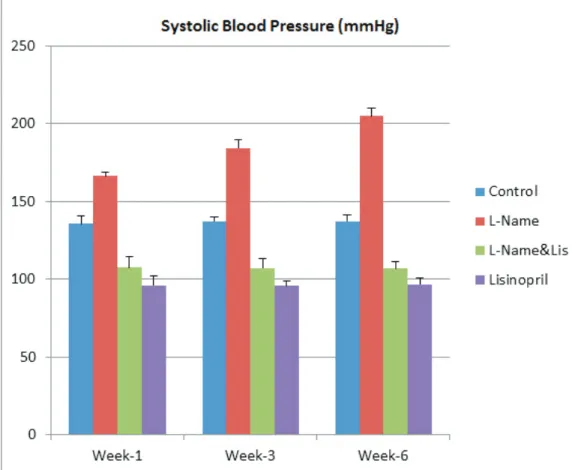
Blood pressure measurements
In Table 1 and Figure, effect of lisinopril on systolic blo-od pressure in rats with L-NAME induced hypertensi-on during first, third and sixth weeks were given. It has been observed that the mean systolic blood pressure was prominently increased in animals treated with L-NAME, whereas lisinopril treatment had significantly reduced the systolic blood pressure and this protective effect was detectable at 10 mg/kg dose. There was a statistically significant difference between only L-NAME given and L-NAME plus lisinopril given groups (p < 0.01).
Lipid peroxidation products
Table 2 shows the mean cerebral MDA (thiobarbituric acid reactive substances, TBARS) levels in L-NAME induced hypertensive rats following lisinopril treatment. Rats solely treated with L-NAME exhibited a signifi-cant increment in mean MDA levels and the established mean values were 2.11 ± 0.28 nmol/mg protein and 1.13 ± 0.19 nmol/mg protein in L-NAME and control groups, respectively (p < 0.001). There was a significant decrea-se in MDA level (1.07 ± 0.21 nmol/mg prot) in L-NAME plus lisinopril treated group (p = 0.001).
Plasma NO concentration
Mean plasma concentrations of NO were given in Table 2. As seen from the table, the mean NO level signifi-cantly decreased in L-NAME given group; whereas the-re was a considerable improvement towards normal ran-ge in L-NAME plus lisinopril treated group. The mean plasma NO levels were 38.3 ± 8.9, 13.6 ± 7.9 and 24.4 ± 6.9 µmol/L in controls, L-NAME given group and L-NAME plus lisinopril administered group, respecti-vely and there was a prominent difference between the treatment groups and controls (p < 0.001). However, the-re was no significant diffethe-rence between L-NAME plus lisinopril and only lisinopril given groups (p = 0.689).
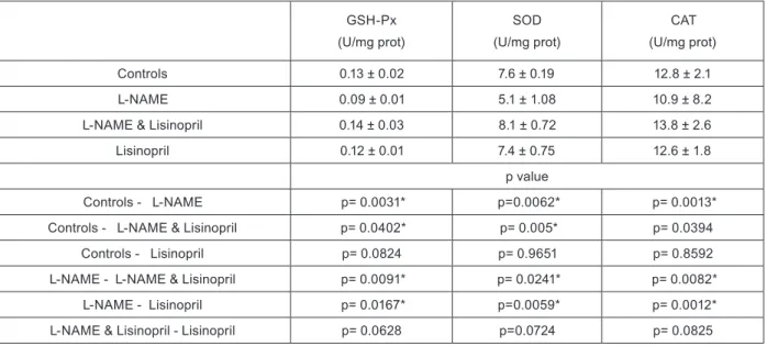
Enzymatic antioxidants
Table 3 shows the effect of lisinopril on mean SOD, CAT and GSH-Px activity levels in tissues of rats with L-NAME induced hypertension. The mean activity le-vels were significantly decreased in only L-NAME gi-ven group. In L-NAME plus lisinopril treated group, there was an increment in these enzymatic antioxidants. Cerebral SOD levels were 5.1 ± 1.08 and 8.1 ± 0.72 U/ mg protein in only L-NAME given group and L-NAME plus lisinopril administered group, respectively (p = 0.0241). Mean CAT levels were found as 10.9 ± 8.2 and 13.8 ± 2.6 U/mg prot in the same treatment groups (p = 0.0082). Mean GSH-Px levels were 0.09 ± 0.01 U/mg
prot in L-NAME group and 0.14 ± 0.03 U/mg prot in
L-NAME plus lisinopril given group (p = 0.0091). Ho-wever, there was no significant difference between these groups (p = 0.0724, p = 0.0825, p = 0.0628, respectively).
Figure. Systolic blood pressure levels of the groups during according to weeks
Table 1. Tail cuff systolic blood pressure (mmHg) values according to weeks (means ± SD)
1st week* 3rd week* 6th week*
Controls 124.5 ± 6.2 122.3 ± 4.8 127.6 ± 5.8
L-NAME 146.5 ± 4.2 163.1 ± 7.9 184.8 ± 8.1
L-NAME&Lisinopril 107.2 ± 8.1 113.4 ± 7.9 117.3 ± 7.6
Lisinopril 92.6 ± 6.5 95.7 ± 5.2 94.1 ± 6.9
* p<0.05 (Control to L-NAME, L-NAME to L-NAME & Lisinopril, and control to lisinopril)
Table 2. Brain tissues MDA levels and plasma NO levels (mean±SD)
MDA* (nmol/mg prot) NO* (µmol/L) Controls 1.13 ± 0.19 38.3 ± 8.9 L-NAME 2.11 ± 0.28 13.6 ± 7.9
L-NAME & Lisinopril 1.07 ± 0.21 24.4 ± 6.9
Lisinopril 1.20 ± 0.16 34.7 ± 5.2
MDA, malondialdehyde; NO, nitric oxide;
and Singh have reported that lisinopril treatment has significantly attenuated the effects of vascular dementia such as impairment of learning and memory, endotheli-al dysfunction and significant changes in various bioc-hemical levels in rats with deoxycorticosterone acetate (DOCA) induced hypertension [23]. According to the-se authors, lisinopril may be considered as a potential pharmacological agent for the management of hyperten-sion-induced vascular dementia. Velayutham et al. have conducted a study to investigate the effect of pretreat-ment with lisinopril on postoperative hypertension in patients undergoing neurosurgery for supratentorial bra-in tumors and the role of oxidative stress bra-in this process [24] and found that perioperative hemodynamic changes were highly associated with oxidative stress parameters in all three groups. It was observed that lisinopril sig-nificantly decreased MDA levels, protein carbonyl con-tent and nitrate during surgical intervention (p < 0.05), an effect which has lasted after the operation. Results established from the study by Velayutham et al. have indicated that pretreatment with angiotensin-converting enzyme inhibitor (lisinopril) could reduce postoperative hypertension in patients undergoing neurosurgery, and inhibition of oxidative stress may be a potential mecha-nism for this effect. Uzar et al. have reported that zofe-nopril significantly reduced cerebral ischemia/reperfu-sion induced oxidative stress as indicated by increased MDA and total oxidant status levels in brain tissues of rats [6]. Zofenopril is more lipophilic and more potent than most other ACE inhibitors. Therefore, the free-ra-dical scavenger effect of zofenopril may be attributed to the robust inhibition mechanism against ACE. The pro-tection may be due to the indirect prevention of oxidati-ve stress and apoptosis. These observations suggest that zofenopril may be a clinically viable protective agent
Discussion
Our study suggests that L-NAME treatment has signifi-cantly raised the arterial systolic blood pressure of rats
and lisinopril may possess robust protective effect in
rats with L-NAME induced hypertension as indicated by significant decrease in mean systolic blood pressure values. We observed that lisinopril has had a protective effect against hypertension-induced oxidative damage, although, according to result of our study, it is somew-hat not possible to say this agent has a direct antioxidant effect.
Hypertension is associated with oxidative stress in the vascularute and is a major risk factor for stroke and cog-nitive abnormalities. Angiotensin II (Ang II) is the main effector peptide of the renin-angiotensin system (RAS) and plays a critical role in promoting oxidative stress in the vasculature. In the cerebral circulation, Ang II has been implicated in reactive oxygen species generation, alterations to vasomotor function, impaired neurovas-cular coupling, inflammation, and vasneurovas-cular remodeling. Furthermore, studies in humans have shown that cereb-ral blood flow is altered during hypertension and the-rapeutically targeting the RAS improves cerebral blood flow. Importantly, many of the aforementioned effects have been shown to be dependent on NADPH oxidases. Thus, Ang II, NADPH oxidases and oxidative stress are likely to play key roles in the pathogenesis of hyperten-sion and associated cerebrovascular disease.
In our literature search, we could not find any related study conducted to reveal the possible protective effect of lisinopril on oxidative stress in brain tissues of rats with L-NAME induced hypertension. However, there were several studies about the protective effect of ACE inhibitors in cerebral injury conditions so far. Sharma
Table 3. Glutathione peroxidase (GSH-Px), superoxide dismutase (SOD), and catalase (CAT) levels of brain tissue (mean±SD), statistical com-parison of all groups (Mann-Whitney U test)
GSH-Px (U/mg prot) SOD (U/mg prot) CAT (U/mg prot) Controls 0.13 ± 0.02 7.6 ± 0.19 12.8 ± 2.1 L-NAME 0.09 ± 0.01 5.1 ± 1.08 10.9 ± 8.2
L-NAME & Lisinopril 0.14 ± 0.03 8.1 ± 0.72 13.8 ± 2.6
Lisinopril 0.12 ± 0.01 7.4 ± 0.75 12.6 ± 1.8
p value
Controls - L-NAME p= 0.0031* p=0.0062* p= 0.0013*
Controls - L-NAME & Lisinopril p= 0.0402* p= 0.005* p= 0.0394
Controls - Lisinopril p= 0.0824 p= 0.9651 p= 0.8592
L-NAME - L-NAME & Lisinopril p= 0.0091* p= 0.0241* p= 0.0082*
L-NAME - Lisinopril p= 0.0167* p=0.0059* p= 0.0012*
L-NAME & Lisinopril - Lisinopril p= 0.0628 p=0.0724 p= 0.0825
against a variety of conditions where cerebral damage is a consequence of oxidative stress and apoptosis. In the present study, we have found that L-NAME tre-atment has led to an increase in cerebral MDA levels. Increased lipid peroxidation appears to be the initial destructive stage for the tissue, which can make it more susceptible to oxidative damage. L-NAME plus lisinop-ril treatment decreases the levels of lipid peroxidation in L-NAME rats. No change has been recorded in MDA levels in only lisinopril given group. Thus, lisinopril has had no toxic effect on brain tissue.
Superoxide dismutase, catalase and glutathione peroxi-dase were decreased in only L-NAME given rats com-pared to the controls, suggesting that the defense against ROS and reactive metabolites was decreased in rats with L-NAME induced hypertension. The observed reducti-on in antioxidant enzyme activities found in L-NAME plus lisinopril administered rats was restored with va-lues closer to those found in normal control rats. The increased activities of superoxide dismutase, catalase and glutathione peroxidase in L-NAME plus lisinopril treated group may be attributed to antioxidant potential of lisinopril against injury caused by free radicals. Ho-wever, no increment could be achieved in antioxidant enzyme activity levels in only lisinopril given group.
In conclusion, further studies are required to find the
full potential and exact mechanism of lisinopril in ma-nagement of cerebrovascular and neurodegenerative di-seases associated with hypertension.
Acknowledgments
This research was part of a project that was financially supported by the Research Foundation of Suleyman De-mirel University (0992-TU-05). We thank them for this support.
Conflict of Interest There is no conflict of interest among the authors who contributed to the present study.
References
[1] Caselli RJ, Dueck AC, Locke DE, Sabbagh MH, Ahern GL, et al. Cerebrovascular risk factors and preclinical memory decline in healthy APOE ε4 homozygotes. Neurology 2011; 76(12):1078-84 [2] Fisher M, Vasilevko V, Cribbs DH. Mixed Cerebrovascular
Disease and the Future of Stroke Prevention.Transl Stroke Res 2012; 3(1):39-51
[3] Rodriguez-Yanez M, Castellanos M, Blanco M, Mosguera E, Castillo J. Vascular protection in brain ischemia. Cerebrovasc Dis 2006; 21(2):21-9
[4] Sierra C, Coca A, Schiffrin EL. Vascular mechanisms in the pat-hogenesis of stroke. Curr Hypertens Rep 2011; 13(3):200-7 [5] Popolo A, Autore G, Pinto A, Marzocco S. Oxidative stress in
cardiovascular disease and chronic renal failure. Free Radic Res 2013; [Epub ahead of print]
[6] Uzar E, Acar A, Evliyaoğlu O, Fırat U, Kamasak K, et al. The anti-oxidant and anti-apoptotic effects of nebivolol and
zofenop-ril in a model of cerebral ischemia/reperfusion in rats. Prog Ne-uropsycopharmacol Biol Psychiatry 2012; 36(1):22-8
[7] Ginelli P, Bella JN. Treatment of diastolic dysfunction in hyper-tension. Nutr Metab Cardiovas Dis 2012; 22(8):613-8
[8] Petkow-Dimitrow P. New therapeutic targets for ACE inhibitors and angiotensin receptor blockers. Pol Arch Med Wewn 2007; 117(4):44-50
[9] Cohuet G, Struijker-Bouder H. Mechanism of target organ da-mage caused by hypertension: Therapeutic potential. Pharmacol Ther 2006; 111(1):81-98
[10] Gokcimen A, Kocak A, Kilbas S, Bayram D, Kilbas A, et al. Ef-fect of lisinopril on rat liver tissues in L-NAME induced hyper-tension model. Mol Cell Biochem 2007; 296(1-2):159-64 [11] Magy L, Vincent F, Faure S, Messerli FH, Wang JG, et al. The
renin-angiotensin systems: evolving pharmacological perspecti-ves for cerebroprotection. Curr Pharma Des 2005; 11(25):3275-3291.
[12] Kurusaki R, Muramatsu Y, Kato H, Watanabe Y, Imai Y, et al. Effect of angiotensin- coverting enzyme inhibitor perin-dopril on interneurons in MPTP-treated mice. Eur Neuropsy-chopharmacol 2005; 15(2):57-67.
[13] Tronvik E, Stovner LJ, Schracer H, Bowim G. Involve-ment of the renin-angiotensin system in migraine. J Hypertens Suppl 2006; 24(1):139-43.
[14] Lopez-Real A, Rey P, Soto-Otero R, Mendez-Alvarez E, La-bandeira-Garcia JL. Angiotensin-converting enzyme inhibition reduces oxidative stress and protects dopaminergic neurons in a 6-hydroxydopamine rat model of Parkinsonism. J Neurosci Res 2005; 81(6): 865-73.
[15] Kumar Sharma D, Manral A, Saini V, Singh A, Srinivasan BP, Tiwari M. Novel diallyldisulfide analogs ameliorate cardio-vascular remodeling in rats with L-NAME-induced hypertensi-on. Eur J Pharmacol 2012; 15: 691(1-3):198-208.
[16] Felix AS, Rocha VN, Nascimento AL, de Carvalho JJ. Ca-rotid body remodelling in l-NAME-induced hypertension in the rat. J Comp Pathol 2012; 146(4):348-56.
[17] Lowry OH, Rosebrough NJ, Farr AL, Randall RJ. Protein measurement with the folin phenol reagent. J Clin Chem 1951; 193(1):265-75.
[18] Draper HH, Hadley M. Malondialdehyde determination as index of lipid peroxidation. Methods Enzymol 1990; 186:421-431. [19] Cortas NK, Wakid NW. Determination of inorganic
nitra-te in serum and urine by a kinetic cadmium-reduction method. Clin Chem 1990; 36(1):1440-3.
[20] Paglia DE, Valentine WN. Studies on the quantitative and quali-tative characterization of erythrocyte glutathione peroxidase. J Lab Clin Med 1967; 70(9):158-169.
[21] Sun Y, Oberley LW, Li Y. A simple method for clinical assay of superoxide dismutase. Clin Chem 1988; 34(3):497-500. [22] Aebi H. Catalase in vitro. Enzymol 1984;105:121-126
[23] Sharma B, Singh N. Defensive effect of natrium diethyldithi-ocarbamate trihydrate (NDDCT) and lisinopril in DOCA-salt hypertension-induced vascular dementia in rats. Psychophar-macology (Berl) 2012; 223(3):307-17.
[24] Velayutham PK, Adhikary SD, Babu SK, Vedantan R, Korula G, et al. Oxidative stress-associated hypertension in surgically induced brain injury patients: effects of β-blocker and angioten-sin-converting enzyme inhibitor. J Surg Res 2013; 179(1):125-31.