https://doi.org/10.1007/s12012-019-09524-x
Therapeutic Effects of Liraglutide, Oxytocin and Granulocyte
Colony‑Stimulating Factor in Doxorubicin‑Induced Cardiomyopathy
Model: An Experimental Animal Study
Emin Taşkıran1 · Mümin Alper Erdoğan2 · Gürkan Yiğittürk3 · Oytun Erbaş4 Published online: 3 May 2019
© Springer Science+Business Media, LLC, part of Springer Nature 2019
Abstract
Doxorubicin-induced (DXR) cardiomyopathy is a serious health issue in oncology patients. Effective treatment of this clinical situation still remains to be discovered. In this experimental animal study, we aimed to define therapeutic effects of liraglutide, oxytocin and granulocyte colony-stimulating factor in DXR-induced cardiomyopathy model. 40 male Sprague–Dawley rats were included to study. 32 rats were given doxorubicin (DXR) for cardiomyopathy model. DXR was administered intraperi-tonally (i.p.) at every other day of 2.5 mg/kg/day at six times. Eight rats were taken as normal group and no treatment was performed. 32 rats given doxorubicin were divided into 4 groups. Group 1 rats were assigned to a placebo group and was given with a 0.9% NaCl saline solution at a dose of 1 ml/kg/day i.p. (DXR + saline), Group 2 rats were given with 1.8 mg/ kg/day of Liraglutide i.p. (DXR + LIR), Group 3 rats were given with 160 μg/kg/day oxytocin i.p. (DXR + OX), Group 4 rats were given with 100 μg/kg/day filgrastim i.p. (DXR + G-CSF). All medications were given for 15 days. On day 16, under anesthesia, ECG was recorded from derivation I. After that, blood samples were taken by tail vein puncture for biochemical analysis. Finally, the animals were euthanized and the heart removed and prepared for immunohistochemical examination. All three treatments were shown to ameliorate the toxic effect of doxorubicin in cardiac tissue with the best results in DXR + OX group. DXR + OX group had the most preserved tissue integrity examined by light microscopy, least immune expression level of CASPASE-3 (5.3 ± 0.9) (p < 0.001) the highest ECG QRS wave voltage amplitude (0.21 ± 0.008 mV) (p < 0.00001) least plasma MDA (115.3 ± 19.8 nm) (p < 0.001), TNF-alpha (26.6 ± 3.05 pg/ml) (p < 0.001), pentraxin-3 (2.7 ± 0.9 ng/ml) (p < 0.001), Troponin T (1.4 ± 0.08 pg/ml) (p < 0.001), pro-BNP (11.1 ± 3.6 pg/ml) (p < 0.001) levels among all three treat-ment groups. Consistent with previous literature, we found that OX treattreat-ment decreased oxidative, apoptotic and inflam-matory activity in DXR-induced cardiomyopathy rat model as well as provided better tissue integrity and better results in clinically relevant measures of ECG assessment, plasma Troponin T and pro-BNP levels. LIR and G-CSF treatment caused similar results with less powerful effects. Our findings suggest that with the best results in OX treatment group, all three agents including LIR and G-CSF attenuates DXR-induced cardiomyopathy in this rat model.
Keywords Doxorubicin · Cardiotoxicity · Liraglutide · Oxytocin · G-CSF · Inflammation
Introduction
Doxorubicin is a well-known chemotherapeutic agent belongs to anthracycline group and is extensively used to treat numerous malignancies such as adenocarcinomas and lymphomas. Survival rates of these malignancies had improved since doxorubicin was introduced to chemo-therapy protocols in 1960s [1]. However, it was discovered that doxorubicin caused acute cardiotoxicity characterized with prolonged QT interval in patients especially older than 65 years old [2]. Later, increased prevalence of heart failure was detected 10–15 years after chronic exposure to
Handling Editor: Dipak K Dube. * Emin Taşkıran
emintaskiran@yahoo.com
1 Department of Geriatrics, Faculty of Medicine, Ege
University, Izmir, Turkey
2 Department of Physiology, Faculty of Medicine, Katip Çelebi
University, Izmir, Turkey
3 Department of Histology and Embryology, Faculty
of Medicine, Sıtkı Koçman University, Muğla, Turkey
4 Department of Physiology, Faculty of Medicine, Bilim
doxorubicin and the severity of heart failure was correlated with the duration and dosage of doxorubicin exposured [3].
Liraglutide is a dipeptidyl peptidase -4 resistant gluca-gon like peptid-1 agluca-gonist that is used to treat type 2 diabe-tes mellitus and acts via enteral glucose intake-dependent incretin effect to lower blood glucose level. Aside from its glucose lowering effect, liraglutide was shown to exert neu-roprotective and anti-oxidative features described by cur-rent literature. Liraglutide blocks lipid peroxidation, reverses AMP-activated protein kinase (AMPK) inhibition, lowers proinflammatory cytokine levels and causes less inflamma-tory state in recent animal studies conducted with different oxidating agents [4, 5].
Oxytocin is a peptide which is produced in supraoptical and paraventricular nuclei of hypothalamus and causes milk ejection during lactation and uterine contractions during labor. On the other hand, recent studies suggest that oxy-tocin has immunomodulatory, anti-inflammatory, anti- oxi-dative, anti-apoptotic features as well as it is shown to inhibit inflammatory cytokines and tissue damage in animal models of sepsis and neuropathy [6, 7].
Granulocyte colony-stimulating factor (G-CSF) is exten-sively used in hematologic malignancies to support myeloid proliferation after chemotherapy or bone marrow trans-plantation. In addition to its proliferative and differentia-tive features, recent animal studies suggest that it may have some beneficial effects in neurological disease. Although the mechanism of this situation is not clearly enlighted, researchers comment that anti-apoptotic, anti-inflammatory features and stem cell proliferative effect of G-CSF may have played important role [8, 9].
Doxorubicin causes myocardial damage by blocking anti-oxidative cellular mechanisms, causing accumulation of reactive oxygen species and increasing apoptosis of myocar-diocytes [10]. According to the current literature we hypoth-esized that liraglutide, oxytocin and G-CSF can ameliorate or prevent doxorubicin-induced cardiomyopathy by blockade of inflammatory pathways, inducing anti-oxidative enzymes and supporting the proliferation of stem cells in heart tissue.
Materials and Methods
Animals
In this study, 40 male Sprague–Dawley albino mature rats, weighing 200–220 g, were used. Animals were fed ad libi-tum and housed inpairs in steel cages having a temperature-controlled environment (22 ± 2 °C) with 12-h light/dark cycles. The experimental procedures employed in present study were approved by Animal Ethics Committee. All experiments were carried out according to the Guide for
the Care and Use of Laboratory Animals, as confirmed by National Institues of Health (U.S.)
Experimental Protocol
40 male Sprague–Dawley rats were included to study. 32 rats were given doxorubicin (DXR) for cardiomyopathy model. DXR were administered intraperitoneally (i.p.) at every other day of 2.5 mg/kg/day at six times. The total doxorubicin dose was 15 mg/kg. Eight rats were taken as normal group and no treatment was performed. When these rats were given doxorubicin, three different medications were admin-istered to prevent cardiomyopathy. For this, 32 rats given doxorubicin were divided into 4 groups. Group 1 rats were assigned to a placebo group and was given with a 0.9% NaCl saline solution at a dose of 1 ml/kg/day i.p. (DXR + saline), Group 2 rats were given with 1.8 mg/kg/day of Liraglutide i.p. (DXR + LIR), Group 3 rats were given with 160 μg/kg/ day oxytocin i.p. (DXR + OX), Group 4 rats were given with 100 μg/kg/day filgrastim i.p. (DXR + G-CSF). All medica-tions was given for 15 days. On day 16, under anesthesia, ECG was recorded from derivation I. After that, blood samples were taken by tail vein puncture for biochemical analysis. Finally, the animals were euthanized and the heart removed.
Assessment of ECG
ECGs were recorded on male Sprague–Dawley rats under anesthesia in the prone position. Electrodes consisted of 26-gauge needles placed subcutaneously for 1 cm. Stand-ard limb leads were constructed from electrodes placed at the paws. Rats were anesthetized by combination of keta-mine hydrochloride at a dose of 80 and 8 mg/kg of xylazine hydrochloric which were administered i.p. under anesthesia ketamine and xylazine are administered ip., ECG is taken in derivation I (D I) (Biopac MP 150). Data were evaluated using Biopac Student Lab Pro version 3.6.7 software, QRS wave amplitude was calculated peak to peak (mV). During the ECG recordings, rectal temperatures of the rats were monitored by a rectal probe and the temperature of each rat was kept at approximately 36–37 °C by heating pad.
Histopathological Examination of Heart Tissue
For histological and immunohistochemical studies, all ani-mals were anesthetized by an i.p. of ketamin/xylazine and perfused with 200 ml of 4% formaldehyde in 0.1 M phos-phate-buffered saline (PBS). Formalin-fixed heart sections (5 μm) were stained with hematoxylin and eosin (H&E). All sections were photographed with Olympus C-5050 digi-tal camera mounted on Olympus BX51 microscope. Mor-phological analysis was assessed by computerized image
analysis system. The degree of damage in cardiac tissue was examined by light microscopy.
CASPASE‑3 Immunoexpression
For immunohistochemistical examination, sections were incubated with H2O2 (10%) for 30 min to eliminate endog-enous peroxidase activity and blocked with 10% normal goat serum for 1 h at room temperature. Subsequently, sections were incubated with primary antibodies for 24 h at 4 °C. Antibody detection was performed with the Histostain-Plus Bulk kit (Invitrogen) against rabbit IgG, and 3,3′ diamin-obenzidine (DAB) was used to visualise the final product. The photos were taken by Olympus C-5050 digital camera mounted on Olympus BX51 microscope. Brown cytoplas-mic staining in cardiomyocytes was scored positive for
CAS-PASE-3. The number of CASPASE-3 (+) cells were assessed
by systematically scoring at least cardiomyocytes 100 cells per field in 10 fields of tissue sections at ×10 magnification objective.
Measurement of Plasma TNF‑ α
Plasma TNF-α levels were measured using commercially available enzyme-linked immunosorbent assay (ELISA) kit. The plasma samples were diluted 1:2 and TNF- α was deter-mined in duplicate according to the manufacturer’s guide.
Measurement of Plasma pro‑BNP, Troponin T levels
Plasma pro-BNP, Troponin T levels were measured by using commercially available (ELISA) kit.
Determination of Lipid Peroxidation (MDA)
Lipid peroxidation was determined in plasma samples by measuring malondialdehyde (MDA) levels as thiobarbituric acid reactive substances (TBARS) [11]. Briefly, trichloro-acetic acid and TBARS reagent were added to the tissue samples, then mixed and incubated at 100 °C for 60 min. After cooling on ice, the samples were centrifuged at 3000 rpm for 20 min and the absorbance of the supernatant was read at 535 nm.
Evaluation of Plasma Pentraxin‑3 Levels
Plasma pentraxin-3 (PTX3) levels were measured in each 100 μl sample by standard ELISA apparatus at 450 nm using a PTX3 kit. PTX3 levels were determined in duplicate according to the manufacturer’s guide.
Statistical Analysis
Data analyses were performed using SPSS version 15.0 for Windows. The groups of parametric variables were com-pared by Student’s t test and analysis of variance (ANOVA). The groups of nonparametric variables were compared by Mann–Whitney U test. Results were given as mean ± stand-ard error of mean (SEM). A value of p < 0.05 was accepted as statistically significant. p < 0.001 was accepted as statisti-cally highly significant.
Results
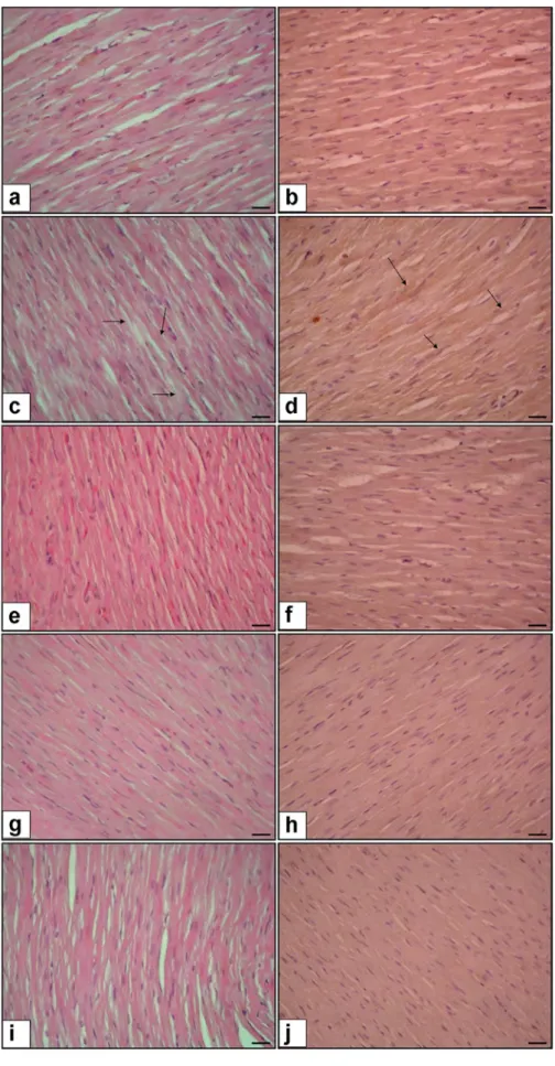
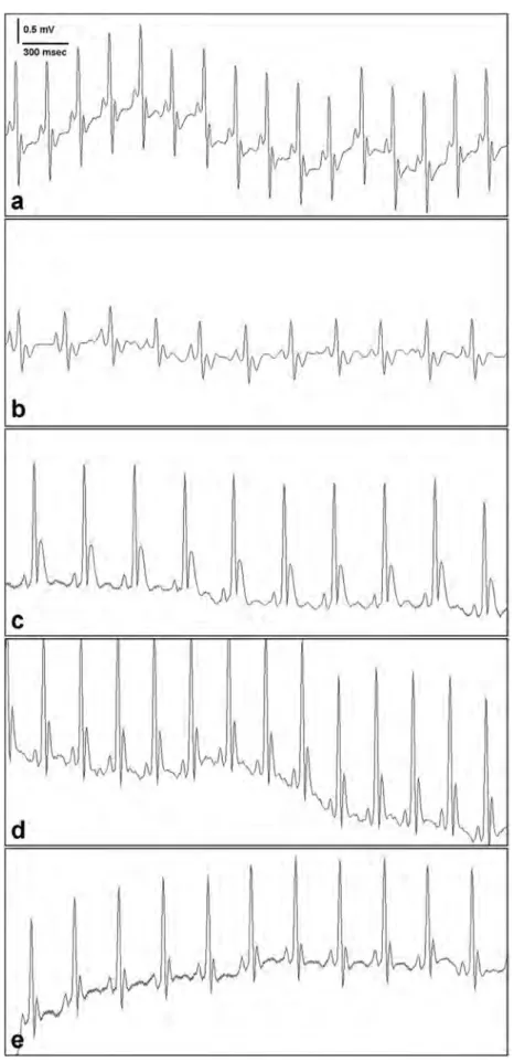
According to findings above, DXR + Saline group has the worst tissue integrity with the highest level of CASPASE-3 activity (14.6 ± 2.8) (p < 0.000001) which is an indicator of apoptosis. As indicators of tissue damage, increased oxida-tive and inflammatory state; ECG QRS voltage amplitude was found to be lowest (0.10 ± 0.004 mV) (p < 0.000001) plasma MDA (328.1 ± 43.2 nm) (p < 0.001), TNF-alpha (46.9 ± 4.1 pg/ml) (p < 0.001), pentraxin-3 (4.7 ± 1.1 ng/ ml) (p < 0.001), Troponin T (3.3 ± 1.02 pg/ml) (p < 0.05), pro-BNP (32.5 ± 6.8 pg/ml) (p < 0.001) levels were found to be highest in this group. All three treatments were shown to ameliorate the toxic effect of doxorubicin in cardiac tis-sue with the best results in DXR + OX group. It is shown that tissue integrity, cellular apoptosis rate, QRS ampli-tude, plasma MDA, TNF-alpha, pentraxin-3, Troponin T, pro-BNP levels were closer to normal group in DXR + OX group than DXR + LIR and DXR + G-CSF groups. Among latter 2 groups, DXR + LIR group exerted better results than DXR + G-CSF group (Figs. 1, 2; Tables 1, 2, and 3).
Discussion
DXR has been used for the treatment of carcinomas and hematologic malignancies since discovery of the drug [12]. After started to be used extensively, physicians detected acute and chronic phase cardiotoxicity related to drug. In acute term, QT interval prolongation and arrhythmias were seem to be frequent. On the other hand with cumulative doses, they discovered that drug caused cardiomyopathy and heart failure with a dose-dependent manner especially in elderly. Chronic DXR-associated cardiotoxicity was found to develop about after 10 years of DXR usage [13]. Swain et al. [14] showed a cumulative dose of 400 mg/m2 of DXR was related with a 5% increased risk of HF. This risk increased by 26% and 48% with the use of 550 and 700 mg/ m2 of DXR, respectively. In another study, at 500–550 mg/ m2 dose of DXR caused cardiomyopathy in 4% of patients, at the dose of 551–600 mg/m2 this rate increased to %18 [15].
Fig. 1 Cardiac tissue histopa-thology H& E (a, c, e, g, i) and CASPASE-3 immunoexpression in cardiomyocytes (b, d, f, h, j) (×40 magnification). a and b Normal cardiomyocytes, c and d DXR + saline group increased CASPASE 3 immunoexpres-sion (arrow) and disintegration of damaged cardiomyocytes (arrow), e and f DXR + LIR Group decreased CASPASE 3 immunoexpression and cardiomyocytes showing regular sequence, g and h DXR + OX Group decreased CASPASE 3 immunoexpression and cardiomyocytes showing regular sequence, i and j DXR + G-CSF Group decreased CASPASE 3 immunoexpression
Fig. 2 ECG results of all groups. Derivation I ECG, a Normal Group, b DXR + saline group, c DXR + LIR group, d DXR + OX group, e DXR + G-CSF Group
I~
300 mseca
b
DXR-induced cardiotoxicity has been investigated since dis-covery of the drug. Several mechanisms including increased oxidation of lipids [16], excessive producement of reactive oxygen species (ROS) [17], increased inflammation, DXR-induced DNA damage [18] and apoptosis [19] have been suggested to enlight this issue.
Among all of them, oxidative damage remains to be the pivotal mechanism. DXR accumulates in mitochondria and causes electron chain impairment which causes excessive production of superoxide radicals O2∙− and hydrogen per-oxide (H2O2). In the presence of Fe+2, O2∙− is converted to hydroxyl radical (OH∙). These reactive oxygen species cause lipid peroxidation and DNA damage which leads cellular apoptosis and autophagy. While ROS damage the cell, anti-oxidant enzymes such as superoxide dismutase (SOD), cata-lase (CAT), glutathione peroxidase (GPx) and glutathione (GSH) try to defence organism by neutralizing these radicals with hydrogen element and producing H2O [17]. We tried to evaluate oxidative stress by measuring plasma MDA level and inflammation by measuring plasma pentraxin-3 and TNF-alpha levels in our study.
A second mechanism may be the trigger of apoptosis by DXR treatment in cardiomyocytes. The extrinsic apoptosis
machinery is valid for rat cardiomyocytes, too. DXR treat-ment was shown to increase expression of death recep-tors like Fas/FasL as well as TNFα/TNFR1 in rat cardio-myocytes by Wu et al. [20] and Niu et al. [21]. We tried to evaluate apoptosis rate in cardiac tissue by measuring microscopic CASPASE-3 immuno-expression level and define microscopic degree of tissue damage by scoring hemotoxilen–eosin staining slides. In addition, we recorded ECG form derivation I to asses clinical reflection of DXR-associated cardiotoxicity and measured QRS wave voltage amplitude as well as plasma Troponin T and pro-BNP levels which were proven to be reliable clinical indicators of car-diac tissue damage.
Oxytocin (OX) treatment caused the best results in our study. DXR + OX group has the most preserved tissue integrity, least immune expression level of CASPASE-3 (5.3 ± 0.9), (p < 0.001) the highest ECG QRS wave volt-age amplitude (0.21 ± 0.008 mV) (p < 0.00001) least plasma MDA (115.3 ± 19.8 nm) (p < 0.001), TNF-alpha (26.6 ± 3.05 pg/ml) (p < 0.001), pentraxin-3 (2.7 ± 0.9 ng/ml) (p < 0.001), Troponin T (1.4 ± 0.08 pg/ml) (p < 0.001), pro-BNP (11.1 ± 3.6 pg/ml) (p < 0.001) levels among all three treatment groups. OX was found to have beneficial effects
Table 1 CASPASE 3 immunoexpression results of all groups
*p < 0.000001, DXR + Saline group compared with Normal group
# p < 0.001, DXR + LIR or DXR + OX or DXR + G-CSF group compared with DXR +Saline group
Normal group DXR + saline group DXR + LIR group DXR + OX group DXR + G-CSF group
CASPASE 3 immunoexpression 1.2 ± 0.1 14.6 ± 2.8* 7.2 ± 1.3# 5.3 ± 0.9# 8.8 ± 2.4#
Table 2 ECG QRS amplitude results of all groups
*p < 0.000001, DXR + Saline group compared with Normal group
# p < 0.00001, DXR + LIR or DXR + OX or DXR + G-CSF group compared with DXR +Saline group
Normal group DXR + Saline group DXR + LIR group DXR + OX group DXR + G-CSF group ECG QRS amplitude (mV) 0.24 ± 0.002 0.10 ± 0.004* 0.19 ± 0.003# 0.21 ± 0.008# 0.16 ± 0.007#
Table 3 Plasma MDA, TNF-alpha, Pentraxin-3, Troponin T and pro-BNP results of all groups
**p < 0.001, DXR + Saline group compared with Normal group *p < 0.05, DXR +Saline group compared with Normal group
## p < 0.001, DXR + LIR or DXR + OX or DXR + G-CSF group compared with DXR +Saline group # p < 0.01, DXR + LIR or DXR + OX or DXR + G-CSF group compared with DXR + Saline group
Normal group DXR + saline group DXR + LIR group DXR + OX group DXR + G-CSF group Plasma MDA (nm) 43.8 ± 6.01 328.1 ± 43.2** 215.7 ± 28.4## 115.3 ± 19.8## 278.8 ± 36.1#
Plasma TNF-alpha (pg/ml) 17.6 ± 2.3 46.9 ± 4.1** 34.1 ± 5.1## 26.6 ± 3.05## 38.9 ± 6.3
Plasma Pentraxin-3 (ng/ml) 1.6 ± 0.08 4.7 ± 1.1** 3.3 ± 1.04# 2.7 ± 0.9## 4.1 ± 1.5
Pro-BNP (pg/ml) 4.15 ± 0.9 32.5 ± 6.8** 16.5 ± 4.2# 11.1 ± 3.6## 19.5 ± 7.2##
on experimental Parkinson rat model [7], on experimental ovarian ischemia–reperfusion rat model [22], on experimen-tal Cisplatin-induced neurotoxicity rat model [23], and on experimental diabetic polyneuropathy rat model [24]. In all these literature, it was suggested that OX exerts its cyto-protective effect via anti-inflammatory features. OX prevents neutrophil infiltration of damaged tissue, decreases TNF-alpha and IL-6 levels, reduces ROS levels and lipid peroxi-dation. In addition, OX attenuates NADPH-dependent super-oxide production and has antioxidant effects on endothelium and vascular smooth muscle. Consistent with the previous literature, we found that OX treatment led decreased oxida-tive, apoptotic and inflammatory activity in DXR-induced cardiomyopathy rat model as well as provided better tissue integrity and better results in clinically relevant measures of ECG assessment, plasma Troponin T and pro-BNP levels.
Liraglutide (LIR) is shown to restore myocardial tis-sue integrity, reduce CASPASE-3 immunoexpression (7.2 ± 1.3) (p < 0.000001), preserve QRS voltage amplitude (0.19 ± 0.003 mV) (p < 0.000001) reduce plasma MDA (215.7 ± 28.4 nm) (p < 0.01), TNF-alpha (34.1 ± 5.1 pg/ ml) (p < 0.01), pentraxin-3 (3.3 ± 1.04 ng/ml) (p < 0.01), Troponin T (2.1 ± 0.09 pg/ml) (p < 0.01), pro-BNP (16.5 ± 4.2 pg/ml) (p < 0.01) levels when compared to DOX +saline group and all of the results were better than DOX + G-CSF group (p < 0.01). Liraglutide (LIR) was proven to have antioxidant effects in previous studies. It was shown that LIR has beneficial effects on non-alcoholic fatty liver disease animal model [25], LIR reduces SOD activity in macrophage derived foam cell culture model [4] and LIR attenuates DXR-induced elevation of proinflam-matory cytokines like IL-1 and TNF-alpha. Li et al. [26] discovered that DXR treatment suppresses AMPK activ-ity which leads p53 accumulation, apoptosis and elevated producing of ROS, eventually cell death. Abbas et al. [27] showed that LIR attenuates DXR-induced AMPK blockage thus ROS production and apoptosis of cell. They found that CASPASE-3 level induced by DXR can be reduced by LIR treatment in rats. Inhibition of apoptosis by reducing CAS-PASE-3 expression, increase in TNF-alpha and ROS levels, and increase in BCl2 expression were suggested compounds of this beneficial mechanism in their paper.
In our study, LIR is proven to reduce proinflammatory cytokines as TNF-alpha, reduce lipid peroxidation prod-ucts as MDA, reduce Troponin T, pentraxin-3 and pro-BNP levels and attenuates QRS voltage amplitude reduction and histopathologically shown cardiac tissue damage which are indicator of myocardium damage and heart failure, reduce CASPASE-3 level which is an indicator of apoptosis in DXR treated rats. Our findings are in consistent with pre-vious findings. Furthermore, we showed pentraxin-3 level reduction which is a novel marker for inflammatory cardiac disease and provided ECG assessment data.
DXR + G-CSF group has the worst preserved tissue integrity, highest immune expression level of CASPASE-3 (8.8 ± 2.4) (p < 0.001) the least ECG QRS wave voltage amplitude (0.16 ± 0.007 mV) (p < 0.00001) the highest plasma MDA (278.8 ± 36.1 nm) (p < 0.001), TNF-alpha (38.9 ± 6.3 pg/ml) (statistically not meaningful), pentraxin-3 (4.1 ± 1.5 ng/ml) (statistically not meaningful), Troponin T (2.04 ± 1.1 pg/ml) (p < 0.001), pro-BNP (19.5 ± 7.2 pg/ml) (p < 0.001) levels among all three treatment groups.
G-CSF is extensively used to treat neutropenia after chemotherapy in hematologic malignancies. Recently, lit-erature remarking cyto-protective and anti-inflammatory effects of G-CSF in some neurological diseases has been published [28].
G-CSF was shown to decrease infarct volume in an ischemic cerebral infarct rat model. Researchers suggested that G-CSF showed this effect by increasing stem cell pro-duction and migration to damaged tissue [8]. Studies of spine injury animal models were conducted to investigate G-CSF effect in damaged tissue and they revealed that G-CSF inhib-ited the apoptosis [9]. In a clinical study, patients with acute ischemic stroke treated with G-CSF for 12 months exhibited better results than patients who were not given G-CSF [29]. It was reported that G-CSF had anti-inflammatory effect by blocking synthesis of IL-1, TNF-alpha and had anti-apoptotic effect by increasing extracellular regulated kinase (ERK) signaling pathway in several other studies [30]. To date, there has been no another study which investigated the effect of G-CSF in DXR-induced cardiomyopathy animal model. Although results of DXR + G-CSF group were infe-rior when compared to DXR + OX and DXR + LIR groups, we found that G-CSF treatment in DXR cardiotoxicity revealed better results than DXR treatment alone and our findings were correlated with the previous literature.
Conclusion
Our findings suggest that with the best results in OX treat-ment group, all three agents including LIR and G-CSF attenuates tissue damage directly assessed by light micros-copy and indirectly measured by plasma Troponin T and pro-BNP levels, inhibits apoptosis, preserves ECG QRS wave amplitude, decreases inflammation and lipid peroxi-dation indirectly measured by plasma MDA, TNF-alpha and pentraxin-3 levels in the rat model of DXR-induced cardiomyopathy. Although our study did not specifically define the exact mechanism of action of selected drugs, we showed their beneficial effect directly in cardiac tissue and indirectly proved their anti-inflammatory effect by measur-ing biochemical markers of inflammation and heart failure. Previous findings in the literature supports these findings in different sets of animal disease models such as diabetes,
Parkinson, neuropathy. Clinical studies to assess effects of these drugs in mentioned diseases still remain to be con-ducted for more accurate data.
Compliance with Ethical Standards
Conflict of interest The authors did not declare any conflict of interest.
References
1. Octavia, Y., Tocchetti, C. G., Gabrielson, K. L., Janssens, S., Crijns, H. J., & Moens, A. L. (2012). Doxorubicin-induced cardiomyopa-thy: From molecular mechanisms to therapeutic strategies. Journal
of Molecular and Cellular Cardiology, 52, 1213–1225.
2. Takemura, G., & Fujiwara, H. (2007). Doxorubicin-induced car-diomyopathy from the cardiotoxic mechanisms to management.
Progress in Cardiovascular Diseases, 49, 330–352.
3. L’Ecuyer, T., Sanjeev, S., Thomas, R., Novak, R., Das, L., Campbell, W., et al. (2006). DNA damage is an early event in doxorubicin-induced cardiacmyocyte death. Am. J. Physiol Heart Circ. Physiol.,
291, H1273–H1280.
4. Wang, Y., & Yang, T. (2015). Liraglutide reduces oxidized DL induced oxidative stress and fatty degeneration in raw 264.7 cells involving the AMPK/SREBP1 pathway. J of Geriatric Cardiology,
12, 410–416.
5. Zhu, H., Zhang, Y., Shi, Z., Lu, D., Li, T., Ding, Y., et al. (2016). The neuroprotection of liraglutide against ischemia-induced apoptosis through the activation of the PI3k/Akt and MAP pathways. Scientific
Reports. https ://doi.org/10.1038/srep2 6859.
6. Biyikli, N. K., Tugtepe, H., Sener, G., Velioglu-Ogunc, A., Cetinel, S., & Midillioglu, S. (2006). Oxytocin alleviates oxidative renal injury in pyelonephritic rats via a neutrophil-dependent mechanism.
Peptides, 27, 2249–2257.
7. Erbas, O., Oltulu, F., & Taskiran, D. (2012). Amelioration of rote-none-induced dopaminergic cell death in the striatum by oxytocin treatment. Peptides, 38, 312–317. https ://doi.org/10.1016/j.pepti des.2012.05.026.
8. Song, S., Sava, V., Rowe, A., Li, K., Cao, C., Mori, T., et al. (2011). Granulocyte-colony stimulating factor (G-CSF) enhances recovery in mouse model of Parkinson’s disease. Neuroscience Letters, 487, 153–157.
9. Ha, Y., Park, H. S., Park, C. W., Yoon, S. H., Park, S. R., Hyun, D. K., et al. (2005). Granulocyte macrophage colony stimulating factor (GM-CSF) prevents apoptosis and improves functional outcome in experimental spinal cord contusion injury. Clinical Neurosurgery,
52, 341–347.
10. Wallace, K. B. (2003). Doxorubicin-induced cardiac mitochondri-onopathy. Pharmacology and Toxicology, 93, 105–115.
11. Demougeot, C., Marie, C., & Beley, A. (2000). Importance of iron location in iron-induced hydroxyl radical production by brain slices.
Life Sciences, 67, 399–410.
12. Dimitrakis, P., Romay-Ogando, M. I., Timolati, F., Suter, T. M., & Zuppinger, C. (2012). Effects of doxorubicin cancer therapy on autophagy and the ubiquitin-proteasome system in long-term cultured adult rat cardiomyocytes. Cell and Tissue Research, 350, 361–372.
13. Von Hoff, D. D., Layard, M. W., Basa, P., Davis, H. L., Jr., Von Hoff, A. L., Rozencweig, M., et al. (1979). Risk factors for doxorubicin-induced congestive heart failure. Annals of Internal Medicine, 91, 710–717.
14. Swain, S. M., Whaley, F. S., & Ewer, M. S. (2003). Congestive heart failure in patients treated with doxorubicin: a retrospective analysis of three trials. Cancer, 97, 2869–2879.
15. Lefrak, E., Pit’ha, J., Rosenheim, S., & Gottlieb, J. (1973). A clin-icopathologic analysis of adriamycin cardiotoxicity. Cancer, 32, 302–314.
16. Aleman, B. M. P., Moser, E. C., Nuver, J., Suter, T. M., Maraldo, M. V., Specht, L., et al. (2014). Cardiovascular disease after cancer therapy. European Journal of Cancer Supplements, 12, 18–28. 17. Aryal, B., Jeong, J., & Rao, V. A. (2014). Doxorubicin-induced
carbonylation and degradation of cardiac myosin binding protein C promote cardiotoxicity. Proceedings of the National Academy of
Sciences of the USA, 111(5), 2011–2016.
18. Sharma, P., Jha, A. B., Dubey, R. S., & Pessarakli, M. (2012). Reac-tive oxygen species, oxidaReac-tive damage, and antioxidaReac-tive defense mechanism in plants under stressful conditions. Journal of Botany,
2012, 26.
19. Kotamraju, S., Konorev, E. A., Joseph, J., & Kalyanaraman, B. (2000). Doxorubicin-induced apoptosis in endothelial cells and cardiomyocytes is ameliorated by nitrone spin traps and ebselen. Role of reactive oxygen and nitrogen species. Journal of Biological
Chemistry, 275, 33585–33592.
20. Wu, S., et al. (2002). Adriamycin-induced cardiomyocyte and endothelial cell apoptosis: In vitro and in vivo studies. Journal of
Molecular and Cellular Cardiology, 34, 1595–1607.
21. Niu, J., Azfer, A., Wang, K., Wang, X., & Kolattukudy, P. E. (2009). Cardiac-targeted expression of soluble fas attenuates doxorubicin-induced cardiotoxicity in mice. Journal of Pharmacology and
Experimental Therapeutics, 328, 740–748. https ://doi.org/10.1124/ jpet.108.14642 3.
22. Akdemir, A., Erbas, O., Gode, F., Ergenoglu, M., Yeniel, O., Oltulu, F., et al. (2014). Protective effect of oxytocin on ovarian ischemia-reperfusion injury in rats. Peptides, 55, 126–130.
23. Akman, T., Akman, L., Erbas, O., Terek, M. C., Taskiran, D., & Ozsaran, A. The preventive effect of oxytocin to cisplatin-induced neurotoxicity: An experimental rat model. BioMed Research
Inter-national, 2015, Article ID 167235
24. Erbas, O., Taşkıran, D., Oltulu, F., Yavaşoğlu, A., Bora, S., Bilge, O., et al. (2017). Oxytocin provides protection against diabetic poly-neuropathy in rats. Neurological Research, 39(1), 45–53. https ://doi. org/10.1080/01616 412.2016.12496 30.
25. Gao, H., Hu, L., Li, D., et al. (2013). Effects of glycagon like pep-tide-1 on liver oxidative stress, TNFα and TGF-β1 in rats with non-alcoholic fatty liver disease. Nan FangYi Ke DaXue Xue Bao, 33, 1661–1664. (In Chinese).
26. Li, Y., Hansotia, T., Yusta, B., Ris, F., Halban, P. A., & Drucker, D. J. (2003). Glycagon-like peptide-1 receptor signaling modulates beta cell apoptosis. Journal of Biological Chemistry, 278, 471–478. 27. Abbas, A. T. N., & Kabil, L. S. (2017). Liraglutide ameliorates
car-diotoxicity induced by doxorubicin in rats through the Akt/GSK-3β signaling pathway. Naunyn-Schmiedeberg’s Archives of
Pharmacol-ogy, 390, 1145–1153.
28. Shyu, W. C., Lin, S. Z., Yang, H. I., Tzeng, Y. S., Pang, C. Y., Yen, P. S., et al. (2004). Functional recovery of stroke rats induced by granulocyte colony-stimulating factor-stimulated stem cells.
Circu-lation, 110, 1847–1854.
29. Shyu, W. C., Lin, S. Z., Lee, C. C., Liu, D. D., & Li, H. (2006). Granulocyte colony-stimulating factor for acute ischemic stroke: A randomized controlled trial. CMAJ, 174, 927–933.
30. Hartung, T. (1998). Anti-inflammatory effects of granulocyte col-ony-stimulating factor. Current Opinion in Hematology, 5, 221–225. Publisher’s Note Springer Nature remains neutral with regard to jurisdictional claims in published maps and institutional affiliations.


