Contents lists available atScienceDirect
Respiratory Medicine Case Reports
journal homepage:www.elsevier.com/locate/rmcrCase report
A case report of reversible bronchiectasis in an adult: Pseudobronchiectasis
Canan Kucuk
a,∗, Mustafa Hamidullah Turkkani
b, Kemal Arda
caAnkara 29 Mayıs Hospital Anesthesiology and Reanimation Department, Turkey bAnkara Dr. Nafiz Körez Sincan Hospital Chest Disease Department, Turkey cSBU Gülhane Education and Research Hospital Radiology Department, Turkey
A B S T R A C T
Bronchiectasis is a sequel to permanent chronic airway inflammation and defined as ‘irreversible bronchial dilatation’. It is increasingly being recognized with the wider availability of high-resolution computed tomography. Children with noncysticfibrosis can be seen with improvement in bronchiectasis even resolution, as opposed to adults. We report a case of reversible bronchial dilatation in an adult as evidenced by high-resolution computed tomography, which demonstrates that, although quite rare in adults, spontaneous resolution may occur.
1. Introduction
Classical definition of bronchiectasis is permanent, irreversible, lo-calized abnormal dilatation of bronchi that ends withfibrosis [1–6]. There is no well-defined guide for diagnosis of bronchiectasis and handling of managing this issue is still difficult by all means. The reason for that may be the variety of causes. One of the causes of bronch-iectasis is transmural lung infections (pulmonary tuberculosis, pneu-monia). Chronic inflammation result with failure to clear mucoid se-cretions and destruction of lung tissue especially elasticfibers [7].
This also can be genetic. In cystic fibrosis there is more viscous sputum. In primary ciliary dyskinesia the problem is inability to clear sputum because of genetic reasons. The pathophysiology of bronch-iectasis is still not well defined.
The other causes are primary and secondary immunodeficiencies, connective tissue diseases, allergic bronchopulmonary aspergillosis. Bronchiectasis has also been associated with chronic obstructive pul-monary disease and asthma. There is high percentage of patients with no identifiable cause making the etiological diagnosis difficult and considered idiopathic.
During pneumonia, sometimes reversible dilatations may develop without destruction of segmental bronchi. These recurrent bronchial dilatations in acute infectious illnesses can be mistakenly defined as bronchiectasis. It is called pseudobronchiectasis for bronchial dilatation that develops frequently following an infection and resolves after the acute infection heals. It is usually cylindrical, rarely cystic [8]. 2. Case report
A 64-year-old female patient with HT and DM type 2, nonsmoker,
presented with acute cough, purulent sputum production and fever. Vitalfindings were stable except for the fever of 37.8 °C.
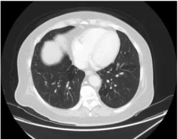
Inspiratory ral was present at the basal area of lung. Posteroanterior (PA) chest X-ray showed infiltration of the left lung including the costophrenic sinus. The patient had clarithromycin and ceftriaxone therapy started. Moxifloxacin treatment was initiated on continuing infiltration at control PA chest X-ray after 15 days. Computed tomo-graphy of thorax was performed. In CT examination, the structure in-cluding the pleura in the basal segment of the left lower lobe of the lung and the localized bronchiectasis appearance in the adjacent area were detected (Fig. 1). However, after 10 months fromfirst CT examination, the appearance of bronchiectasis was not detected in second CT ex-amination (Fig. 2).
3. Discussion
Bronchiectasis is a progressive, irreversible bronchial dilatation that has been shown to be resistant to long-term follow-up. Inflammation and infectious damage to the bronchial and bronchial walls result in a vicious cycle and ultimately bronchiectasis. In pneumonia, dilatations may occur in bronchi. However, these dilatations are temporary and return to normal with treatment. This type of bronchiectasis is called pseudobronchiectasis. It disappears in the following months of pneu-monia. Reversible bronchiectasis with bronchography is reported. But reversible bronchiectasis with CT/HRCT is rarely reported [9].
The definition of pseudobronchiectasis in early recycling remains uncertain in the pathogenesis of bronchiectasis; either it is a different clinical situation ora misinterpretation of radiology [10]. Pseudo-bronchiectasis may represent an early stage in the pathogenesis of bronchiectasis. On the contrary, pseudobronchiectasis may be an
over-https://doi.org/10.1016/j.rmcr.2019.03.002
Received 27 November 2018; Received in revised form 22 February 2019; Accepted 2 March 2019 ∗Corresponding author.
E-mail address:[email protected](C. Kucuk).
Respiratory Medicine Case Reports 26 (2019) 315–316
2213-0071/ © 2019 Published by Elsevier Ltd. This is an open access article under the CC BY-NC-ND license (http://creativecommons.org/licenses/BY-NC-ND/4.0/).
T
Downloaded for Anonymous User (n/a) at Baskent University from ClinicalKey.com by Elsevier on February 25, 2021. For personal use only. No other uses without permission. Copyright ©2021. Elsevier Inc. All rights reserved.
read of radiology [11].
‘Irreversible dilatation of the bronchi’ is the definition of bronch-iectasis and reversible form has been seen rarely but in 6 non-cystic fibrosis patients bronchiectasis is reversed and in 8 patients improve-ment was seen in pediatric group that is reviewed by Gaillard et al. Of course a single study would not be sufficient for explanation of com-plete reversal [12].
We think that careful interpretation of bronchiectasis in the as-sessment of CT/HRCT of patients with brief symptoms of pneumonia and it should be mentioned in the report that may be pseudo-bronchiectasis. All the broncheactesis are not permanent after infection and should be followed up.
Conflict of interest statement There is no conflict of interest.
Appendix A. Supplementary data
Supplementary data to this article can be found online athttps:// doi.org/10.1016/j.rmcr.2019.03.002.
References
[1] L.M. Reid, Reduction in bronchial subdivision in bronchiectasis, Thorax 5 (3) (1950) 233–247.
[2] M.A. Morechi, S.B. Fiel, An update on bronchiectasis, Curr. Opin. Pulm. Med. 1 (1995) 119–124.
[3] E.Y. Kang, R.R. Miller, N.L. Müller, Bronchiectasis: comparison of preoperative thin-section CT and pathologicfindings in resected specimens, Radiology 195 (3) (1995) 649–654.
[4] A.F. Barker, Bronchiectasis, N. Engl. J. Med. 346 (2002) 1383–1393. [5] N. Müller, R. Fraser, K. Lee, T. Johkoh, Large airway disease, in: N. Müller,
R. Fraser, K. Lee, T. Johkoh (Eds.), Diseases of the Lung: Radiologic and Pathologic Correlations,first ed., Lippincott Williams and Wilkins, Philadelphia, 2003, pp. 266–286.
[6] P.T. King, S.R. Holdsworth, N.J. Freezer, E. Villanueva, P.W. Holmes, Characterisation of the onset and presenting clinical features of adult bronch-iectasis, Respir. Med. 100 (2006) 2183–2189.
[7] P.J. Cole, Inflammation: a two-edged sword–the model of bronchiectasis, Eur. J. Respir. Dis. Suppl. 147 (1986) 6–15.
[8] N.E. Wiseman, The diagnosis of foreign body aspiration in childhood, J. Pediatr. Surg. 19 (1984) 531–535.
[9] V.L. Yap, M.L. Metersky, Reversible bronchiectasis in an adult: a case report, J. Bronchol. Interv. Pulmonol. 19 (4) (2012) 336–337.
[10] A.K. Aung, P.J. Thompson, B.M. Teh, C. McGrath, Pseudobronchiectasis after per-tussis and mycoplasma infection, Am. J. Respir. Crit. Care Med. 1 (3) (2012) 292–294 186.
[11] R. Agarwal, Bronchiectasis in acute pneumonia…pseudobronchiectasis, Chest 132 (2007) 2054–2055.
[12] E.A. Gaillard, H. Carty, D. Heaf, R.L. Smyth, Reversible bronchial dilatation in children: comparison of serial high-resolution computer tomography scans of the lungs, Eur. J. Radiol. 47 (3) (2003) 215–220.
Fig. 1. First CT examination reveals bronchiectasis and peribronchial in filtra-tion on left lower lobe of the lung (arrow).
Fig. 2. After 10 months on control CT examination, bronchiectasis appearance could not be seen.
C. Kucuk, et al. Respiratory Medicine Case Reports 26 (2019) 315–316
316
Downloaded for Anonymous User (n/a) at Baskent University from ClinicalKey.com by Elsevier on February 25, 2021. For personal use only. No other uses without permission. Copyright ©2021. Elsevier Inc. All rights reserved.