Address for Correspondence: Dr. Uğur Abbas Bal, Başkent Üniversitesi Tıp Fakültesi, Kardiyoloji Anabilim Dalı, Fevzi Çakmak Cad. 10. Sok. No: 45, 06490, Bahçelievler, Ankara-Türkiye
Phone: +90 312 212 68 68 Fax: +90 312 223 86 97 E-mail: [email protected] Accepted Date: 08.04.2014 Available Online Date: 03.06.2014
©Copyright 2015 by Turkish Society of Cardiology - Available online at www.anakarder.com DOI:10.5152/akd.2014.5473
A
BSTRACTObjective: In healthy women, there is a progressive age-related increase in myocardial mass that is not seen in their male counterparts and occurs primarily in postmenopausal women. Raloxifene is a selective estrogen receptor modulator that has estrogenic actions on bone and the cardiovascular system. The aim of this study was to investigate the effect of raloxifene on myocardial hypertrophy in postmenopausal patients. Methods: A total of 22 postmenopausal osteoporotic women were included in this open-label, randomized, prospective, controlled study. Patients were randomized into two groups: 11 of the patients (group 1) were treated with raloxifene 60 mg/day, and the other 11 patients (group 2) were defined as the control group. Quantitative 2-dimensional and M-mode echocardiographic examination was performed in all patients at the beginning and repeated at the end of the 6-month follow-up period. Left ventricle mass (LVM) and left ventricle mass index (LVMI) were calculated for all patients.
Results: The mean age of the patients was 57.2±3.9 years, and baseline clinical characteristics and echocardiographic parameters were similar between the two groups. After 6 months of raloxifene treatment, there was no difference in echocardiographic parameters of LVM and LVMI compared with the control group (201.2±25.9 gr vs. 169.7±46.2 gr, p=0.14 and 120.4±25.9 gr/m2 vs. 105.5±26.3 gr/m2, p=0.195, respectively). There
was also no significant difference in LVM and LVMI in the within-group analysis of both groups.
Conclusion: Raloxifene therapy does not affect myocardial hypertrophy in postmenopausal women after 6 months of treatment. (Anatol J Cardiol 2015; 15: 480-4)
Keywords: raloxifene, echocardiography, hormone replacement therapy, left ventricle mass, left ventricle hypertrophy
Uğur Abbas Bal, İlyas Atar, Mesut Öktem*, Hulusi B. Zeyneloğlu*, Aylin Yıldırır, Esra Kuşcu*, Haldun Müderrisoğlu
Departments of Cardiology and *Obstetrics and Gynecology, Faculty of Medicine, Başkent University; Ankara-Turkey
The effect of raloxifene on left ventricular hypertrophy in postmenopausal
women: A prospective, randomized, and controlled study
Introduction
In postmenopausal women, left ventricular hypertrophy is common and is likely to be a strong cardiovascular risk factor compared with men, suggesting that estrogen has potential pro-tective effects in the cardiovascular system (1). There are sub-stantial gender differences in myocardial remodeling due to the influence of sex hormones. Left ventricular hypertrophy is asso-ciated with an increased risk of cardiovascular morbidity and mortality in women (1).
In the general population, there is a progressive age-related increase in myocardial mass in healthy women that is not seen in their male counterparts (2). These findings were confirmed in a healthy, non-obese, normotensive subset of the Framingham Heart Study (3). These studies demonstrate that the increase in myocardial mass occurs primarily in postmenopausal women.
Indeed, premenopausal women with essential hypertension have thinner posterior left ventricular walls, smaller left ven-tricular mass, and better cardiac function than age-matched men (4).
Elderly (postmenopausal) women with systolic hypertension or aortic stenosis have more concentric remodeling and better preserved left ventricular systolic function than their male coun-terparts both at rest and with exercise (5, 6). These data suggest that the female sex hormones estrogen and progesterone influ-ence myocardial hypertrophy and myocardial remodeling by suppressing cardiac hypertrophy and preserving myocardial function.
Selective estrogen receptor modulators (SERMs) have been approved as hormone replacement therapy (HRT) to prevent osteoporosis and improve lipid profiles in postmenopausal women without producing uterine proliferation (7). Raloxifene is
a second-generation SERM and exerts estrogen-agonistic effects on the cardiovascular system and bone (7). The benefi-cial effects of raloxifene on cardiovascular risks and events have been investigated in the MORE study, and it has been shown that raloxifene decreased the risk of cardiovascular events among the subset of 1035 women with increased car-diovascular risk at baseline, compared with placebo (8). However, in the RUTH study, raloxifene did not significantly affect the risk of coronary heart disease in patients with high risk for major coronary events based on established cardiovas-cular disease (9).
The knowledge about the effects of raloxifene on myocardial hypertrophy in postmenopausal women is scant in the literature, and we aimed to investigate the effect of raloxifene on myocar-dial hypertrophy in this study.
Methods
This study was designed as a prospective-randomized-con-trolled study according to the CONSORT statement, and institu-tional review board approval was obtained from the Ethics Committee of Başkent University. Subjects were recruited from the Menopause Clinic of the Department of Obstetrics and Gynecology. All postmenopausal [at least 12 months elapsed from the last spontaneous menstrual bleeding. and each partici-pant exhibited a serum follicle-stimulating hormone (FSH) level >40 U/L and an estradiol (E2) level <30 pg/mL] and osteoporotic [T-score for femoral neck or lumbar spine bone mineral density (BMD) measurements ≤2.5 standard deviation (SD) according to the results of dual-energy x-ray absorptiometry] women with endometrial thickness ≤5 mm in ultrasound imaging were evalu-ated for the study. Exclusion criteria were defined as: history of gynecologic malignancy, ischemic heart disease, heart failure, hypertrophic cardiomyopathy, aortic stenosis, thromboembo-lism, abnormal liver function, renal dysfunction, epilepsy, severe migraines, non-treated thyroid dysfunction, and patients treated with hormone therapy during the 3 months before the initiation of the study. A total of 22 postmenopausal osteoporotic women (age range, 51-70 years) were included in the study. A table of randomly generated numbers was used for randomization. Patients were randomized into two groups: group 1 (n: 11) was treated with raloxifene 60 mg/day, and group 2 (n: 11) was defined as the control group; all of the patients received 600 mg calcium daily + 400 IU vitamin D, an over-the-counter product. All subjects signed an informed consent form.
Hypertension was defined as systolic blood pressure ≥140 mm Hg, diastolic blood pressure ≥90 mm Hg, or both or the use of antihypertensive medication for the study purpose according to the 2013 guidelines of the European Society of Hypertension (ESH) and the European Society of Cardiology (ESC) task force for the management of arterial hypertension (10). Diabetes was defined as fasting glucose level >126 mg on at least 2 occasions or current use of anti-diabetic agents (11). Patients who were currently smoking cigarettes were considered smokers. BMI
was calculated as weight (kilogram or kg) divided by height (meter or m) squared (kg/m2).
Echocardiographic examination
All patients underwent an echocardiographic examination, including quantitative 2-dimensional and M-mode echocardio-graphic examination by means of an echocardiography unit with a 3.5-MHz transducer (Siemens Acuson Sequa c256, Munich, Germany). Echocardiograms were obtained by the same cardi-ologist after each patient was placed in the left lateral decubitus position. To reduce bias, the cardiologist was blinded to the patient groups. Measurements for M-mode-guided calculation of left ventricular mass (LVM) were taken in the parasternal short-axis view and were recorded at the papillary level. Left ventricular internal dimensions at end-diastole (LVIDd) and sep-tal (SWTd) and posterior wall thickness at end-diastole (PWTd) were measured. LVM was calculated by the American Society of Echocardiography (ASE)-recommended formula (12) LVM(g) = 0.8 × {1.04 × [(LVIDd + PWTd + SWTd)3 - (LVIDd)3]} + 0.6 and standardized to body size by dividing the raw LVM by body sur-face area as left ventricular mass index (LVMI). A second echo-cardiographic evaluation was conducted after the sixth month of the treatment, and all measurements were repeated.
Statistical analysis
The statistical package SPSS (Statistical Package for the Social Sciences, version 17.0, SSPS Inc, Chicago, Ill, USA) was used for statistical analyses. Continuous variables are expressed as mean±SD. All continuous variables were checked with Kolmogorov-Smirnov normality test to show their distributions. Continuous variables with normal distributions were compared using the unpaired student t-test. Continuous variables with abnormal distributions were compared using the Mann-Whitney U test. The chi-square test was used for categorical variables. P values of less than .05 were considered statistically significant. Changes in the echocardiographic measurements from baseline to those recorded at the end of the sixth month of the study were evaluated by means of the paired-sample t-test.
Results
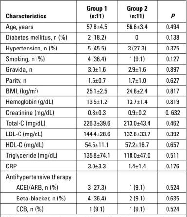
The mean age of the two groups was 57.8±4.5 vs. 56.6±3.4 years (p=0.494). Gravidity and parity of the patients were not dif-ferent between the groups (p=0.897 and p=0.627, respectively). Baseline laboratory parameters and clinical features were simi-lar in both groups (Table 1). All patients completed the follow-up period of 6 months.
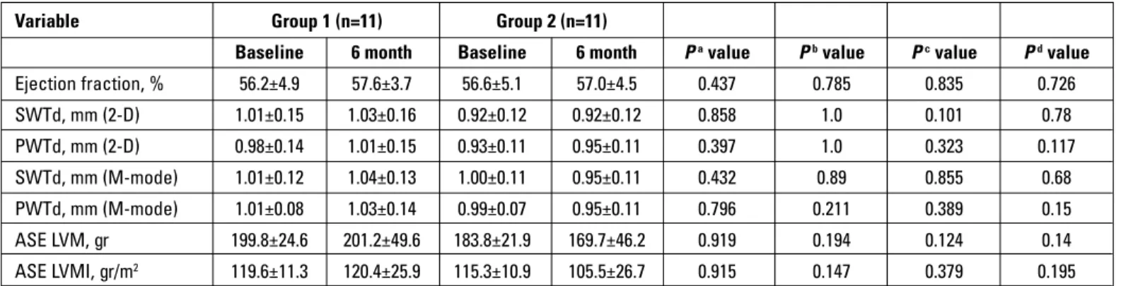
The baseline echocardiographic parameters were similar, and ejection fraction was normal in both groups (p=0.835). There was no significant within-group changes in echocardiographic parameters of LVM and LVMI at the end of the 6-month follow-up period in both grofollow-ups (grofollow-up 1: p=0.919 and p=0.915, respec-tively; group 2: p=0.194 and p=0.147, respectively). Also, these echocardiographic parameters (LVM and LVMI) did not differ
between the raloxifene group compared with control group at baseline and the 6th month (p=0.14 and p=0.195, respectively).
The other echocardiographic parameters were also similar in the between- and within-group analyses at the end of the study (Table 2).
Discussion
In this prospective, randomized, controlled study, we studied the association between raloxifene use and LVMI determined by echocardiography in postmenopausal women, and we did not find any effect of raloxifene on left ventricle hypertrophy in the 6-month follow-up period.
It is known that estrogen receptors are present in the myo-cardium (13), and there are controversial data in the literature about the effect of HRT on LVM and LVMI. In contrast with our study, Lim et al. (14) showed that women who were using HRT for more than 10 years had a significant reduction in septal and posterior left ventricular wall thickness when compared with controls. In an autopsy study, Olivetti et al. (15) demonstrated that myocyte number and volume are better preserved in women than in men. This intriguing study is supported by findings that estrogen may be important in the activation of the antiapoptotic
AKT (protein kinase B) kinase in the heart (16). Thus HRT, and particularly estrogen, positively affects myocardial hypertrophy and remodeling, and this mechanism seems to be the most plau-sible explanation for the improved survival with HRT in women with advanced heart failure (17).
However, some studies were consistent with our findings. Snabes et al. (18) examined the effect of E2 replacement ther-apy on cardiac structure and function in healthy postmeno-pausal women, and they found that E2 replacement therapy, which results in physiological serum concentrations, does not affect cardiac structure or function in normal postmenopausal women after 12 weeks of treatment. In a prospective, con-trolled study, Kessel et al. (19) studied the short- and interme-diate-term effects of low-dose HRT on echocardiographic parameters of cardiac function in healthy postmenopausal women, and they did not find any clinically relevant differenc-es in M-mode, quantitative 2-dimensional, and Doppler echo-cardiographic parameters within 15 months of 17 beta-estradi-ol and dydrogesterone treatment. Recently, Schwarz et al. (20) analyzed the association between HRT and either LVMI, deter-mined echocardiographically, in a cross-sectional study, and they did not find any significant associations with LVH after full adjustment. However, all of these studies were performed by using non-selective estrogens or did not consider what the patients were treated with.
Selective estrogen receptor modulators bind to and alter estrogen receptor function by inhibiting the binding of endoge-nous estrogens and can be conveniently divided into different categories (21). Tamoxifen is the prototype of SERMs, and it has been shown that tamoxifen can cause severe dilated cardiomy-opathy in animal models (22, 23). Raloxifene is a second-gener-ation SERM that is used for the same indicsecond-gener-ation but has a differ-ent mechanism of action by comparison with tamoxifen. Conversely, raloxifene increases myocyte contraction and was not associated with an increased risk of heart failure (24, 25). However, the knowledge in the literature about the effect of ral-oxifene on myocardial hypertrophy is scant.
Two studies performed with raloxifene were consistent with our study. In a mouse model, Westphal et al. (26) showed that raloxifene did not reduce myocardial hypertrophy but improved cardiac function in ovariectomized mice. Similarly, Vogelvang et al. (27) performed two studies with a follow-up period of 2 years. They found significant within-group changes in some parame-ters (LVMI, aortic peak flow velocity, and aortic velocity integral) from baseline, but interestingly, the same changes occurred in the placebo group; so, they found no effect on echocardiograph-ic parameters of left ventrechocardiograph-icular systolechocardiograph-ic function within 2 years of raloxifene treatment in healthy postmenopausal women. In our study, we also did not find any effect of raloxifene treatment on LVM and LVMI, but unlikely, we did not find any difference in within-group echocardiographic parameters. A small number of our patients were hypertensive and diabetic, and the distribution was similar between groups. Antihypertensive and antidiabetic treatment of patients during follow-up was unchanged, and all
Group 1 Group 2 Characteristics (n:11) (n:11) P Age, years 57.8±4.5 56.6±3.4 0.494 Diabetes mellitus, n (%) 2 (18.2) 0 0.138 Hypertension, n (%) 5 (45.5) 3 (27.3) 0.375 Smoking, n (%) 4 (36.4) 1 (9.1) 0.127 Gravida, n 3.0±1.6 2.9±1.6 0.897 Parity, n 1.5±0.7 1.7±1.0 0.627 BMI, (kg/m2) 25.1±2.5 24.8±2.4 0.817 Hemoglobin (g/dL) 13.5±1.2 13.7±1.4 0.819 Creatinine (mg/dL) 0.8±0.3 0.9±0.2 0. 632 Total-C (mg/dL) 226.3±39.6 213.0±43.4 0.462 LDL-C (mg/dL) 144.4±28.6 132.8±33.7 0.392 HDL-C (mg/dL) 54.5±11.1 57.2±16.7 0.657 Triglyceride (mg/dL) 135.8±74.1 118.0±47.0 0.511 CRP 3.0±3.3 1.4±1.4 0.176 Antihypertensive therapy ACEI/ARB, n (%) 3 (27.3) 1 (9.1) 0.524 Beta-blocker, n (%) 4 (36.4) 2 (9.1) 0.635 CCB, n (%) 1 (9.1) 1 (9.1) 0.524
ACEI - angiotensin-converting enzyme inhibitor; ARB - angiotensin receptor blocker; BMI - body mass index; CCB - calcium channel blocker; CRP - C-reactive protein; HDL-C - high-density lipoprotein cholesterol; LDL-C - low-density lipoprotein cholesterol; Total-C - total cholesterol.
Independent t-test and Mann-Whitney U-test for baseline characteristics. Parametric values given as mean±standard deviation
Table 1. Baseline clinical characteristics and laboratory parameters of the groups
patients’ blood pressure and blood glucose values did not change significantly. Therefore, the possible effect of these fac-tors on LVH was neglected during the study.
Study limitations
The small number of the subjects and the short follow-up period were the major limitations of our study.
Conclusion
Based on our results, we may conclude that raloxifene therapy has no effect on left ventricle hypertrophy in healthy postmenopausal women. But, there is still a need for larger and long-term studies to draw firm conclusions.
Conflict of interest: None declared. Peer-review: Externally peer-reviewed.
Authorship contributions: Concept - U.B., M.Ö., İ.A.; Design - A.Y., H.B.Z.; Supervision - E.K., H.M.; Resource - H.B.Z., A.Y., E.K., H.M.; Materials - M.Ö., İ.A.; Data collection &/or processing - U.B., İ.A., M.Ö.; Analysis and/or Interpretation - İ.A., A.Y.; Literature search - U.B., İ.A., M.Ö.; Writing - U.B., İ.A.; Critical review - H.B.Z., A.Y., E.K., H.M.
References
1. Agabiti-Rosei E, Muiesan ML. Left ventricular hypertrophy and heart failure in women. J Hypertens Suppl 2002; 20: 34-8.
2. Marcus R, Krause L, Weder AB, Dominguez-Meja A, Schork NJ, Julius S. Sex-specific determinants of increased left ventricular mass in the Tecumseh Blood Pressure Study. Circulation 1994; 90: 928-36. [CrossRef]
3. Dannenberg AL, Levy D, Garrison RJ. Impact of age on echocardio-graphic left ventricular mass in a healthy population. (The Framingham Study). Am J Cardiol 1989; 64: 1066-8. [CrossRef]
4. Garavaglia GE, Messerli FH, Schmieder RE, Nunez BD, Oren S. Sex differences in cardiac adaptation to essential hypertension. Eur Heart J 1989; 10: 1110-4.
5. Krumholz HM, Larson M, Levy D. Sex differences in cardiac adaptation to isolated systolic hypertension. Am J Cardiol 1993; 72: 310-3. [CrossRef]
6. Legget ME, Kuusisto J, Healy NL, Fujioka M, Schwaegler RG, Otto CM. Gender differences in left ventricular function at rest and with exercise in asymptomatic aortic stenosis. Am Heart J 1996; 131: 94-100. [CrossRef]
7. Delmas PD, Bjarnason NH, Mitlak BH, Ravoux AC, Shah AS, Huster WJ, et al. Effects of raloxifene on bone mineral density, serum cholesterol concentrations, and uterine endometrium in post-menopausal women. N Engl J Med 1997; 337: 1641-7. [CrossRef]
8. Barrett-Connor E, Grady D, Sashegyi A, Anderson PW, Cox DA, Hoszowski K, et al; MORE Investigators (Multiple Outcomes of Raloxifene Evaluation). Raloxifene and cardiovascular events in osteoporotic postmenopausal women: four-year results from the MORE (Multiple Outcomes of Raloxifene Evaluation) randomized trial. JAMA 2002; 287: 847-57. [CrossRef]
9. Barrett-Connor E, Mosca L, Collins P, Geiger MJ, Grady D, Kornitzer M, et al. Effects of raloxifene on cardiovascular events and breast cancer in postmenopausal women. Raloxifene Use for The Heart (RUTH) Trial Investigators. N Engl J Med 2006; 355: 125-37. [CrossRef]
10. Mancia G, Fagard R, Narkiewicz K, Redon J, Zanchetti A, Böhm M. Task Force Members. 2013ESH/ESC guidelines for the manage-ment of arterial hypertension: the Task Force for the Managemanage-ment of Arterial Hypertension of the European Society of Hypertension (ESH) and of the European Society of Cardiology (ESC). Eur Heart J 2013; 34: 2159-219. [CrossRef]
11. American Diabetes Association. Diagnosis and classification of diabetes mellitus. Diabetes Care 2014; 37: 81-90. [CrossRef]
12. Devereux RB, Alonso DR, Lutas EM, Gottlieb GJ, Campo E, Sachs I, et al. Echocardiographic assessment of left ventricular hypertrophy: com-parison to necropsy findings. Am J Cardiol 1986; 57: 450-8. [CrossRef]
13. Grohe C, Kahlert S, Lobbert K, Stimpel M, Karas RH, Vetter H, et al. Cardiac myocytes and fibroblasts contain functional estrogen receptors. FEBS Lett 1997; 416: 107-12. [CrossRef]
14. Lim WK, Wren B, Jepson N, Roy S, Caplan G. Effect of hormone replacement therapy on left ventricular hypertrophy. Am J Cardiol 1999; 83: 1132-4. [CrossRef]
Variable Group 1 (n=11) Group 2 (n=11)
Baseline 6 month Baseline 6 month Pa value Pb value Pc value Pd value
Ejection fraction, % 56.2±4.9 57.6±3.7 56.6±5.1 57.0±4.5 0.437 0.785 0.835 0.726 SWTd, mm (2-D) 1.01±0.15 1.03±0.16 0.92±0.12 0.92±0.12 0.858 1.0 0.101 0.78 PWTd, mm (2-D) 0.98±0.14 1.01±0.15 0.93±0.11 0.95±0.11 0.397 1.0 0.323 0.117 SWTd, mm (M-mode) 1.01±0.12 1.04±0.13 1.00±0.11 0.95±0.11 0.432 0.89 0.855 0.68 PWTd, mm (M-mode) 1.01±0.08 1.03±0.14 0.99±0.07 0.95±0.11 0.796 0.211 0.389 0.15 ASE LVM, gr 199.8±24.6 201.2±49.6 183.8±21.9 169.7±46.2 0.919 0.194 0.124 0.14 ASE LVMI, gr/m2 119.6±11.3 120.4±25.9 115.3±10.9 105.5±26.7 0.915 0.147 0.379 0.195
ASE - American Society of Echocardiography; LVM - left ventricular mass; LVMI - left ventricular mass index; PWTd - posterior wall thickness at end-diastole; SWTd - septal wall thickness at end-diastole
a. Paired sample t-test for within-group differences in group 1 b. Paired sample t-test for within-group differences in group 2 c. Independent t-test for baseline levels; group 1 vs. group 2 d. Independent t-test for 6-month levels; group 1 vs. group 2
15. Olivetti G, Giogano G, Corradi D, Melissari M, Lagrasta C, Gambert SR, et al. Gender differences and aging: effects on the human heart. J Am Coll Cardiol 1995; 26: 1068-79. [CrossRef]
16. Camper-Kirby D, Welch S, Walker A, Shiraishi I, Setchell KD, Schaefer E, et al. Myocardial Akt activation and gender: increased nuclear activity in females versus males. Circ Res 2001; 88: 1020-7.
[CrossRef]
17. Lindenfeld J, Ghali JK, Krause-Steinrauf HJ, Khan S, Adams K, Goldman S, et al; BEST Investigators. Hormone replacement thera-py is associated with improved survival in women with advanced heart failure. J Am Coll Cardiol 2003; 42: 1238-45. [CrossRef]
18. Snabes MC, Payne JP, Kopelen HA, Dunn JK, Young RL, Zoghbi WA. Physiologic estradiol replacement therapy and cardiac structure and function in normal postmenopausal women: a randomized, double-blind, placebo-controlled, crossover trial. Obstet Gynecol 1997; 89: 332-9. [CrossRef]
19. Kessel H, Kamp O, Kenemans P, Mijatovic V, van Baal WM, Visser CA, et al. Effects of 15 months of 17 beta-estradiol and dydroges-terone on systolic cardiac function according to quantitative and Doppler echocardiography in healthy postmenopausal women. Am J Obstet Gynecol 2001; 184: 910-6. [CrossRef]
20. Schwarz S, Obst A, Schwahn C, Völzke H, Schmidt CO, Dörr M, et al. Menopausal hormone therapy does not play a major role in left ventricular hypertrophy. Maturitas 2010; 66: 212-8. [CrossRef]
21. Osborne CK, Zhao H, Fuqua SA. Selective estrogen receptor modula-tors: structure, function and, clinical use. J Clin Oncol 2000; 18: 3172-86.
22. Hougen K, Aronsen JM, Stokke MK, Enger U, Nygard S, Andersson KB, et al. Cre-loxP DNA recombination is possible with only mini-mal unspecific transcriptional changes and without cardiomyopa-thy in Tg(alphaMHC-MerCreMer) mice. Am J Physiol Heart Circ Physiol 2010; 299: 1671-8. [CrossRef]
23. Chen PP, Patel JR, Powers PA, Fitzsimons DP, Moss RL. Dissociation of structural and functional phenotypes in cardiac myosin-binding protein C conditional knockout mice. Circulation 2012; 126: 1194-205.
[CrossRef]
24. Asp ML, Martindale JJ, Metzger JM. Direct, differential effects of tamoxifen, 4-hydroxytamoxifen, and raloxifene on cardiac myocyte contractility and calcium handling. PLoS One 2013; 8: e78768.
[CrossRef]
25. Grove EL, Abrahamsen B, Vestergaard P. Heart failure in patients treated with bisphosphonates. J Intern Med 2013; 274: 342-50.
[CrossRef]
26. Westphal C, Schubert C, Prelle K, Penkalla A, Fliegner D, Petrov G, et al. Effects of estrogen, an ERa agonist and raloxifene on pressure overload induced cardiac hypertrophy. PLoS One 2012; 7: e50802.
[CrossRef]
27. Vogelvang TE, Mijatovic V, Kamp O, Netelenbos JC, Neele SJ, Pines A, et al. Neither long-term treatment with raloxifene nor hormone replacement therapy modulate cardiac function in healthy post-menopausal women: two randomized, placebo-controlled, 2-year studies. Am J Obstet Gynecol 2002; 186: 729-36. [CrossRef]